Carotid Cavernous Fistula: Red Eye, Pulsing Noise, and Endovascular Treatment

Carotid cavernous fistula is an abnormal blood flow connection between the carotid artery system and the cavernous sinus. Common signs include a red eye, eye bulging, double vision, headache, and a pulsating whooshing sound.
Key Takeaways
- Carotid cavernous fistula is an abnormal blood flow connection between the carotid artery system and the cavernous sinus.
- Common signs include a red eye, eye bulging, double vision, headache, and a pulsating whooshing sound.
- Diagnosis usually relies on eye examination, brain and vessel imaging, and often cerebral angiography.
- Endovascular treatment is the main treatment approach for many patients.
- Prompt medical evaluation is important, especially if vision changes, severe pain, or neurological symptoms occur.
Carotid cavernous fistula is an abnormal connection between arteries and veins near the base of the skull, close to the eye socket. It often causes a red, bulging eye, a pulsing sound in the head, and visual symptoms, and it is commonly treated with minimally invasive endovascular techniques.
Overview
Carotid cavernous fistula is a rare vascular condition in which blood flows abnormally between the carotid artery system and the cavernous sinus, a vein-filled space located behind the eyes at the base of the skull. Because this area is closely connected to the veins of the eye socket and to nerves that control eye movement, the condition can affect both eye appearance and eye function.
In normal circulation, blood travels from arteries into smaller vessels and then into veins. In carotid cavernous fistula, this pattern is disrupted. High-pressure arterial blood enters the venous system too directly, raising pressure in the veins around the eye. This can lead to redness, swelling, eye bulging, and changes in vision.
Some carotid cavernous fistulas develop suddenly, while others appear gradually. Symptoms may affect one eye or, less commonly, both. Although the condition can sound alarming, effective diagnosis and treatment are available, and many people improve with timely specialist care.
Symptoms
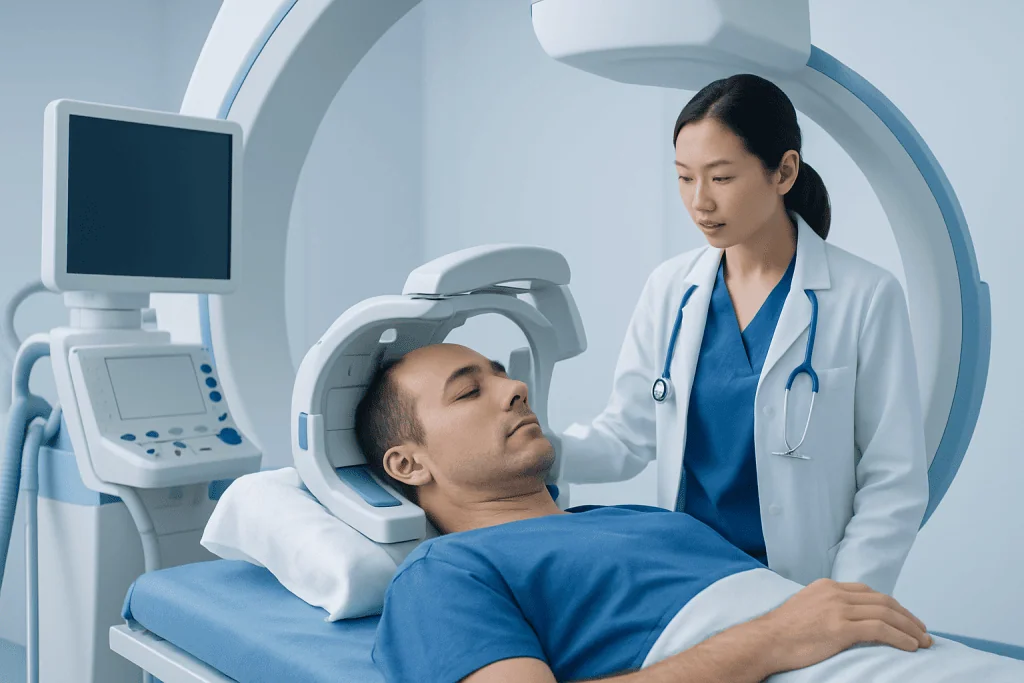
The symptoms of carotid cavernous fistula often involve the eye because the abnormal blood flow increases pressure in the veins that drain the orbit. One of the best-known signs is a red eye that does not behave like a typical eye irritation or infection. The eye may also look swollen or more prominent than usual.
People may notice a pulsing or whooshing sound in the head, sometimes described as a heartbeat-like noise in the ear. Doctors call this pulsatile tinnitus. This symptom can be especially noticeable at night or in quiet environments and may occur on the same side as the affected eye.
Other symptoms can include:
- Eye bulging or prominence
- Double vision
- Blurred vision
- Eye pain or pressure
- Headache
- Drooping eyelid
- Difficulty moving the eye normally
- Increased tearing or eye irritation
Symptoms vary depending on the size and type of the fistula. Direct, high-flow fistulas usually cause more sudden and obvious symptoms. Indirect, lower-flow fistulas may begin more subtly and can sometimes be mistaken for other eye conditions.
Causes and Risk Factors

Carotid cavernous fistulas are often grouped into direct and indirect types. A direct fistula is usually a straightforward connection between the internal carotid artery and the cavernous sinus. This form is often high-flow and commonly follows trauma, such as a facial injury, skull base injury, or, less often, rupture of an aneurysm.
Indirect fistulas involve smaller abnormal connections between branches of the carotid arteries and the cavernous sinus. These are often lower-flow and may occur spontaneously, especially in older adults. They may be associated with underlying weakness in blood vessel walls, high blood pressure, atherosclerosis, or connective tissue disorders.
Risk factors can include head trauma, previous vascular abnormalities, aneurysms, and conditions that affect the structure of blood vessels. However, some people develop this condition without a clear trigger. Because symptoms may overlap with other disorders, doctors may also consider related vascular causes of eye and brain symptoms, including brain aneurysm or other abnormalities of the blood vessels.
Diagnosis
Diagnosis begins with a careful medical history and physical examination. Doctors ask when symptoms started, whether there has been recent trauma, and whether the person hears a pulsating sound. An eye examination is important to assess redness, swelling, eye pressure, eye movements, and vision.
Imaging tests help confirm the diagnosis and define the blood vessel anatomy. These may include CT, MRI, CT angiography, or MR angiography. Such tests can show enlargement of the veins around the eye, swelling of eye muscles, or changes in the cavernous sinus that suggest abnormal blood flow.
The most detailed test is cerebral angiography, also called digital subtraction angiography. In many cases, this is considered the gold standard because it shows exactly where the fistula is, how blood is flowing, and which vessels are involved. This information is essential when planning endovascular treatment or deciding whether observation is appropriate.
Because symptoms can resemble other problems, doctors may also rule out conditions such as conjunctivitis, thyroid eye disease, orbital inflammation, glaucoma, or other vascular disorders affecting the brain and eye.
Treatment Options
Treatment depends on the type of fistula, the severity of symptoms, the effect on vision, and the overall pattern of blood flow. Some low-flow indirect fistulas may close on their own or may be monitored if symptoms are mild and vision is stable. However, many patients need active treatment, especially if there is visual decline, severe eye congestion, increased eye pressure, or troublesome neurological symptoms.
The main treatment for carotid cavernous fistula is minimally invasive endovascular therapy. In this approach, a specialist guides very thin tubes called catheters through the blood vessels to reach the fistula from inside the body. The abnormal connection can then be closed using devices or materials such as coils, balloons, or liquid embolic agents. This approach aims to stop the abnormal flow while preserving normal circulation as much as possible.
Depending on the anatomy, treatment may be performed through an arterial route, a venous route, or a combination of both. The exact technique is individualized after angiography. In some cases, care is coordinated with specialists in neurology and eye care to monitor nerve function, visual symptoms, and recovery.
Supportive care may include managing eye pressure, protecting the eye surface if the eyelids do not close fully, and treating discomfort. When a fistula is related to another vascular condition, further assessment may be needed, sometimes with input from teams experienced in brain and nerve surgery if complex anatomy or associated lesions are present.
Recovery, Follow-Up, and Self-Care
After treatment, many patients notice gradual improvement in eye redness, swelling, and the pulsating sound. Some symptoms improve quickly, while others, especially double vision or nerve-related eye movement problems, may take longer. Recovery depends on how long the fistula was present, how severe the venous congestion was, and whether vision or cranial nerves were affected before treatment.
Follow-up appointments are important to confirm that the fistula has closed and that symptoms are improving. Doctors may recommend repeat imaging or follow-up angiography in selected cases. Ongoing eye examinations help monitor visual acuity, eye pressure, and healing of the tissues around the eye.
At home, patients should follow their doctor’s advice closely. Practical self-care may include using prescribed eye drops, avoiding eye rubbing, resting as advised after procedures, and reporting any return of pulsating noise, worsening redness, or visual changes. It is also helpful to manage general vascular health by controlling blood pressure, not smoking, and attending routine medical care.
When to See a Doctor
A person should seek medical evaluation if a red eye is accompanied by eye bulging, double vision, a pulsing sound in the head, or unusual swelling around the eye. These features are not typical of simple eye irritation and deserve professional assessment. A timely diagnosis can help protect vision and reduce the chance of complications.
Urgent care is especially important if there is sudden vision loss, severe eye pain, rapidly increasing swelling, new weakness of the eye muscles, severe headache, or symptoms after head trauma. Emergency assessment is also important if neurological symptoms appear, such as confusion, facial weakness, or other sudden changes.
Carotid cavernous fistula is usually managed by a multidisciplinary team that may include interventional neuroradiologists, neurologists, neurosurgeons, and ophthalmologists. Acibadem International’s multidisciplinary specialists and JCI-accredited hospitals diagnose and treat this condition for international patients, with care tailored to the person’s symptoms and imaging findings.
Frequently asked questions
Is carotid cavernous fistula an emergency?
It can be urgent, especially if vision is changing, eye pressure is rising, symptoms started after trauma, or neurological symptoms are present. Not every case requires emergency treatment, but prompt medical assessment is important to decide the safest next step.
Can carotid cavernous fistula go away on its own?
Some low-flow indirect fistulas may close spontaneously, particularly if symptoms are mild. However, many cases need treatment, and a doctor should monitor the condition because ongoing abnormal blood flow can affect the eye and surrounding nerves.
What does the pulsing noise mean?
The pulsing or whooshing noise usually reflects abnormal blood flow near the cavernous sinus and veins around the eye. People often notice it in rhythm with their heartbeat, and it may lessen after successful treatment.
Will carotid cavernous fistula affect vision permanently?
Many people improve when the condition is diagnosed and treated in time. Permanent visual problems are more likely if there has been prolonged pressure on the eye, high eye pressure, or delayed treatment, which is why early evaluation matters.
How is carotid cavernous fistula treated without open surgery?
The most common treatment is endovascular therapy, a minimally invasive procedure performed from inside the blood vessels using catheters. The specialist closes the abnormal connection with materials such as coils or other embolic devices, often without the need for traditional open surgery.
What kind of doctor treats carotid cavernous fistula?
This condition is often treated by an interventional neuroradiologist or endovascular neurospecialist, working with ophthalmologists, neurologists, and sometimes neurosurgeons. Team-based care is helpful because the condition can affect blood vessels, cranial nerves, and vision at the same time.
References
- National Institute of Neurological Disorders and Stroke
- American Association of Neurological Surgeons
- American Academy of Ophthalmology
- Radiological Society of North America
- National Health Service
This article is for general information only and is not a substitute for professional medical advice. Please consult a qualified doctor about your individual situation.
Explore treatments in Turkey — costs, top hospitals & a free quote
JCI-accredited · board-certified surgeons · reply within 24h
Add us as a Preferred Source to see more of our trusted health content across Google Search, AI Overviews and Discover.
Check your numbers in seconds
BMI, calories, due date, blood pressure and 30+ more clinical calculators — free, instant, doctor-reviewed ranges.

Neurosurgery
Surgical treatment of the brain, spine and nervous system, including minimally invasive and functional procedures.
70 specialists in this unit