Cavernoma: Symptoms, Causes, and Treatment Options

A cavernoma is a blood vessel malformation that can occur in the brain or spinal cord. Many cavernomas are found incidentally and may never cause symptoms.
Key Takeaways
- A cavernoma is a blood vessel malformation that can occur in the brain or spinal cord.
- Many cavernomas are found incidentally and may never cause symptoms.
- Symptoms depend on the lesion’s size, location, and whether it has bled.
- MRI is the main imaging test used to diagnose and monitor cavernoma.
- Treatment may include observation, medicines for symptoms, or surgery in selected cases.
- Urgent medical attention is needed for sudden neurological symptoms, severe headache, or seizure.
A cavernoma is a cluster of fragile, abnormal blood vessels, most often found in the brain or spinal cord. Some cavernomas never cause problems, while others can lead to headaches, seizures, neurological symptoms, or bleeding and may need monitoring or treatment.
What is a cavernoma?
A cavernoma, also called a cerebral cavernous malformation, is a cluster of enlarged, irregular blood vessels with thin walls. These vessels form a small lesion that can look like a mulberry on imaging or under the microscope. Cavernomas most often develop in the brain, but they can also occur in the spinal cord.
Some people have a single cavernoma, while others have multiple lesions. A cavernoma may remain stable for years and never cause symptoms. In other cases, it can leak small amounts of blood or bleed more significantly, irritating nearby brain or spinal tissue and leading to symptoms.
Unlike some other blood vessel abnormalities, cavernomas are not usually visible on routine angiography because blood flow through them is slow. They are most commonly identified on magnetic resonance imaging. Depending on the individual case, a doctor may also discuss related conditions such as brain aneurysm or other vascular lesions when evaluating symptoms and imaging findings.
How a cavernoma can affect the body
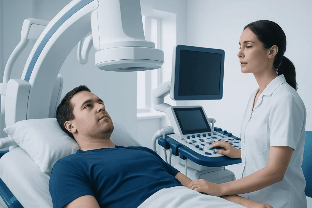
The effects of a cavernoma depend mainly on where it is located. A lesion in the outer part of the brain may be more likely to trigger seizures, while one deeper in the brain or in the brainstem may affect balance, vision, speech, swallowing, or limb strength. Cavernomas in the spinal cord can cause pain, numbness, weakness, or bowel and bladder symptoms if surrounding nerve pathways are involved.
A key feature of cavernoma is that symptoms can be absent, gradual, or sudden. Slow, repeated microscopic bleeding may cause irritation and leave deposits of old blood products around the lesion. This can increase the chance of seizures or persistent neurological symptoms, even if there has not been a large hemorrhage.
Not every cavernoma needs treatment. The overall approach is based on symptoms, location, bleeding history, the person’s age and general health, and how likely intervention is to improve safety or quality of life. Care is often planned with input from neurology, neuroradiology, and neurosurgery specialists.
Cavernoma symptoms

Many cavernomas cause no symptoms at all and are discovered during scans done for another reason. When symptoms do occur, they can vary widely. Some people experience one brief event, while others have recurrent or progressive problems.
Possible cavernoma symptoms include headaches, seizures, dizziness, balance problems, weakness, numbness, tingling, visual changes, speech difficulty, memory or concentration changes, and nausea or vomiting if bleeding causes pressure in surrounding tissue. A cavernoma in the spinal cord may cause back pain, limb weakness, sensory changes, or difficulty with walking.
Symptoms may appear after bleeding, but they can also occur without a major hemorrhage. For example, a cavernoma can trigger seizures because nearby brain tissue becomes irritated. It is important to remember that these symptoms can also occur in many other neurological conditions, so medical assessment is needed for an accurate diagnosis.
- Some cavernomas are completely silent.
- Seizures are a common presenting symptom for brain cavernomas near the cortex.
- Sudden new neurological symptoms may suggest recent bleeding and require urgent evaluation.
Causes and risk factors
The exact reason a cavernoma forms is not always known. Some are sporadic, meaning they occur without a clear inherited cause and often appear as a single lesion. Others are familial, which means they are linked to inherited gene changes and may be associated with multiple cavernomas in the brain or spinal cord.
A family history of cavernoma can increase the likelihood of the condition. In familial cases, more than one family member may be affected, and MRI may show multiple lesions. Genetic counseling or testing may be considered when there are several cavernomas, a strong family history, or diagnosis at a younger age.
Prior radiation treatment to the brain has also been associated with the later development of cavernous malformations in some patients. However, many people with a cavernoma have no identifiable risk factor. Having a cavernoma does not mean someone has done anything to cause it, and most daily activities are not the underlying reason it formed.
How cavernoma is diagnosed
MRI is the most important test for diagnosing a cavernoma. It can show the lesion’s size, location, and signs of old or recent bleeding. Specialized MRI sequences, including susceptibility-sensitive imaging, are especially helpful because they can detect blood products that may not be obvious on other scans.
In some situations, a CT scan may be done first, especially in emergency settings when bleeding is suspected. However, CT is less sensitive than MRI for many cavernomas. Conventional angiography often does not show a cavernoma clearly because these lesions are low-flow malformations.
Diagnosis also involves a neurological examination and a careful review of symptoms, seizure history, prior bleeding, medications, and family history. If seizures are part of the presentation, the doctor may arrange further neurological testing. When surgery is being considered, more detailed imaging and specialist evaluation help determine whether neurosurgical treatment is appropriate and safe.
Treatment options for cavernoma
Treatment for cavernoma is individualized. If a cavernoma is found incidentally and has not caused symptoms or bleeding, doctors may recommend observation with follow-up MRI scans. This approach helps track whether the lesion changes over time while avoiding unnecessary intervention.
When symptoms are present, treatment may focus first on symptom control. Anti-seizure medicines may be used for seizures, and supportive therapies can help with balance, weakness, or recovery after a neurological event. If bleeding has occurred, or if the cavernoma is causing repeated seizures or progressive deficits, surgery may be considered in selected patients.
Surgical removal is generally considered when the cavernoma is accessible and the expected benefit outweighs the risks. This decision depends heavily on location, especially if the lesion is in a sensitive area such as the brainstem. In some cases, the care team may use advanced MRI evaluation and related imaging to plan treatment and reduce risk. If the lesion has caused pressure on surrounding structures or ongoing neurological symptoms, surgery may offer the best chance of preventing further events.
Management often overlaps with the evaluation of other structural brain conditions, and specialists may compare findings with disorders such as brain tumor or different vascular abnormalities before confirming the treatment plan. For international patients, Acibadem International’s multidisciplinary specialists and JCI-accredited hospitals diagnose and treat neurological conditions including cavernoma using coordinated neurology and neurosurgery care.
Living with cavernoma: monitoring, self-care, and prevention
There is no guaranteed way to prevent a cavernoma from forming, especially when it is inherited. However, good follow-up care can help reduce complications and support quality of life. People with a diagnosed cavernoma should keep regular appointments, report any new symptoms promptly, and ask their doctor how often imaging is needed.
Self-care focuses on staying safe and managing symptoms. If seizures have occurred, the person should follow medical guidance about medication use, driving, swimming, heights, and other activities that could become dangerous during a seizure. Keeping a record of headaches, seizures, sensory changes, or episodes of weakness can help the care team assess whether the condition is stable.
General health habits also matter. Adequate sleep, stress management, avoiding missed medications, and discussing any blood-thinning medicines with a doctor may all be relevant depending on the person’s medical history. Patients should not stop prescribed medicines on their own, but they should tell clinicians about all medicines and supplements they use before any procedure, including brain surgery if it becomes part of care.
When to seek medical care
Anyone with symptoms that could suggest a neurological problem should seek medical care promptly. A doctor should evaluate new headaches that are unusual for the person, a first seizure, repeated vomiting with neurological symptoms, unexplained weakness or numbness, balance changes, new vision problems, or changes in speech.
Emergency care is especially important for sudden severe headache, loss of consciousness, a prolonged seizure, sudden one-sided weakness, facial drooping, confusion, or difficulty speaking. These symptoms can occur with cavernoma bleeding, but they can also signal stroke or another urgent brain condition and should never be ignored.
People who already know they have a cavernoma should contact their doctor if symptoms become more frequent, severe, or different from usual. Early reassessment can help determine whether new imaging or changes in treatment are needed.
Frequently asked questions
Is cavernoma a tumor?
No. A cavernoma is not a cancer and not a typical tumor. It is a vascular malformation, meaning a cluster of abnormal blood vessels.
Can a cavernoma go away on its own?
A cavernoma usually does not disappear on its own. Many remain stable for long periods, while some change over time or bleed. Follow-up imaging helps doctors monitor its behavior.
Does every cavernoma need surgery?
No. Many cavernomas can be monitored without surgery, especially if they are not causing symptoms. Surgery is usually considered only when the lesion is accessible and the expected benefits outweigh the risks.
Can cavernoma cause seizures?
Yes. Cavernomas in the brain can irritate nearby tissue and trigger seizures, particularly if they are near the brain’s surface or have bled. Seizures may be managed with medication, and some patients may need further treatment.
Is cavernoma hereditary?
Sometimes. Some cavernomas are sporadic and occur as a single lesion, while others are inherited and may appear as multiple lesions. A family history or multiple cavernomas may lead the doctor to discuss genetic testing or counseling.
Can people with cavernoma live normal lives?
Many people with cavernoma live full and active lives, especially when the lesion causes no symptoms or is well managed. The outlook depends on the lesion’s location, bleeding history, and whether seizures or neurological problems occur. Regular follow-up and individualized medical advice are important.
References
- National Institute of Neurological Disorders and Stroke
- National Organization for Rare Disorders
- Mayo Clinic
- NHS
- American Association of Neurological Surgeons
This article is for general information only and is not a substitute for professional medical advice. Please consult a qualified doctor about your individual situation.
Joint and spine care in Turkey — expert assessment & treatment
JCI-accredited · board-certified surgeons · reply within 24h
Add us as a Preferred Source to see more of our trusted health content across Google Search, AI Overviews and Discover.

More from the Health Library



Related Specialists

Dr. Aygun Babayeva
Pediatrics
Dr. Erald Bakıu
Inpatient Clinic Physicians
Dilara Nur Kara
Physical Medicine & Rehabilitation
Dr. Canan Genim Erdem
Gynecology & Obstetrics