Cavernous Malformation Surgery: Procedure, Recovery and Results

Surgery is not necessary for every cavernous malformation; observation is often appropriate for stable, symptom-free lesions. The decision depends on the lesion’s location, prior bleeding, symptoms, accessibility, and the person’s overall health.
Key Takeaways
- Surgery is not necessary for every cavernous malformation; observation is often appropriate for stable, symptom-free lesions.
- The decision depends on the lesion’s location, prior bleeding, symptoms, accessibility, and the person’s overall health.
- Recovery varies widely, but hospital recovery often takes days and rehabilitation may be needed after surgery near important brain or spinal cord pathways.
- Surgery can prevent future bleeding from the removed lesion, but it cannot eliminate risks from other cavernomas in people with multiple or familial lesions.
- A neurosurgical team uses detailed MRI planning and microsurgical techniques to protect nearby brain, spinal cord, and blood vessel structures.
Cavernous malformation surgery is a microsurgical procedure to remove a cavernoma from the brain or spinal cord. It may be considered when a lesion has bled, causes difficult-to-control seizures or progressive neurological symptoms, or can be reached with an acceptable level of risk.
Overview: what cavernous malformation surgery involves
Cavernous malformation surgery, also called cavernoma resection, is an operation to remove a cluster of abnormal, thin-walled blood vessels from the brain or spinal cord. These lesions are usually diagnosed on magnetic resonance imaging (MRI). Some cavernous malformations never cause symptoms and can be monitored safely, while others may bleed or affect nearby tissue.
The purpose of surgery is to remove the lesion while preserving neurological function. It is most often discussed after a symptomatic bleed, repeated bleeding, seizures that remain difficult to control with medicine, or steadily worsening symptoms caused by the cavernoma’s location. The decision is individualized because the natural risks of the lesion must be carefully weighed against the risks of operating.
A detailed understanding of the underlying condition is useful before making treatment decisions. Patients can learn more about cavernous malformations and how their location and symptoms may affect care planning.
Who may be a candidate for surgery?
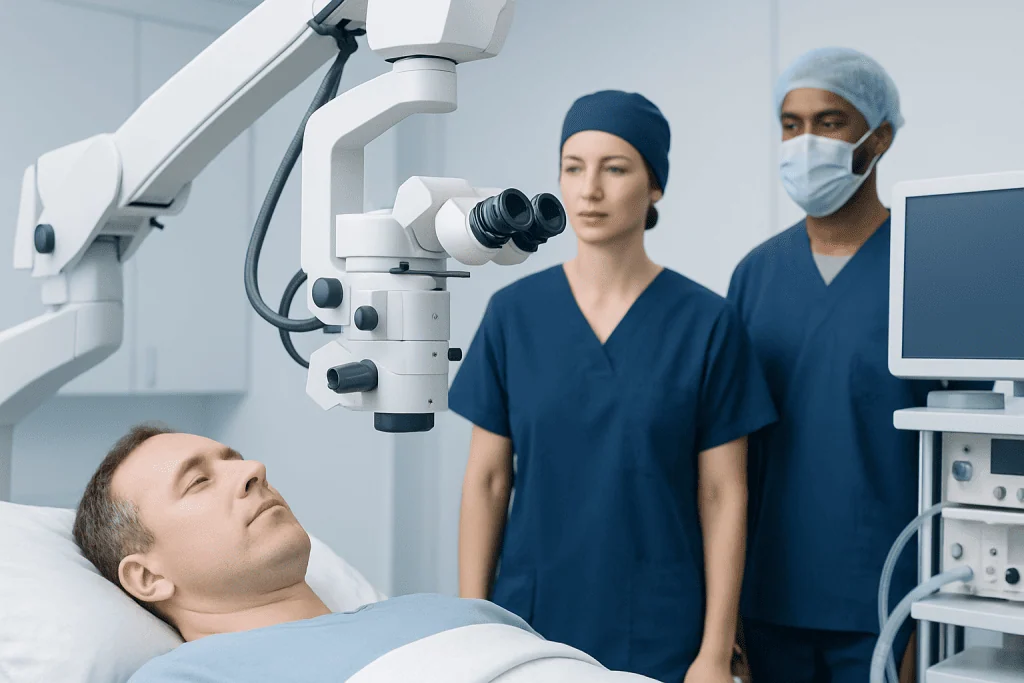
Not every person with a cavernoma needs an operation. For an incidental lesion that has not bled and is not causing symptoms, clinicians commonly recommend periodic clinical review and MRI monitoring instead. This approach avoids surgical risk when the expected benefit of removal is low.
Surgery may be more reasonable when a cavernoma has caused one or more symptomatic hemorrhages, produces seizures that do not respond adequately to anti-seizure medicines, or creates neurological problems such as weakness, altered sensation, balance difficulty, speech changes, or vision changes. A lesion that lies near the surface of the brain or in another surgically accessible area may be more suitable for removal than one deep within highly functional tissue.
The team considers MRI findings, prior bleeding events, symptom pattern, age, overall health, use of blood-thinning medicines, and the possible consequences of leaving the lesion untreated. For cavernomas in the brainstem, deep brain structures, or spinal cord, surgery requires particularly careful discussion because these areas contain densely packed pathways important for movement, sensation, breathing, swallowing, and coordination.
How cavernous malformation surgery is performed

Before surgery, the neurosurgical team reviews high-quality MRI scans and may use specialized imaging to map the lesion in relation to critical brain or spinal cord structures. Depending on the site, planning may also involve functional mapping, neurophysiological monitoring, and consultation with neurologists, neuroradiologists, anesthesiologists, and rehabilitation specialists.
During the operation, the patient receives general anesthesia. For a brain cavernoma, the surgeon performs a craniotomy, temporarily removing a small piece of skull to reach the lesion through the safest possible surgical corridor. For a spinal cavernoma, an approach to the relevant part of the spine is used. The surgeon works with an operating microscope and fine instruments to separate and remove the cavernoma while minimizing disruption to normal tissue.
Navigation technology and monitoring of nerve signals may help guide the procedure in selected cases. Once the lesion is removed, the surgeon controls bleeding, closes the protective tissue layers, and replaces the bone flap when a craniotomy has been performed. brain surgery planning is tailored to the individual lesion rather than following a single standard approach.
Removed tissue may be examined by a pathologist to confirm the diagnosis. After surgery, patients are closely monitored for neurological changes, pain control needs, seizures, and signs of complications.
Benefits and risks of cavernoma surgery
The principal potential benefit of complete removal is that the treated cavernoma should no longer bleed in the future. Surgery may also reduce or stop symptoms caused by pressure or repeated small hemorrhages. In people with cavernoma-related epilepsy, removal may improve seizure control, although some people still need anti-seizure medicine for a time after the operation.
As with any brain or spinal operation, cavernoma surgery carries risks. These can include bleeding, infection, blood clots, fluid leakage, seizures, stroke-like neurological deficits, headache, and anesthesia-related complications. Depending on the lesion’s location, there may be temporary or permanent changes in strength, sensation, balance, speech, vision, memory, swallowing, or coordination.
The level of risk is strongly influenced by anatomy. A small lesion near the brain surface may have a different risk profile from a lesion in the brainstem, thalamus, basal ganglia, or spinal cord. The surgeon should explain the likely benefits, the specific risks for that location, alternatives such as observation or medication, and the expected recovery needs before a decision is made.
How long does it take to recover from cavernous malformation surgery?
Recovery from cavernous malformation surgery varies according to the lesion’s location, the type of surgical approach, whether bleeding or neurological symptoms occurred before surgery, and the person’s baseline health. Many patients remain in hospital for several days, although a longer stay may be needed after complex surgery or if rehabilitation support is required.
During the first days and weeks, tiredness, headaches, scalp or incision discomfort, and reduced stamina are common. The care team may arrange follow-up brain imaging, wound review, and neurological assessments. Return to desk-based activities may be possible within several weeks for some people, while recovery after surgery in a critical brain or spinal cord region can take months.
Physical therapy, occupational therapy, speech and language therapy, or neuropsychological support may help patients regain function and adapt to temporary or lasting changes. It is important to follow individualized advice about driving, exercise, work, alcohol, anti-seizure medication, and return to travel. neurological rehabilitation can be an important part of recovery when weakness, balance changes, or cognitive symptoms are present.
What is the success rate of cavernoma surgery?
There is no single success rate that applies to all cavernoma operations. Outcomes differ substantially depending on whether surgery is performed for bleeding, seizures, or another symptom, as well as the lesion’s size, location, depth, and relationship to important neurological pathways.
When a cavernoma is completely removed, the risk of future hemorrhage from that particular lesion is generally eliminated. For seizure-related cavernomas, many patients experience improved seizure control after surgery, but the degree of improvement cannot be predicted with certainty. Seizure outcome may be influenced by how long seizures have been present, whether surrounding brain tissue has become epileptic, and whether the lesion can be fully removed safely.
For complex or deep lesions, success also means protecting function and achieving the best possible balance between removal and safety. A neurosurgeon can provide an individualized outlook after reviewing the MRI scans, symptoms, and results of relevant neurological testing.
How risky is cavernoma surgery? Can a cavernoma come back after surgery?
Cavernoma surgery can be low risk in some accessible locations and higher risk in others. Surgery near areas that control movement, speech, vision, sensation, balance, swallowing, or breathing may carry a greater possibility of neurological complications. Previous bleeding and the person’s existing symptoms may also affect both operative planning and recovery expectations.
A completely removed cavernoma does not usually grow back in the same location. However, an MRI may show a small remnant if complete removal was not safe or technically possible, and that residual tissue may still require monitoring. In people with familial cavernous malformation, new lesions can develop elsewhere over time; surgery on one lesion does not prevent this possibility.
Follow-up appointments and MRI scans help the team assess the surgical site and identify any other lesions that need observation. Ongoing care may also include seizure management or treatment for neurological symptoms. Acibadem International’s multidisciplinary specialists and JCI-accredited hospitals diagnose and treat cavernous malformations for international patients, with care plans coordinated across neurosurgery, neurology, imaging, and rehabilitation.
When to seek medical care
Urgent medical assessment is needed for sudden or severe headache, a new seizure, fainting, new weakness or numbness, facial drooping, confusion, trouble speaking, loss of balance, double vision, or a significant new change in vision. These symptoms can have several causes, but they need prompt evaluation, particularly in someone known to have a cavernous malformation.
Patients who have already been diagnosed should contact their clinician if they develop new or worsening neurological symptoms, a change in seizure frequency, medication side effects, or concerns during recovery after surgery. They should seek urgent advice for fever, increasing wound redness or drainage, severe worsening headache, persistent vomiting, or a sudden neurological change after an operation.
Regular follow-up is important even when symptoms are stable. A qualified neurologist or neurosurgeon can advise whether MRI surveillance, medication, rehabilitation, or further treatment is appropriate.
Frequently asked questions
Is cavernous malformation surgery always necessary?
No. Many cavernous malformations do not require surgery, especially when they are found incidentally and have not caused bleeding, seizures, or neurological symptoms. A neurologist or neurosurgeon may recommend observation with clinical follow-up and MRI scans instead.
How long does it take to recover from cavernous malformation surgery?
Hospital recovery often lasts several days, but the overall recovery period ranges from weeks to months. Recovery is usually longer when surgery involves the brainstem, spinal cord, or areas controlling important functions, or when rehabilitation is needed.
What is the success rate of cavernoma surgery?
A single success rate is not meaningful because surgical outcomes depend greatly on the lesion’s location and the reason for surgery. Complete removal generally prevents future bleeding from that specific lesion, while seizure improvement is possible but varies between individuals.
How risky is cavernoma surgery?
Risks include bleeding, infection, seizures, and new or worsened neurological symptoms. The individual risk may be relatively low for an accessible lesion but can be higher for cavernomas in deep or highly functional areas of the brain or spinal cord.
Can a cavernoma come back after surgery?
A cavernoma that has been completely removed does not usually recur at the same site. If a small part remains, it may need follow-up, and people with familial cavernous malformation can develop additional lesions elsewhere.
Will surgery cure cavernoma-related seizures?
Surgery may substantially improve seizure control, particularly when the cavernoma is the clear source of seizures and can be removed safely. However, some people continue to have seizures or need anti-seizure medicine after surgery, so follow-up with a neurologist remains important.
References
- American Association of Neurological Surgeons
- National Institute of Neurological Disorders and Stroke
- Alliance to Cure Cavernous Malformation
- Mayo Clinic
- European Stroke Organisation
This article is for general information only and is not a substitute for professional medical advice. Please consult a qualified doctor about your individual situation.
Joint and spine care in Turkey — expert assessment & treatment
JCI-accredited · board-certified surgeons · reply within 24h
Add us as a Preferred Source to see more of our trusted health content across Google Search, AI Overviews and Discover.