Cerebrospinal Fluid Leak MRI: Preparation, Procedure and Results

MRI uses magnetic fields and radio waves rather than ionizing radiation to assess possible CSF leaks. A brain MRI may reveal indirect signs of low CSF pressure, while spine MRI can help identify fluid collections and possible leak sites.
Key Takeaways
- MRI uses magnetic fields and radio waves rather than ionizing radiation to assess possible CSF leaks.
- A brain MRI may reveal indirect signs of low CSF pressure, while spine MRI can help identify fluid collections and possible leak sites.
- Not every CSF leak is visible on a routine MRI, so further specialized imaging may sometimes be needed.
- Preparation is usually simple, but patients should report implants, pregnancy, kidney concerns, and contrast allergies before the scan.
- Prompt medical assessment is important for suspected leaks after head trauma, surgery, or with symptoms of infection.
A cerebrospinal fluid leak MRI is a noninvasive scan that can show signs of leaked spinal fluid, its likely location, and related changes in the brain or spine. It is commonly used when symptoms such as clear nasal drainage, positional headache, hearing changes, or symptoms after injury or surgery raise concern for a CSF leak.
Overview: What Is a Cerebrospinal Fluid Leak MRI?
A cerebrospinal fluid leak MRI is an imaging examination used to evaluate whether cerebrospinal fluid (CSF) may be escaping from its normal enclosed space around the brain and spinal cord. CSF cushions the central nervous system, helps maintain a stable environment for nerve tissue, and carries away waste products. A leak can occur through a defect in the tissues surrounding the brain or spinal cord.
MRI, or magnetic resonance imaging, creates detailed pictures using a powerful magnet and radio waves. For a suspected cranial leak, a cerebrospinal fluid leak in brain MRI may look for fluid near the skull base and for brain changes associated with low CSF pressure. For suspected spinal leaks, MRI of the spine can assess for fluid outside the normal CSF space, meningeal diverticula, and other clues that guide further testing.
The scan is one part of a clinical assessment. Symptoms, medical history, laboratory analysis of nasal or ear fluid when present, and sometimes additional imaging are considered together. MRI is particularly valuable because it can evaluate the brain, soft tissues, spinal canal, and surrounding structures without radiation exposure.
Who May Need This Scan and How It Works
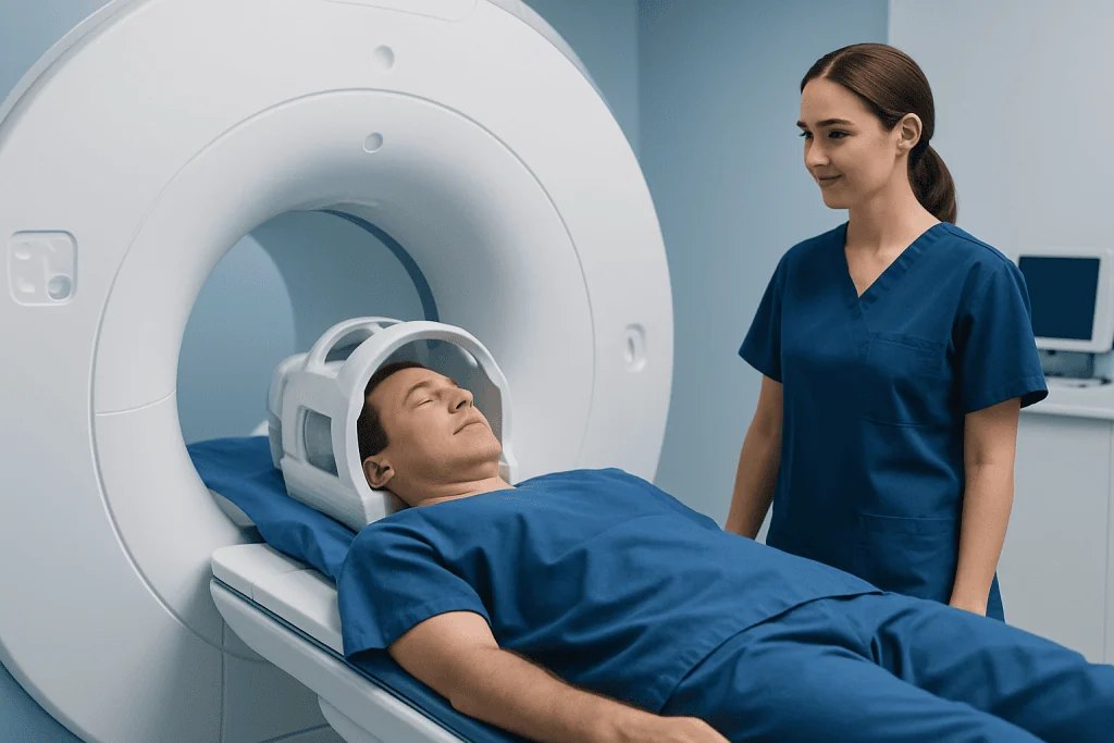
Doctors may recommend MRI when a person has symptoms that could be linked to a CSF leak. Cranial leaks may cause persistent clear, watery drainage from one side of the nose or an ear, especially after head injury, sinus surgery, or skull-base surgery. Spinal leaks often cause a headache that becomes worse when upright and improves when lying down, sometimes with neck pain, nausea, ringing in the ears, muffled hearing, or dizziness.
Some leaks arise after trauma, surgery, a lumbar puncture, or an epidural procedure. Others occur spontaneously, often in association with a small weakness in the dura, the protective membrane around the brain and spinal cord. Connective-tissue conditions and structural changes in the spine can increase susceptibility in some people, but a clear cause is not always found.
During MRI, different sequences highlight fluid, soft tissue, bone-related anatomy, and blood vessels in different ways. A csf leak MRI protocol is tailored to the symptoms and suspected location. The radiologist may recommend a brain MRI with and without contrast, a heavily T2-weighted MRI of the whole spine, or both. Contrast is not required for every examination, but it can help identify patterns associated with intracranial hypotension.
Preparation and Step-by-Step Procedure

Most people can eat, drink, and take usual medicines before a cerebrospinal fluid leak MRI unless their care team provides different instructions. Before the appointment, patients should tell the imaging team about any implanted medical device, metal fragment, cochlear implant, previous surgery, pregnancy, kidney disease, or prior reaction to MRI contrast. MRI staff check whether devices are MRI-safe before the scan begins.
Patients remove jewelry, hearing aids, credit cards, removable dental items, and other metal-containing belongings. They may change into a gown. If contrast is planned, a small intravenous line may be placed in the arm or hand. People with claustrophobia, severe pain, or difficulty remaining still can discuss supportive options with the referring doctor before the appointment.
For the scan, the patient lies on a padded table that moves slowly into the MRI scanner. A head coil may be used for brain imaging, and spine coils are used for spinal imaging. The examination is painless, although the machine makes repeated loud tapping or knocking sounds; ear protection is provided. The technologist can communicate throughout the scan, and it is important to remain as still as possible for clear images.
Timing varies with the imaging area and whether contrast is needed, but many examinations take approximately 30 to 60 minutes. Afterward, most patients can return to normal daily activity immediately. If sedating medication was used, someone else should arrange transport home and follow the post-sedation instructions provided.
Will a CSF Leak Show Up on MRI?
A CSF leak may show up on MRI, but not every leak is directly visible. MRI can identify fluid collections outside the normal spinal fluid space, changes in the tissues around the brain, and signs of low CSF volume or pressure. In cranial cases, it may reveal fluid signal in the sinuses or nearby structures, though CT may be better for defining a small bony defect in the skull base.
For a spinal leak, whole-spine MRI may show an epidural fluid collection or other indirect evidence of leakage. However, the precise site can remain difficult to identify, particularly when the leak is slow, intermittent, or caused by a CSF-venous fistula. A normal MRI therefore does not always exclude a leak when symptoms and clinical findings are strongly suggestive.
When MRI findings are inconclusive, specialists may consider other tests, such as CT myelography, dynamic myelography, digital subtraction myelography, or cisternography in selected cases. The choice depends on whether the suspected source is cranial or spinal and on the imaging findings already available. Results should be reviewed with a neuroradiologist, neurologist, neurosurgeon, or ENT specialist experienced in CSF leak assessment.
What Is the Protocol for MRI Spine CSF Leak Diagnosis?
An MRI spine CSF leak assessment commonly begins with imaging of the entire spine because a leak can occur away from the area where pain is felt. The protocol often includes sagittal and axial T1- and T2-weighted images, with heavily T2-weighted sequences that make fluid especially conspicuous. Fat-suppressed sequences may improve detection of fluid in the epidural space and surrounding soft tissues.
The exact csf leak MRI protocol varies by hospital, clinical question, scanner capability, and whether there is concern for a fast leak, slow leak, meningeal diverticulum, or CSF-venous fistula. In people with positional headache and concern for spontaneous intracranial hypotension, clinicians commonly combine brain MRI findings with whole-spine MRI. A csf leak MRI brain protocol may include pre- and post-contrast sequences to look for diffuse dural enhancement, venous changes, pituitary enlargement, or downward displacement of brain structures.
MRI generally serves as an important first-line, noninvasive study. If it shows a large epidural collection, the findings can help plan more targeted myelography. If it does not reveal a source but clinical suspicion remains, specialized dynamic imaging may still be appropriate. The imaging plan should be individualized rather than relying on a single test for every person.
Results, Benefits, Limitations and Possible Risks
A radiologist interprets the images and sends a report to the referring clinician, often within a few days, although timing varies by facility. The report may describe whether there are imaging features of intracranial hypotension, an epidural fluid collection, a suspected skull-base defect, or no evidence of a leak on that examination. The clinician explains what these findings mean in the context of the person’s symptoms and other test results.
The main benefits of MRI are its detailed soft-tissue images, absence of radiation, and ability to evaluate the brain and spine in a coordinated way. It can support a diagnosis, identify complications, and help determine whether observation, targeted treatment, or additional imaging is needed. MRI may also help distinguish CSF leak symptoms from other causes of headache or nasal drainage.
MRI is generally very safe when standard safety screening is followed. Some people find the confined space or noise uncomfortable. Gadolinium contrast, when used, has a low risk of allergic reaction and is considered carefully in people with severe kidney impairment or during pregnancy. The principal limitation is that MRI cannot detect every leak or always pinpoint the exact leak site.
How Long Does It Take for a Spinal CSF Leak to Heal?
Healing time for a spinal CSF leak varies considerably. Some small leaks improve with conservative measures such as rest, hydration, and avoiding activities that increase pressure or strain, under a clinician’s guidance. Symptoms may settle over days to weeks, but persistent symptoms should not be managed with prolonged self-treatment alone.
When a leak does not resolve or significantly affects daily life, doctors may recommend an epidural blood patch, which places a patient’s own blood into the epidural space to encourage sealing of the leak. Some people improve after one treatment, while others need repeat or targeted procedures. Identified structural causes may require specialized repair.
Recovery also depends on the type, location, and cause of the leak. A clinician may recommend temporary limits on heavy lifting, straining, or vigorous activity after treatment. Follow-up is important because symptoms can recur, and imaging or additional treatment may be needed when recovery is incomplete.
Acibadem International’s multidisciplinary neurology, neuroradiology, ENT, and neurosurgery specialists assess suspected CSF leaks and coordinate diagnostic imaging and treatment planning for international patients in JCI-accredited hospitals.
What Is the Paper Towel Test for CSF?
The paper towel test is an informal home observation sometimes discussed for clear nasal drainage. A person may notice that watery fluid forms a relatively clear spot on a tissue or paper towel, sometimes with a surrounding ring after drying. This appearance is not a reliable way to diagnose a CSF leak.
Normal nasal mucus, tears, saline sprays, allergies, and irritation can also produce clear fluid. The test cannot identify the source of fluid or confirm whether it contains CSF. Relying on it may delay appropriate medical assessment, particularly after a head injury or surgery.
If a clinician suspects a cranial CSF leak, a sample of drainage may be sent for a specific laboratory test, such as beta-2 transferrin or beta-trace protein testing, depending on local practice. Imaging is then used to investigate the possible source. People should avoid inserting tissue, swabs, or other objects deeply into the nose or ear to collect a sample.
When to Seek Medical Care
Medical assessment is advisable for persistent clear, watery drainage from the nose or ear, particularly if it begins after a head injury, sinus or ear surgery, or a procedure involving the spine. It is also important to seek care for a new severe positional headache, especially when it is accompanied by neck pain, nausea, hearing symptoms, or neurological changes.
Urgent medical care is needed for fever, stiff neck, confusion, worsening severe headache, repeated vomiting, seizure, weakness, altered vision, or loss of consciousness. These symptoms have many possible causes, but they warrant prompt evaluation. After significant head trauma, emergency assessment should not be delayed if fluid is leaking from the nose or ears.
Until medical advice is received, people with a possible leak should avoid forceful nose blowing, heavy lifting, and straining when possible. A qualified clinician can determine whether imaging, laboratory testing, referral, or treatment is appropriate and can explain the next steps based on the likely location and cause of the leak.
Frequently asked questions
Does an MRI confirm a cerebrospinal fluid leak?
MRI can provide strong evidence of a CSF leak and its effects, but it does not confirm every leak. Some leaks are intermittent, slow, or difficult to localize. Doctors may combine MRI with laboratory testing and specialized imaging when necessary.
Is contrast always used for a CSF leak MRI?
No. Contrast is not needed for every suspected CSF leak. It may be used for brain MRI when clinicians are looking for imaging signs associated with low CSF pressure or other conditions that can mimic a leak.
Can a brain MRI show a spinal CSF leak?
A brain MRI can show indirect effects of a spinal leak, such as signs of intracranial hypotension. It usually cannot identify the exact spinal location of the leak. Spine MRI and, when needed, specialized myelography are used to investigate the source.
Should a person fast before a CSF leak MRI?
Most MRI examinations do not require fasting. The imaging center may provide different instructions if sedation, contrast, or another procedure is planned. Patients should follow the instructions given with their appointment.
Is a suspected CSF leak an emergency?
It depends on the symptoms and cause. Clear drainage after a head injury, or symptoms such as fever, stiff neck, confusion, seizures, or worsening neurological symptoms, require urgent medical assessment. Ongoing positional headache or clear drainage without emergency symptoms should still be evaluated promptly.
Can a CSF leak heal without surgery?
Some CSF leaks heal with conservative care or minimally invasive treatment, such as an epidural blood patch for selected spinal leaks. Others persist because of an underlying structural opening and may need targeted repair. The appropriate approach depends on the leak's location, cause, and severity.
References
- American College of Radiology
- Radiological Society of North America
- National Institute of Neurological Disorders and Stroke
- American Association of Neurological Surgeons
- International Headache Society
This article is for general information only and is not a substitute for professional medical advice. Please consult a qualified doctor about your individual situation.
Joint and spine care in Turkey — expert assessment & treatment
JCI-accredited · board-certified surgeons · reply within 24h
Add us as a Preferred Source to see more of our trusted health content across Google Search, AI Overviews and Discover.

More from the Health Library



Related Specialists

Dyt. Petek Arı Turacı
Nutrition & Diet
Dr. Metehan Bağış
Emergency Service
Prof. Dr. Sarper Karaküçük
Ophthalmology
Prof. Dr. Salih Marangoz
Orthopedic Surgery & Traumatology