Chorioangioma Ultrasound: Preparation, Procedure and Results

Ultrasound, especially color Doppler ultrasound, is central to diagnosing and monitoring placental chorioangioma. Most chorioangiomas are small, cause no symptoms, and do not affect the baby.
Key Takeaways
- Ultrasound, especially color Doppler ultrasound, is central to diagnosing and monitoring placental chorioangioma.
- Most chorioangiomas are small, cause no symptoms, and do not affect the baby.
- Large or blood-flow-rich chorioangiomas can occasionally contribute to fetal anemia, excess amniotic fluid, or pregnancy complications.
- Follow-up scans focus on the size and blood flow of the lesion, fetal growth, amniotic fluid, and signs of fetal strain.
- A high-risk obstetric or maternal-fetal medicine team can tailor monitoring and treatment when needed.
A chorioangioma ultrasound is the main test used to identify a chorioangioma, a usually benign blood-vessel growth in the placenta, and to assess whether it is affecting the pregnancy. Most small chorioangiomas need monitoring only, while larger or highly vascular lesions may require closer specialist follow-up.
Overview: What a Chorioangioma Ultrasound Shows
A chorioangioma ultrasound is a pregnancy scan used to find and assess a chorioangioma, which is a non-cancerous growth arising from blood vessels in the placenta. It may be noticed during a routine anatomy scan or during imaging performed for another reason. Ultrasound helps the care team determine the lesion’s location, size, internal features, and blood supply.
In many pregnancies, a small chorioangioma does not interfere with placental function and is simply monitored. If the growth is larger or has substantial blood flow, repeated ultrasound and Doppler assessments can help identify early signs that the fetus or pregnancy may need additional care.
A chorioangioma is different from many other placental findings because its blood vessels may connect functionally with the fetal circulation. The scan is therefore not only used to confirm the placental lesion but also to assess fetal well-being over time.
How Does a Chorioangioma Appear on an Ultrasound?
On a standard chorioangioma placenta ultrasound, the lesion often appears as a well-defined mass within the placenta, commonly near the side where the umbilical cord attaches. Its appearance can vary according to its size, composition, and blood supply. It may look darker, brighter, or mixed in texture compared with nearby placental tissue.
Color Doppler ultrasound is especially useful because it can show blood moving through vessels within or around the mass. A vascular pattern that resembles fetal blood flow can support the diagnosis. Doppler measurements may also be used to assess blood flow in fetal vessels, particularly when clinicians are checking for possible fetal anemia.
Chorioangioma ultrasound images are interpreted in context rather than in isolation. A specialist may compare the finding with previous scans and consider other possible placental masses or areas of altered placental tissue. Occasionally, magnetic resonance imaging may be considered if ultrasound does not provide enough detail, although it is not routinely necessary.
Who May Need the Scan and How It Works

There is no special screening test required before a chorioangioma ultrasound. The scan may be recommended after a routine pregnancy ultrasound identifies a placental mass, when the uterus measures larger than expected, or when there is excess amniotic fluid. It may also be arranged if there are concerns about fetal growth, fetal anemia, or changes in fetal well-being.
Ultrasound works by sending harmless high-frequency sound waves through the abdomen and receiving returning echoes. A computer converts these echoes into moving images of the uterus, placenta, fetus, and amniotic fluid. Doppler ultrasound uses the same technology to display and measure blood flow.
Most pregnant people can have this examination safely. It does not use ionizing radiation, injections, or contrast dye. The exact type and frequency of follow-up depend on the ultrasound findings, gestational age, and whether there are any signs that the chorioangioma is influencing fetal circulation or placental function.
Preparation and Step-by-Step Procedure
Preparation is usually simple. The person having the scan can generally eat, drink, and take prescribed medicines as usual unless the imaging center gives different instructions. In early pregnancy, a moderately full bladder may sometimes improve imaging, but later pregnancy scans usually do not require this. Comfortable clothing that allows access to the abdomen can be helpful.
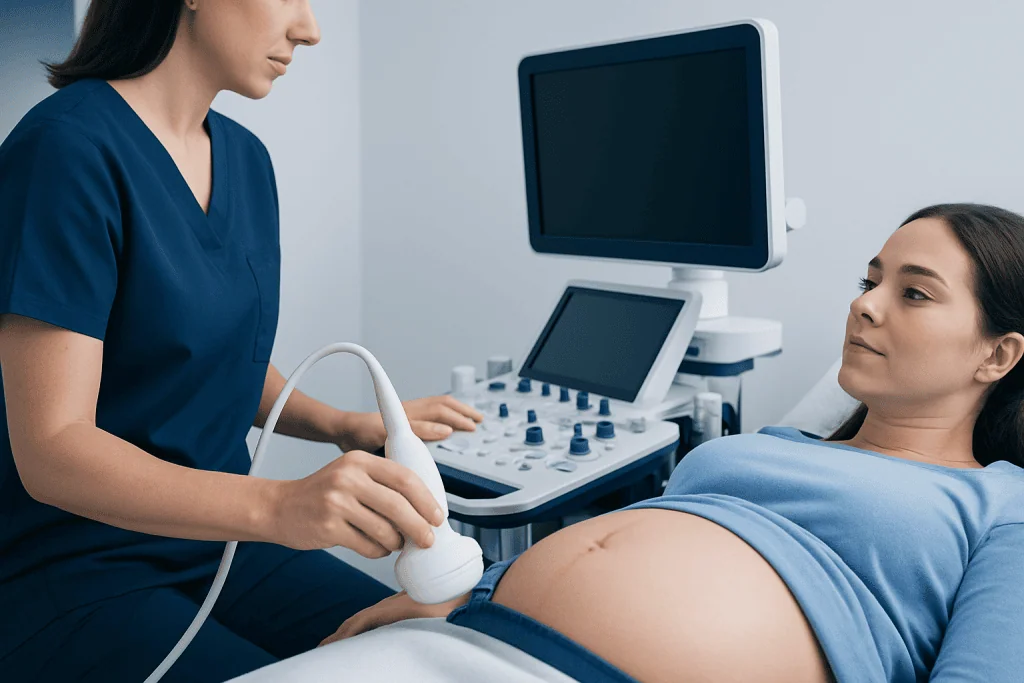
During the examination, the patient lies on an examination couch, usually on the back with a slight tilt if needed for comfort. A sonographer applies gel to the abdomen and moves a handheld probe across the skin. The sonographer records measurements of the fetus, placenta, amniotic fluid, and the suspected chorioangioma.
When Doppler imaging is performed, the screen displays color and waveform information about blood movement. The scan is painless, although gentle pressure from the probe may be felt. A typical assessment may take 20 to 45 minutes, with additional time sometimes needed for a detailed fetal evaluation or specialist review.
There is no recovery period after an abdominal ultrasound. Normal daily activities can usually resume immediately. The results may be discussed on the same day or reviewed by the obstetrician or maternal-fetal medicine specialist shortly afterward, depending on local practice.
Does Chorioangioma Affect the Baby?
Most small chorioangiomas do not affect the baby. The likelihood of complications is generally related to the size of the lesion and how much blood flows through it. A larger, highly vascular chorioangioma can sometimes act as an additional pathway for fetal blood flow, which may place extra demand on the fetal heart.
Potential concerns in more significant cases include fetal anemia, increased cardiac workload, fluid accumulation in the fetus, reduced fetal growth, excess amniotic fluid, preterm labor, or problems related to placental function. These outcomes are not inevitable, and careful surveillance is intended to detect concerning changes as early as possible.
Follow-up may include fetal growth scans, amniotic fluid measurements, Doppler studies of fetal blood vessels, and assessment of fetal heart function. When monitoring indicates a substantial risk, care is usually coordinated through a maternal-fetal medicine service with neonatal and fetal therapy expertise.
Some pregnancies with severe complications may be considered for specialist fetal procedures intended to reduce blood flow through the lesion. The suitable approach depends on the pregnancy stage, lesion characteristics, fetal condition, and local expertise. Decisions are individualized after detailed discussion of potential benefits and risks.
What Is the Prognosis for Chorioangioma?
The prognosis for chorioangioma is often favorable, particularly when the lesion is small and no fetal or maternal complications are present. Many are discovered incidentally and remain stable during pregnancy. In these circumstances, the main plan is usually periodic ultrasound monitoring and routine obstetric care adapted to the individual pregnancy.
Prognosis is more guarded when a chorioangioma is large, grows rapidly, or shows significant internal blood flow. The presence of fetal anemia, hydrops, heart strain, excess amniotic fluid, or preterm labor can affect management and pregnancy outlook. Early recognition and close monitoring help the clinical team plan care and respond to changes.
Serial imaging also assists with delivery planning. Depending on the pregnancy course, a specialist may recommend birth in a hospital able to provide advanced obstetric, neonatal, and pediatric support. After delivery, the placenta is often examined to confirm the diagnosis.
What Are the Symptoms of Chorioangioma of the Placenta?
Chorioangioma of the placenta often causes no noticeable symptoms. It is commonly identified during a routine ultrasound rather than because of a symptom. A pregnant person cannot feel the chorioangioma itself, and its presence does not mean they have done anything to cause it.
When a larger lesion affects the pregnancy, possible associated findings may include unusually rapid abdominal enlargement caused by excess amniotic fluid, uterine discomfort from stretching, contractions before term, or shortness of breath related to a very enlarged uterus. These symptoms have many possible causes and do not by themselves diagnose chorioangioma.
Ultrasound may show findings before symptoms develop, such as increased amniotic fluid, changes in fetal blood flow, or signs of fetal cardiac strain. This is why attending recommended prenatal appointments and follow-up scans is important when a placental mass has been identified.
Risks, Benefits and When to Seek Medical Care
The key benefit of a chorioangioma ultrasound is that it provides real-time information without radiation exposure. It can guide the timing of follow-up, identify pregnancies that remain low risk, and help specialists recognize complications that may benefit from closer observation or treatment. Ultrasound is widely used in pregnancy and is considered safe when medically indicated and performed appropriately.
There are no known direct physical risks from a routine abdominal ultrasound, though the examination may cause brief discomfort from probe pressure. An important limitation is that ultrasound findings may not always be definitive, particularly with small lesions or when imaging conditions are difficult. Further specialist assessment may be needed to clarify the result.
Prompt medical advice is appropriate for vaginal bleeding, leaking fluid, regular painful contractions before term, severe or persistent abdominal pain, sudden marked swelling, severe headache, vision changes, or reduced fetal movements after movements have become established. Reduced movements should be assessed urgently according to the maternity team’s instructions.
Acibadem International’s multidisciplinary specialists and JCI-accredited hospitals support international patients who need assessment and care for complex placental conditions. Ongoing care should always be guided by a qualified obstetrician or maternal-fetal medicine specialist familiar with the individual pregnancy.
Frequently asked questions
Is a chorioangioma ultrasound painful?
No. An abdominal ultrasound is generally painless and noninvasive. Some people may notice mild pressure as the probe is moved across the abdomen, especially during a detailed scan.
How often is a chorioangioma monitored by ultrasound?
The follow-up schedule depends on the lesion’s size, blood flow, gestational age, and whether there are any pregnancy concerns. Small uncomplicated lesions may need less frequent review, while larger or vascular lesions may require regular specialist scans.
Can ultrasound confirm that a placental mass is a chorioangioma?
Ultrasound with color Doppler can often strongly suggest chorioangioma by showing a placental mass with characteristic blood flow. In some cases, the final confirmation comes from examination of the placenta after delivery.
Does a small chorioangioma need treatment?
Small chorioangiomas without signs of fetal or maternal effects usually do not require direct treatment. The care team may recommend periodic scans to ensure the lesion remains stable and the pregnancy continues normally.
Can a chorioangioma disappear during pregnancy?
A chorioangioma may change in appearance over time, particularly if areas within it lose blood supply or undergo degeneration. However, it should not be assumed to disappear, and recommended follow-up imaging remains important.
Will a chorioangioma determine how the baby is delivered?
Not necessarily. Delivery planning is based on the overall pregnancy, including fetal well-being, gestational age, lesion size, and any complications. Many people can have delivery plans similar to those for other pregnancies, while complex cases may need delivery in a specialist center.
References
- American College of Obstetricians and Gynecologists
- International Society of Ultrasound in Obstetrics and Gynecology
- Society for Maternal-Fetal Medicine
- Royal College of Obstetricians and Gynaecologists
This article is for general information only and is not a substitute for professional medical advice. Please consult a qualified doctor about your individual situation.
Ultrasound in Turkey — costs, top hospitals & a free quote
JCI-accredited · board-certified surgeons · reply within 24h
Add us as a Preferred Source to see more of our trusted health content across Google Search, AI Overviews and Discover.

Check-up & Preventive Medicine
Comprehensive health screenings designed for early detection and prevention.
2 specialists in this unitMore from the Health Library



Related Specialists

Dr. Esra Akbulut
Neonatal Intensive Care Unit
Dr. Zehra Karataş
Nuclear Medicine
Dr. Nıgar Alıyeva
Pediatric & Adolescent Psychiatry
Dr. Remzi Özgür Özay
Anesthesiology