Cranioplasty: Benefits, Risks, and Recovery — A Complete Guide

Cranioplasty rebuilds an area of missing or damaged skull using the patient’s own bone or a synthetic implant. The procedure is often considered after decompressive craniectomy, head injury, infection treatment, or tumor surgery.
Key Takeaways
- Cranioplasty rebuilds an area of missing or damaged skull using the patient’s own bone or a synthetic implant.
- The procedure is often considered after decompressive craniectomy, head injury, infection treatment, or tumor surgery.
- Potential benefits include protection of the brain, restoration of skull shape, and possible improvement in symptoms related to the skull defect.
- Recovery varies, but follow-up imaging, wound care, and activity restrictions are important parts of healing.
- Like any neurosurgical procedure, cranioplasty carries risks such as infection, bleeding, seizures, and implant-related problems.
- Patients should contact a doctor promptly for fever, worsening headache, confusion, drainage from the wound, or new weakness.
Cranioplasty is an operation that repairs a defect in the skull, usually after trauma, stroke-related surgery, infection, or tumor treatment. It can improve brain protection and head contour, and in some people it may also support comfort, function, and recovery.
Overview: what cranioplasty is and why it is done
Cranioplasty is surgery to repair a hole or defect in the skull. The defect may develop after a serious head injury, a previous brain operation, treatment for swelling after a stroke or trauma, or removal of infected or damaged bone. The operation places a bone flap or custom implant over the opening to restore the skull’s structure.
The main goals of cranioplasty are to protect the brain, improve the shape of the head, and close the skull defect once the underlying condition is stable. In some patients, doctors also observe improvement in headaches, discomfort at the site, dizziness, or symptoms linked to altered pressure and blood flow around the brain. Recovery and results depend on the reason for the defect, the patient’s overall health, and whether there are any complications such as infection.
Cranioplasty is not the same as emergency surgery to relieve brain swelling. It is usually a planned reconstructive step that happens later, after the urgent problem has been treated. This is why doctors spend time deciding the safest timing, the best material to use, and whether there are conditions that need to be corrected before surgery.
Who may need cranioplasty
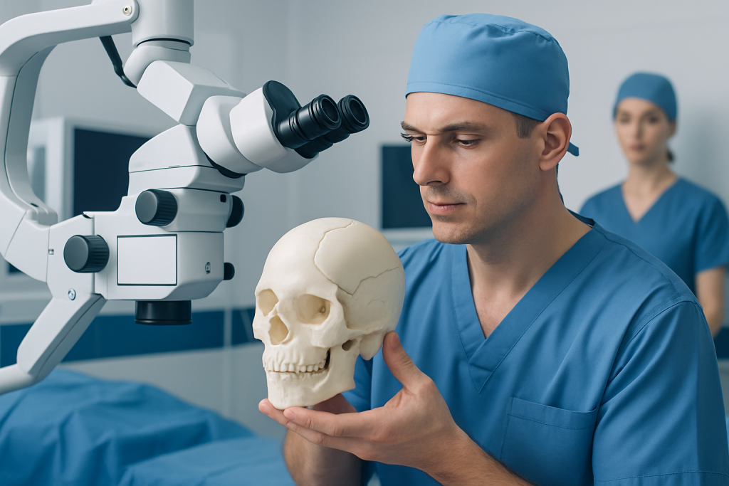
People may be candidates for cranioplasty if they have a skull defect that leaves part of the brain less protected or causes cosmetic and functional concerns. A common reason is a previous decompressive craniectomy, a procedure in which part of the skull is temporarily removed to allow a swollen brain to expand safely. This may be done after severe head trauma, bleeding, or major stroke. Patients recovering from stroke may later be assessed for skull reconstruction once swelling has settled.
Other reasons include loss of skull bone after infection, tumor surgery, congenital defects, or fractures that cannot be repaired at the time of injury. Some patients seek care because they feel tenderness, pulsation, headache, or self-consciousness related to the skull shape. Others are evaluated because their neurosurgical team believes replacing the bone may help rehabilitation or day-to-day comfort.
Not everyone is a candidate right away. Doctors may delay cranioplasty if there is active infection, uncontrolled swelling, unstable medical status, poor wound healing, or a need for further brain treatment. The best candidate is someone whose original condition has stabilized and whose surgical team believes the benefits of repair outweigh the risks.
How cranioplasty works: planning, materials, and the procedure step by step

Before surgery, the team reviews brain scans, the size and location of the skull defect, and the condition of the skin and soft tissue over the area. Imaging helps surgeons decide whether the patient’s original bone flap can be reused or whether a synthetic implant is a better option. Common implant materials include titanium, polyetheretherketone (PEEK), polymethyl methacrylate, and other biocompatible materials designed to fit the skull precisely.
If the patient’s own bone was preserved from the earlier operation and remains suitable, it may be reimplanted. In other cases, a custom-made implant is created from CT imaging to match the defect. The choice depends on factors such as infection history, the condition of the original bone, defect size, patient age, and surgeon preference.
During the operation, the patient receives general anesthesia. The surgeon reopens or uses the previous scalp incision, carefully exposes the edges of the skull defect, and prepares the area. The bone flap or implant is positioned into place and secured, often with plates, screws, or fixation devices. If needed, the surgeon may place a temporary drain to reduce fluid buildup before the incision is closed. Patients who need broader neurosurgical evaluation or reconstructive planning may undergo care through services such as neurosurgery and brain surgery.
Although the steps sound straightforward, cranioplasty is highly individualized. The surgeon must account for scar tissue, previous surgeries, pressure changes, and the health of the surrounding scalp. For this reason, the operation is best planned by an experienced multidisciplinary team.
Benefits and expected outcomes
The most direct benefit of cranioplasty is renewed protection for the brain. Replacing the missing section of skull can reduce vulnerability to accidental impact and may allow patients to feel safer and more confident in daily activities. Restoring the normal contour of the head can also improve appearance and help with emotional well-being after major illness or trauma.
Some patients report functional improvements after surgery. These can include reduced discomfort around the defect, less sensitivity to pressure changes, and progress in mobility or concentration during rehabilitation. Doctors sometimes describe symptom improvement in people who develop changes associated with a large skull defect after craniectomy, although results vary and cannot be guaranteed.
Expected outcomes depend on the cause of the defect, the patient’s neurological condition, and the presence of any complications before surgery. Cranioplasty does not reverse the original brain injury, but it may be an important step in overall recovery. The surgical team explains realistic goals based on each person’s imaging, symptoms, and rehabilitation plan.
Risks and possible complications
Cranioplasty is a major surgical procedure, so it carries real but carefully managed risks. These include infection, bleeding, fluid collection under the scalp, wound healing problems, seizures, blood clots, and reactions related to anesthesia. There can also be problems specific to the implant or bone flap, such as loosening, resorption of stored bone, poor fit, or the need for revision surgery.
In some cases, swelling, pressure changes, or collections of cerebrospinal fluid can occur after surgery. Patients with previous infection, multiple operations, large defects, poor nutrition, or significant medical illness may face a higher risk of complications. Children and adults may also differ in the materials and long-term monitoring they need.
These risks are one reason careful timing matters. Surgeons usually prefer to operate when there is no active infection, the wound is healthy, and the patient is medically stable. Open discussion with the care team helps patients understand why a particular material, technique, and timeline have been chosen.
If the skull defect followed treatment for trauma or other neurological disease, the team may coordinate with specialists in neurology and rehabilitation. This helps align surgical plans with seizure management, mobility goals, and recovery from the original condition.
Recovery timeline and aftercare
After cranioplasty, patients are usually monitored in the hospital so the team can watch for swelling, changes in neurological status, pain control needs, and wound-related issues. Follow-up imaging may be performed to confirm the position of the implant or bone flap and to check for fluid collection or bleeding. The length of stay varies according to the complexity of surgery and the patient’s overall condition.
In the first days to weeks, common recovery steps include keeping the incision clean and dry, taking medicines exactly as prescribed, avoiding strenuous activity, and following instructions about sleeping position, lifting, and return to work or school. Some scalp numbness, tenderness, and fatigue are common early on. The doctor will advise when hair washing, exercise, and travel are appropriate.
Recovery is often gradual rather than immediate. Many people need several weeks before energy improves and normal routines feel comfortable again. If the patient is already in rehabilitation after brain injury or stroke, cranioplasty may become one part of a broader recovery plan that can include physical, occupational, or speech therapy.
Because every case is different, there is no single universal timeline. A small, uncomplicated repair may heal more quickly than a large reconstruction after multiple prior operations. Follow-up visits are important to check wound healing, neurological function, and long-term implant stability.
Self-care, preparation, and questions to ask before surgery
Good preparation can support a smoother recovery. Patients are often advised to attend all preoperative visits, share a full list of medications and supplements, and ask about smoking cessation, nutrition, and management of conditions such as diabetes or high blood pressure. These steps can help reduce the chance of wound problems and other complications.
At home, it is helpful to arrange support for daily tasks during the first part of recovery. Having someone available for transportation, meals, medication reminders, or wound observation can make healing safer and less stressful. Patients should also ask the team what symptoms are expected and which ones require immediate attention.
Useful questions before surgery may include:
- Why is cranioplasty recommended in this case, and why now?
- Will the repair use my own bone or a synthetic implant?
- What are the main risks based on my health history?
- How long is the expected hospital stay and recovery period?
- What restrictions will I have after surgery?
- What follow-up imaging or rehabilitation might be needed?
Near the end of treatment planning, some patients seek care in specialized centers able to coordinate imaging, neurosurgery, and rehabilitation in one setting. Acibadem International’s multidisciplinary specialists and JCI-accredited hospitals diagnose and treat patients needing cranioplasty and related neurological care.
When to seek medical care
Anyone with a known skull defect should seek medical advice if they develop worsening headaches, new weakness, changes in speech, confusion, repeated vomiting, fever, or drainage from the scalp wound. These symptoms do not always mean a serious complication, but they should be assessed promptly. New seizures or loss of consciousness require urgent medical attention.
After cranioplasty, patients should contact their surgical team right away if the incision becomes red, swollen, increasingly painful, or starts leaking fluid. A new soft bulge, sudden drowsiness, severe headache, or worsening balance can also signal a problem that needs evaluation. It is safer to call early than to wait and hope symptoms pass.
Emergency care is especially important after any significant head impact in a person with a recent cranioplasty or an unrepaired skull defect. Even when external injuries look minor, the brain and reconstruction site may need medical review.
Frequently asked questions
Is cranioplasty the same as craniectomy?
No. A craniectomy removes part of the skull, usually to relieve dangerous brain swelling. Cranioplasty is the later operation that repairs the skull defect once the patient is stable.
How long after a craniectomy is cranioplasty done?
The timing varies from person to person. Doctors consider brain swelling, infection risk, wound condition, and overall recovery before deciding when surgery is safest.
What materials are used in cranioplasty?
Surgeons may use the patient’s original bone flap if it is still suitable. If not, they may use a synthetic implant such as titanium or a custom-made biocompatible material shaped to fit the skull.
How painful is cranioplasty recovery?
Some pain, scalp tightness, and fatigue are common after surgery, especially in the first days. The care team usually provides pain control and instructions to make recovery more comfortable and safe.
Can cranioplasty improve neurological symptoms?
In some patients, yes. Repairing the skull defect may improve comfort and may support better function in selected cases, but it does not reverse the original brain injury and results vary.
What are the warning signs of complications after cranioplasty?
Patients should watch for fever, increasing redness or drainage at the incision, worsening headache, confusion, vomiting, new weakness, or seizures. These symptoms should be reported promptly because they may need urgent medical assessment.
References
- National Institute of Neurological Disorders and Stroke
- American Association of Neurological Surgeons
- National Health Service
- MedlinePlus
- World Health Organization
This article is for general information only and is not a substitute for professional medical advice. Please consult a qualified doctor about your individual situation.
Oncology care in Turkey — second opinion and treatment plan
JCI-accredited · board-certified surgeons · reply within 24h
Add us as a Preferred Source to see more of our trusted health content across Google Search, AI Overviews and Discover.