CT Scan — Explained by Medical Evidence, Not Myths
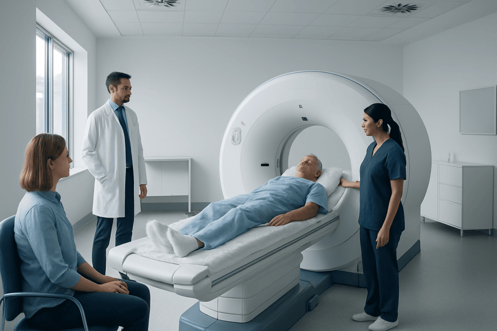
A CT scan combines X-rays and computer technology to produce detailed images of internal organs, bones, blood vessels, and soft tissues. It is often used when doctors need fast, clear information about injury, bleeding, infection, tumors, or unexplained symptoms.
Key Takeaways
- A CT scan combines X-rays and computer technology to produce detailed images of internal organs, bones, blood vessels, and soft tissues.
- It is often used when doctors need fast, clear information about injury, bleeding, infection, tumors, or unexplained symptoms.
- Some CT scans use contrast dye to improve image detail, but this may not be suitable for everyone.
- CT scans involve radiation, yet the benefit usually outweighs the risk when the test is medically appropriate.
- Preparation depends on the body area being scanned and whether contrast will be used.
- Results should always be interpreted by a qualified doctor in the context of symptoms, examination, and other tests.
A CT scan is an imaging test that uses X-rays and computer processing to create detailed cross-sectional pictures of the body. It is commonly used to evaluate symptoms quickly, guide treatment decisions, and monitor many medical conditions with a strong evidence base behind its use.
Overview: what a CT scan is and why it is used
A CT scan, short for computed tomography scan, is a medical imaging test that uses a series of X-ray images and computer processing to create detailed cross-sectional views of the inside of the body. In practical terms, it helps doctors look beyond what a physical exam can show, often with far more detail than a standard X-ray.
CT scans are widely used because they are fast, accurate, and especially helpful in urgent situations. They can show bones, organs, blood vessels, soft tissues, and signs of disease or injury. This makes CT useful in emergency care, cancer assessment, infection evaluation, surgical planning, and follow-up after treatment.
Unlike some myths found online, a CT scan is not a treatment and it does not automatically mean a serious diagnosis is expected. It is simply a tool that helps answer a medical question. A doctor may order it to rule out dangerous conditions, confirm a suspected diagnosis, or decide whether another test would be more helpful.
Depending on the clinical problem, a CT scan may focus on the head, chest, abdomen, pelvis, spine, joints, or blood vessels. It may also support the evaluation of conditions such as lung cancer or stroke, where rapid and reliable imaging can strongly influence the next steps in care.
How a CT scan works
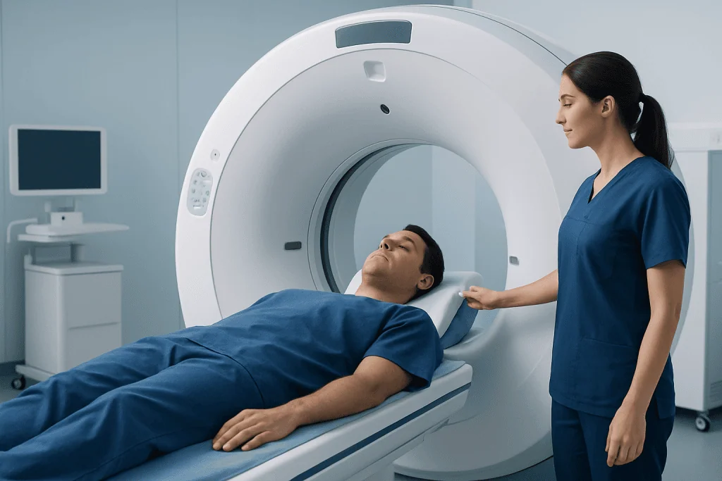
During a CT scan, the patient usually lies on a narrow table that moves through a large ring-shaped machine. An X-ray tube rotates around the body and captures images from many angles. A computer then combines these images into thin slices, which can be reviewed individually or reconstructed into more complete views.
This cross-sectional approach is one of the main reasons CT is so useful. Instead of seeing structures overlap on a flat image, doctors can assess specific layers of tissue. In many cases, this improves the ability to detect bleeding, inflammation, tumors, fractures, blocked blood vessels, or organ damage.
Some CT scans are performed without contrast, while others use a contrast material to make certain tissues or blood vessels easier to see. Contrast may be swallowed, injected into a vein, or occasionally given another way depending on the body part being studied. For example, contrast-enhanced imaging may be helpful when evaluating abdominal pain, tumors, or vascular problems.
CT technology continues to develop, with many modern scanners designed to shorten scan time and reduce radiation exposure while maintaining image quality. Even so, the value of the test still depends on using it for the right reason and interpreting the findings alongside the person’s symptoms and medical history.
Common reasons doctors order a CT scan
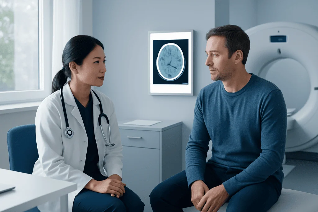
Doctors may recommend a CT scan for many different symptoms and medical concerns. In emergency settings, it is often used to assess head injury, severe pain, suspected internal bleeding, sudden neurological symptoms, or major trauma. Its speed can be critical when rapid treatment decisions are needed.
In non-emergency care, CT can help investigate ongoing or unexplained symptoms such as chest pain, persistent cough, abdominal pain, recurrent infections, kidney stones, sinus problems, unexplained weight loss, or suspected masses. It is also frequently used to monitor known conditions over time and to evaluate how well treatment is working.
CT is important in cancer care because it can help detect a suspicious growth, assess its size and spread, and guide biopsies or treatment planning. When appropriate, specialists may combine CT findings with other imaging methods and procedures, including robotic surgery or radiation oncology, depending on the diagnosis.
Doctors also use CT to guide certain interventions. In some cases, imaging helps place a needle for biopsy or drainage with greater precision. If a CT scan suggests a problem in the digestive tract or chest, additional tests such as endoscopy may be recommended to examine tissue directly.
Safety, radiation, and contrast dye: separating facts from myths
One of the most common concerns about CT scans is radiation exposure. A CT scan does use more radiation than a standard X-ray because it gathers many images. However, this does not mean it is unsafe when medically justified. Doctors order CT when the expected benefit of clear diagnostic information outweighs the potential risk.
Radiation risk depends on factors such as age, body area scanned, and how often scans are repeated. Medical teams aim to use the lowest radiation dose that can still answer the clinical question. Patients should tell their doctor if they are pregnant or may be pregnant, because special precautions or different imaging tests may be preferred in some situations.
Another common question involves contrast dye. Contrast can improve image quality, but it may cause problems in a small number of people. The main concerns are allergy-like reactions and kidney-related issues in selected patients, especially those with significant kidney disease. Before the scan, the care team may ask about previous reactions, kidney function, diabetes, asthma, or other relevant health conditions.
It is also a myth that every abnormality on CT means serious disease. CT is sensitive and may detect incidental findings that are minor, harmless, or simply require follow-up. For this reason, results should be discussed calmly with a qualified doctor, who can explain what matters clinically and what may not.
How to prepare and what happens during the procedure
Preparation for a CT scan depends on what part of the body is being examined and whether contrast will be used. Some people can eat and drink normally beforehand, while others may be asked not to eat for several hours. If oral contrast is needed, instructions are usually given in advance. Patients should bring a list of medications and mention allergies, pregnancy, kidney disease, or prior contrast reactions.
Before the scan, the person may be asked to change into a gown and remove metal objects such as jewelry, glasses, belts, or hearing aids, since these can affect image quality. If intravenous contrast is planned, a small cannula may be placed in a vein. During injection, some people notice a warm sensation or a metallic taste for a brief time.
The scan itself is usually quick and painless. The table moves slowly through the scanner while the person remains as still as possible. The radiographer may give simple instructions such as holding the breath for a few seconds. Although the machine can seem unfamiliar, it is open rather than tunnel-like, and staff can communicate throughout the procedure.
After the scan, most people return to normal activities the same day. If contrast was used, drinking fluids may be advised unless a doctor has recommended otherwise. The images are then reviewed by a radiologist, and the referring doctor explains the results in the context of the person’s overall health picture.
Understanding results and possible next steps
CT scan results are not simply labeled normal or abnormal without context. A radiologist studies the images and describes any findings, such as inflammation, fracture, bleeding, fluid, enlarged organs, masses, or blood vessel changes. The referring doctor then combines the imaging report with symptoms, physical examination, blood tests, and medical history.
A normal CT scan can be very helpful because it may rule out dangerous causes of symptoms and reduce the need for more invasive testing. At the same time, a normal result does not always end the diagnostic process. Some problems are better detected with other tests such as MRI, ultrasound, laboratory studies, or specialist evaluation.
If the CT scan shows a clear cause of symptoms, next steps may include medication, observation, referral to a specialist, or a procedure. For example, findings that suggest a vascular emergency may lead to interventional radiology or surgery, while signs of infection may support targeted medical treatment.
When findings are uncertain, follow-up imaging may be advised after a period of time. This does not always indicate danger. In many situations, interval imaging is simply the safest and most evidence-based way to determine whether a small or unclear finding is stable, resolving, or changing.
When to seek medical care
A person should seek medical care promptly if they have symptoms that could signal an urgent problem, especially after injury or if symptoms begin suddenly. Examples include severe headache, confusion, weakness on one side, chest pain, shortness of breath, severe abdominal pain, persistent vomiting, high fever with worsening symptoms, or signs of significant trauma.
Medical advice is also important when symptoms are less dramatic but do not improve as expected. Ongoing cough, unexplained weight loss, recurrent pain, blood in urine or stool, repeated fainting, or new neurological symptoms may all require assessment. In these situations, a doctor can decide whether CT imaging is the right next step or whether another test is more appropriate.
After a CT scan with contrast, urgent medical attention is needed if a person develops trouble breathing, swelling of the face or throat, severe rash, or other signs of a serious allergic reaction. Patients with reduced kidney function should follow their doctor’s instructions carefully after contrast studies.
For people seeking coordinated assessment, Acibadem International’s multidisciplinary specialists and JCI-accredited hospitals diagnose and treat conditions that may require CT imaging for international patients. The most appropriate test and timing should always be decided with a qualified healthcare professional.
Frequently asked questions
What is the difference between a CT scan and an X-ray?
Both use X-rays, but a CT scan takes many images from different angles and combines them into detailed cross-sectional pictures. This gives much more information about organs, soft tissues, blood vessels, and internal injuries than a standard X-ray.
Is a CT scan painful?
A CT scan is usually not painful. The main part of the test involves lying still on a table while the scanner takes images. If contrast is injected, there may be brief discomfort from the needle and a temporary warm feeling.
How long does a CT scan take?
The scan itself is often completed within minutes, although the full appointment may take longer if preparation or contrast is needed. Timing varies depending on the body area being examined and the reason for the test.
Is a CT scan safe?
A CT scan is generally considered safe when it is used appropriately for a clear medical reason. It does involve radiation, so doctors aim to order it only when the expected diagnostic benefit outweighs the potential risk.
What is contrast dye and why might it be needed?
Contrast dye is a substance used to make certain tissues, organs, or blood vessels show more clearly on the images. It can improve the ability to detect inflammation, tumors, bleeding, or vascular problems, but it is not necessary for every CT scan.
Can someone eat or drink before a CT scan?
It depends on the type of CT scan. Some require no special preparation, while others, especially those using contrast, may require fasting for a few hours. The imaging team usually provides specific instructions in advance.
Will a CT scan always find the cause of symptoms?
Not always. A CT scan is a powerful diagnostic tool, but some conditions are better evaluated with MRI, ultrasound, lab tests, or direct examination. Doctors use the result together with other medical information to decide the next step.
References
- World Health Organization
- Radiological Society of North America
- American College of Radiology
- National Institute of Biomedical Imaging and Bioengineering
- U.S. Food and Drug Administration
This article is for general information only and is not a substitute for professional medical advice. Please consult a qualified doctor about your individual situation.
Explore treatments in Turkey — costs, top hospitals & a free quote
JCI-accredited · board-certified surgeons · reply within 24h
Add us as a Preferred Source to see more of our trusted health content across Google Search, AI Overviews and Discover.

More from the Health Library



Related Specialists

Prof. Dr. Bülent Özçelik
Gynecology & Obstetrics
Prof. Dr. İftahar Köksal
Infectious Diseases & Clinical Microbiology
Asst. Prof. Dr. Ebru Kazancı
Neonatal Intensive Care Unit
Prof. Dr. Tansel Erol
Cardiology