CT Scan for Adrenal Glands: Preparation, Procedure and Results
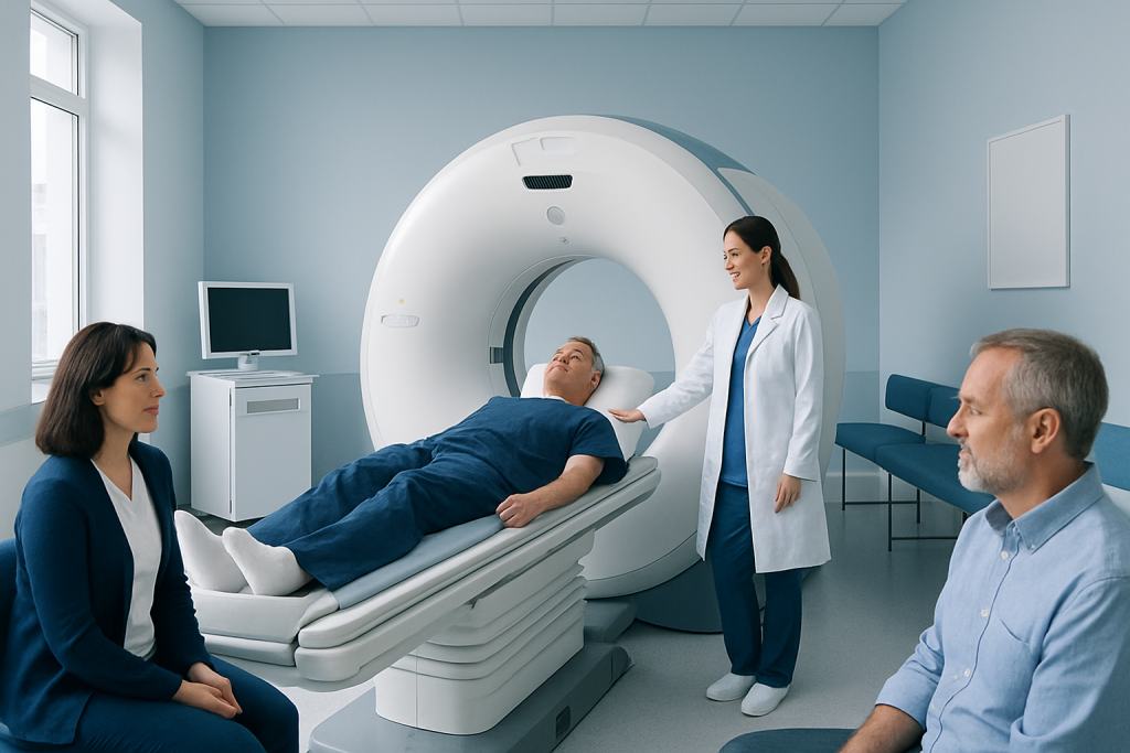
Adrenal CT uses X-rays and computer processing to create cross-sectional images of the adrenal glands and nearby organs. Contrast dye may be used to better characterize an adrenal mass and evaluate how it enhances and washes out over time.
Key Takeaways
- Adrenal CT uses X-rays and computer processing to create cross-sectional images of the adrenal glands and nearby organs.
- Contrast dye may be used to better characterize an adrenal mass and evaluate how it enhances and washes out over time.
- Most adrenal findings are benign, but scan results are interpreted alongside symptoms, hormone blood or urine tests, and medical history.
- Preparation may include temporary fasting and checking kidney function or contrast allergy history.
- The scan is usually quick, and most people return to normal activities soon afterward.
A CT scan for adrenal glands is an imaging test that produces detailed pictures of the small glands above the kidneys. It can help clinicians assess an adrenal lump, gland enlargement, bleeding, injury, or features that may require hormone testing or further care.
CT Scan for Adrenal Glands: Overview
A CT scan for adrenal glands is a detailed imaging examination of the two adrenal glands, which sit above the kidneys and produce hormones involved in stress response, blood pressure, salt balance and metabolism. The scan may be requested after an adrenal abnormality is found on another test, when symptoms suggest excess hormone production, or to assess a known adrenal mass.
Computed tomography (CT) combines a series of X-ray images with computer processing to create thin, cross-sectional views of the body. These views can show the size, shape, density and location of the adrenal glands, as well as surrounding structures such as the kidneys, liver, pancreas, blood vessels and lymph nodes.
An adrenal finding on imaging is not automatically cancer or a cause of symptoms. Small adrenal masses are often discovered incidentally during scans performed for another reason. The next steps depend on the imaging appearance, size, growth over time, personal medical history and whether the gland is making excess hormones.
How the Scan Works and Who May Need It
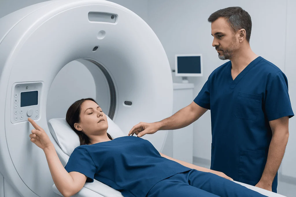
During CT, a rotating X-ray tube takes images from multiple angles while the person lies still on a moving table. A radiologist interprets the images. In many cases, images are obtained before and after an intravenous contrast agent is given, because the way a lesion takes up and releases contrast can provide useful clues about its nature.
A clinician may recommend adrenal CT for a newly identified adrenal mass, unexplained adrenal enlargement, suspected bleeding after trauma, follow-up of a previously seen lesion, or staging when there is a known cancer elsewhere in the body. It may also support evaluation of hormone-related conditions, although imaging alone cannot determine whether a mass produces hormones.
CT may not be the best choice for every person. Pregnancy, prior severe reaction to iodinated contrast, reduced kidney function and cumulative radiation exposure are important considerations. Depending on the clinical question, magnetic resonance imaging (MRI), ultrasound in selected settings, laboratory testing or nuclear medicine imaging may be considered instead.
- People with an incidentally found adrenal mass may need imaging characterization.
- People with high blood pressure, low potassium or unusual hormone symptoms may need both imaging and endocrine testing.
- People with a history of cancer may need evaluation of an adrenal lesion as part of wider assessment.
What Is the Protocol for a CT Adrenal Gland Scan?

The protocol for a CT adrenal gland scan is tailored to the reason for the test and the local radiology service. A dedicated adrenal protocol commonly includes a non-contrast scan first, followed by contrast-enhanced images and, in some cases, delayed images taken after a set interval. Comparing these phases helps radiologists measure attenuation and contrast washout, which may help distinguish common benign adenomas from lesions that need additional assessment.
Before the appointment, the imaging team may ask about pregnancy, kidney disease, diabetes medicines, thyroid conditions, asthma, previous contrast reactions and all current medicines. If contrast is planned, blood tests may be requested to assess kidney function. Instructions about food and drink vary, but temporary fasting for several hours is often advised when intravenous contrast will be used.
Patients should wear comfortable clothing without metal near the abdomen or change into a gown. They should bring prior scan reports or images when available, since comparing a lesion with older studies can be particularly helpful. The radiology team should be told promptly about any history of a significant contrast reaction.
Step-by-Step: What Happens During an Adrenal CT Scan
On arrival, a radiographer confirms identity, reviews safety questions and explains the examination. If contrast is required, a small intravenous cannula is placed, usually in the arm. The person lies on their back on the CT table, often with arms positioned above the head to improve image quality.
The table moves through the doughnut-shaped scanner while images are taken. The scanner is open at both ends and does not enclose the entire body. The radiographer can see and speak with the patient from the control area. Short breath-holds may be requested, as remaining still helps prevent blurred images.
When contrast is injected, a temporary warm sensation, metallic taste or feeling of needing to urinate can occur. These sensations typically pass quickly. The image acquisition itself may take only minutes, though check-in, preparation and any delayed imaging make the total visit longer.
Afterward, a radiologist reviews the images and sends a report to the referring clinician. The timing for results varies by service and by whether urgent findings are suspected. The referring clinician explains what the findings mean in the context of symptoms, examination and laboratory results.
What Will a CT of the Adrenal Gland Show?
A CT of the adrenal gland can show whether an adrenal gland is normal in appearance, enlarged, injured, bleeding or affected by a mass. It can describe a lesion’s size, borders, internal density, calcification, fat content and relationship to nearby organs or blood vessels. These details help guide whether observation, hormone testing, specialist review or additional imaging is appropriate.
A common reason for dedicated imaging is to characterize an adrenal incidentaloma, meaning a mass found unexpectedly. Many adrenal incidentalomas are noncancerous adenomas. CT features, including the density on non-contrast images and the pattern of contrast washout, can often help estimate whether an adenoma is likely, although no scan result should be interpreted in isolation.
CT can also identify findings that may require prompt assessment, such as a large or irregular mass, evidence of local invasion, enlarged lymph nodes, adrenal hemorrhage or changes suggesting infection in the appropriate clinical setting. If imaging suggests a potentially concerning lesion, an endocrinologist, radiologist, surgeon and oncology specialists may work together to plan further evaluation.
Imaging does not directly measure adrenal hormone activity. Blood and urine tests are often used to assess cortisol, catecholamine-related hormones, aldosterone and other hormones when clinically indicated. A clinician may also discuss related information on adrenal cancer when a suspicious adrenal lesion needs specialist assessment.
Benefits, Risks and Recovery After the Scan
The main benefit of CT is that it provides fast, detailed information about adrenal anatomy and nearby structures. Dedicated imaging can help avoid unnecessary procedures when a lesion has reassuring features, while also identifying people who may benefit from closer follow-up or specialist care.
CT involves ionizing radiation. Radiology teams use protocols designed to obtain diagnostic images with the lowest practical radiation exposure. The potential value of the examination is balanced against this risk, especially for younger people and those who have had multiple imaging studies.
Iodinated contrast can occasionally cause side effects. Mild effects may include warmth, nausea or itching. Serious allergic-like reactions are uncommon but require immediate treatment. Contrast can also be a concern for people with significant kidney impairment, which is why the team reviews medical history and may check kidney function beforehand.
Recovery is usually immediate. People can generally eat, drink and resume usual activities after the scan unless they receive different instructions. Drinking fluids may be encouraged after contrast if it is medically appropriate. Anyone who develops breathing difficulty, facial swelling, widespread rash, persistent vomiting or other concerning symptoms after leaving should seek urgent medical care.
What Are the First Signs of Adrenal Problems?
Early signs of adrenal problems vary because the glands can produce too much or too little of different hormones, and some adrenal masses do not cause symptoms at all. Symptoms may be gradual and nonspecific, so they should not be used alone to diagnose an adrenal condition.
Possible signs of excess cortisol include unexpected weight gain around the trunk, easy bruising, muscle weakness, new or worsening high blood pressure, high blood sugar and mood changes. Episodes of headache, palpitations, sweating, tremor or sudden spikes in blood pressure may suggest excess catecholamine-related hormones and should be assessed by a clinician.
Low adrenal hormone production can cause fatigue, weakness, poor appetite, nausea, weight loss, dizziness on standing, salt craving or skin darkening in some forms of adrenal insufficiency. These symptoms can have many causes. Testing should be guided by a qualified clinician, particularly when symptoms are persistent, worsening or associated with low blood pressure or dehydration.
Assessment may involve an endocrinologist alongside radiology and other specialties. Where surgery is needed for an appropriate adrenal lesion, adrenalectomy may be considered after careful hormonal and imaging evaluation.
What Are the First Signs of Adrenal Cancer? When to Seek Medical Care
Adrenal cancer is uncommon, and early symptoms may be absent. When symptoms occur, they can result from a growing abdominal mass or from excess hormone production. Possible signs include persistent abdominal, side or back discomfort; a feeling of fullness; unexplained weight change; or new symptoms of hormone excess, such as easy bruising, muscle weakness, abnormal hair growth, menstrual changes, deepening of the voice, or marked blood-pressure changes.
These symptoms are not specific to adrenal cancer and are more often caused by other conditions. However, a clinician should assess persistent or unexplained symptoms, especially when an adrenal mass has been identified. New symptoms should also be discussed promptly by people with a personal history of cancer.
Urgent medical care is appropriate for severe or sudden abdominal or flank pain, fainting, severe weakness, confusion, chest pain, difficulty breathing, or very high blood pressure accompanied by severe headache, neurological symptoms or vision changes. These symptoms need timely assessment regardless of their cause.
Acibadem International’s multidisciplinary specialists and JCI-accredited hospitals diagnose and treat adrenal conditions for international patients, coordinating endocrine evaluation, radiology and surgical care when appropriate.
Frequently asked questions
How long does a CT scan for adrenal glands take?
The scanning portion is often completed within minutes. The full appointment may take longer because of registration, safety checks, intravenous access for contrast and delayed images if a dedicated adrenal protocol is used. The imaging center can explain the expected timing before the appointment.
Do I need contrast for an adrenal CT scan?
Not every adrenal CT scan requires contrast, but contrast is commonly useful when characterizing an adrenal mass. Images before and after contrast, sometimes including delayed images, can provide information about the lesion’s density and enhancement pattern. The decision depends on the clinical question and individual safety considerations.
Can a CT scan tell if an adrenal mass is cancerous?
CT can identify features that make a lesion more or less likely to be benign, but it may not give a definite diagnosis in every case. Size, density, contrast behavior, margins and evidence of spread are all considered. Hormone tests, previous imaging, MRI or specialist review may also be needed.
Can I eat before an adrenal CT scan?
Instructions differ depending on whether contrast is planned and on the imaging center’s protocol. Some people are asked not to eat for several hours before a contrast-enhanced CT. Patients should follow the instructions provided by their care team and ask if they are unsure.
What should I tell the CT team before the scan?
Patients should tell the team about pregnancy or possible pregnancy, kidney disease, prior contrast reactions, diabetes medicines, thyroid conditions and all current medicines. They should also mention previous imaging studies and any known adrenal or cancer diagnosis. This information helps the team choose the safest and most useful protocol.
What happens if an adrenal mass is found?
The next step depends on the lesion’s imaging appearance, size, whether it has changed over time and whether it produces hormones. Many lesions only need observation or planned follow-up. Others may require blood or urine testing, repeat imaging, referral to endocrinology or discussion of treatment options.
References
- American College of Radiology
- Endocrine Society
- National Cancer Institute
- Radiological Society of North America
- European Society of Endocrinology
This article is for general information only and is not a substitute for professional medical advice. Please consult a qualified doctor about your individual situation.
Medical weight-loss options in Turkey — costs & eligibility
JCI-accredited · board-certified surgeons · reply within 24h
Add us as a Preferred Source to see more of our trusted health content across Google Search, AI Overviews and Discover.
Check your numbers in seconds
BMI, calories, due date, blood pressure and 30+ more clinical calculators — free, instant, doctor-reviewed ranges.