Cyst in Uterus on Ultrasound: Preparation, Procedure and Results

Pelvic ultrasound is the main first test for assessing a suspected cyst in the pelvic area. Many ovarian and cervical cysts are benign and may resolve or need only monitoring.
Key Takeaways
- Pelvic ultrasound is the main first test for assessing a suspected cyst in the pelvic area.
- Many ovarian and cervical cysts are benign and may resolve or need only monitoring.
- A transvaginal ultrasound usually provides the clearest view of the ovaries, uterus and endometrium.
- Ultrasound can identify features that need further assessment, but it cannot alone confirm whether every cyst is cancerous.
- Sudden severe pelvic pain, fainting, fever or heavy bleeding require prompt medical assessment.
A cyst in uterus on ultrasound does not always mean a cyst is actually inside the uterus. Pelvic ultrasound can show fluid-filled areas in or near the uterus, including ovarian cysts, cervical cysts or changes in the uterine lining, and the next steps depend on the location and appearance.
Overview: What Does a Cyst in Uterus on Ultrasound Mean?
A cyst in uterus on ultrasound is a phrase that may be used when a scan detects a fluid-filled area within the uterus or somewhere nearby in the pelvis. The ultrasound report should clarify the exact site: the uterine lining (endometrium), cervix, ovary, fallopian tube region or surrounding pelvic tissue. This distinction matters because these findings have different causes and follow-up plans.
Most cyst-like findings are not an emergency and are not cancer. For example, functional ovarian cysts are common during reproductive years, while small cervical nabothian cysts are usually harmless. Some findings require a repeat scan, specialist review or additional tests when they are large, persistent, complex in appearance, associated with symptoms, or found after menopause.
Ultrasound uses sound waves rather than radiation. It is often the first imaging test used for pelvic symptoms such as pain, bloating, changes in menstrual bleeding or a feeling of pressure. Results are interpreted alongside age, menstrual status, symptoms, examination findings and, when relevant, pregnancy status.
How Pelvic Ultrasound Works and Who May Need It
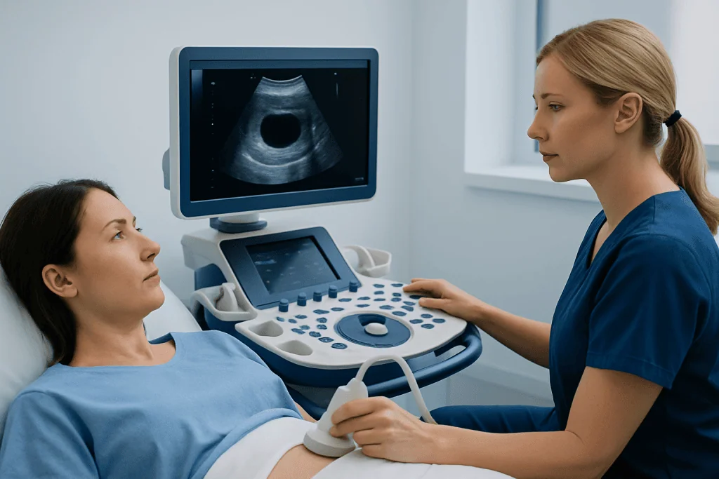
A pelvic ultrasound creates real-time images of the uterus, ovaries, cervix and nearby pelvic structures. A sonographer performs the examination, and a radiologist or treating clinician interprets the images. The scan can show whether a finding is filled with clear fluid, contains internal material, has partitions called septations, or includes solid areas that may need closer evaluation.
A clinician may recommend an ultrasound for pelvic pain, irregular or heavy menstrual bleeding, unexplained abdominal fullness, suspected ovarian cysts, fertility concerns, follow-up of a previous finding, or symptoms after menopause. It can also help assess conditions such as endometriosis, which may be associated with ovarian endometriomas.
Ultrasound is generally suitable for people of all ages, including those who may be pregnant. A transabdominal scan is performed across the lower abdomen. A transvaginal scan, performed with a slender covered probe placed gently in the vagina, often gives more detailed pictures of the uterus and ovaries in adults who consent to the examination. A person may decline the internal scan; the care team can discuss alternatives and the limits of abdominal imaging.
Preparation and Step-by-Step Procedure

Preparation depends on the type of pelvic ultrasound. For a transabdominal examination, the person may be asked to drink water beforehand and avoid urinating so that a comfortably full bladder can improve the view of pelvic organs. For a transvaginal examination, an empty bladder is usually preferred. The imaging center will provide specific instructions before the appointment.
During a transabdominal scan, the person lies on an examination couch while gel is applied to the lower abdomen. The sonographer moves a handheld probe over the skin to obtain images. Pressure may be felt, particularly if the bladder is full, but the scan should not be painful.
For a transvaginal scan, privacy is provided and the person is positioned comfortably. The probe is covered, lubricated and inserted only a short distance into the vagina. It is moved gently to capture detailed views. The person can ask questions, request a pause or stop the examination at any time. The imaging portion commonly takes around 15 to 30 minutes, although timing varies.
Ultrasound may be combined with a pelvic examination, pregnancy test, blood tests or other imaging depending on the clinical question. If a cyst needs closer assessment, a gynecologist may discuss follow-up imaging, tumor-marker blood tests in selected situations, or a procedure such as laparoscopy when diagnosis or treatment cannot be achieved with observation alone.
Understanding Ultrasound Results and Follow-Up
Reports usually describe the location, size and features of the cyst or cyst-like area. A simple ovarian cyst has thin, smooth walls and clear fluid and is commonly benign. A complex cyst may contain blood, debris, septations or solid-looking components. Complex does not automatically mean cancer; it means the result may need a more individualized assessment.
In the uterus, an ultrasound may identify fluid in the cavity, a focal change in the endometrium, a polyp-like lesion or a cystic area associated with fibroids or prior procedures. In the cervix, small nabothian cysts are common benign mucus-retention cysts. The report should be reviewed by the clinician who requested the scan so it can be matched to symptoms and medical history.
Follow-up can range from reassurance alone to a repeat ultrasound after a defined interval. A repeat scan can show whether an ovarian cyst has resolved, changed or remained stable. Depending on the findings, clinicians may suggest a gynecology consultation, magnetic resonance imaging (MRI), blood tests, or evaluation of the uterine lining. Management of persistent or symptomatic ovarian cysts may include ovarian cyst surgery when clinically appropriate.
Benefits of ultrasound include its lack of ionizing radiation, accessibility and ability to assess pelvic organs in real time. Risks are minimal; some people experience temporary discomfort from probe pressure or an internal scan. Ultrasound has limits, including reduced detail in some body types or when bowel gas obscures the view, so further tests are sometimes needed.
Can an Ultrasound Determine if an Ovarian Cyst Is Cancerous?
Ultrasound can identify features that make an ovarian cyst more or less likely to be benign, but it cannot by itself definitively diagnose cancer. Features that may prompt further investigation include a solid component, irregular walls, thick septations, papillary projections, fluid in the abdomen, enlargement over time, or increased blood flow in certain areas. These features must be interpreted by an experienced clinician.
Age and menopausal status are important. Most ovarian cysts before menopause are benign, especially when they have the appearance of a simple functional cyst. A newly detected ovarian mass after menopause is assessed more carefully because the balance of possible causes differs.
If ultrasound findings are indeterminate or concerning, the clinician may recommend repeat imaging, MRI, selected blood tests or referral to a gynecologist or gynecologic oncology team. A diagnosis of cancer requires appropriate clinical assessment and, when indicated, tissue examination after surgery or biopsy. It is important not to assume the meaning of a report before discussing it with the treating clinician.
What Are Red Flags for Ovarian Cysts?
Many ovarian cysts cause no symptoms. When symptoms occur, they can include one-sided pelvic discomfort, pressure, bloating, pain during sex, or changes in periods. These symptoms are common and may have many causes, so they do not necessarily indicate a serious cyst.
Urgent assessment is advisable for sudden severe pelvic or abdominal pain, especially when accompanied by nausea or vomiting. These symptoms can occur with ovarian torsion, in which an ovary twists, or with a ruptured cyst and internal bleeding. Fainting, marked dizziness, shoulder-tip pain, fever, a rigid abdomen, or very heavy vaginal bleeding also need prompt medical care.
Less urgent but important reasons to arrange a medical review include persistent pelvic pain, increasing abdominal size or bloating, feeling full unusually quickly, unexplained weight change, bleeding after menopause, or symptoms that continue despite routine care. A clinician can determine whether an ovarian cyst or another gynecologic condition is responsible.
Is an Endometrial Cyst Serious?
The term “endometrial cyst” can mean different things, so it is important to ask the clinician what structure is involved. It may describe a cystic area in the endometrium, the lining inside the uterus, or it may be used informally for an endometrioma, an ovarian cyst linked with endometriosis. These are different conditions and should not be assumed to have the same significance.
A cystic change in the uterine lining may be related to benign hormonal changes, a polyp, fluid within the uterine cavity or other conditions. Its importance depends on symptoms, the thickness and appearance of the endometrium, age and whether the person has reached menopause. Abnormal uterine bleeding, particularly bleeding after menopause, should always be assessed promptly.
An endometrioma is usually benign but can be associated with pelvic pain, painful periods, pain during sex and fertility difficulties. Treatment may include symptom management, hormonal therapies or surgery in selected cases. Decisions should be individualized because preserving ovarian tissue and fertility may be important. A gynecology team can discuss options, including endometriosis treatment, when relevant.
How Long Does It Take to Get Ultrasound Results for an Ovarian Cyst?
The timing of ovarian cyst ultrasound results varies by clinic, hospital and the urgency of the request. In some settings, the sonographer or clinician may share initial observations immediately after the scan. A formal report from the radiologist may be available the same day or within several days.
If the scan was requested through a primary care clinician or gynecologist, results may be reviewed at a scheduled follow-up appointment or communicated by phone or a patient portal. Urgent or unexpected findings are usually escalated more quickly according to local clinical processes.
It can be helpful to ask when and how results will be provided before leaving the imaging appointment. People should seek medical advice sooner if symptoms worsen while they are waiting. At Acibadem International, multidisciplinary specialists at JCI-accredited hospitals can assess pelvic ultrasound findings and coordinate gynecologic care for international patients.
Frequently asked questions
Can a cyst in uterus on ultrasound go away on its own?
Some cysts, particularly functional ovarian cysts, can resolve without treatment over one or more menstrual cycles. Whether observation is appropriate depends on the cyst's size, appearance, symptoms and the person's age. A clinician may recommend a follow-up ultrasound to confirm it has resolved.
Is a transvaginal ultrasound painful?
A transvaginal ultrasound may cause pressure or mild discomfort, but it is usually brief and should not cause significant pain. The person can tell the sonographer if they are uncomfortable and can request that the scan be paused or stopped. An abdominal ultrasound may be used when an internal scan is not suitable or not desired, although it may provide less detail.
What is the difference between an ovarian cyst and a uterine cyst?
An ovarian cyst develops on or within an ovary, while a uterine cyst-like finding involves the uterus, its lining or cervix. The phrase "cyst in uterus" is sometimes used loosely before the exact location is clear. The ultrasound report and clinician's explanation should identify the structure involved.
Do simple ovarian cysts need surgery?
Most simple ovarian cysts do not need surgery, especially in people who have not reached menopause and have no concerning symptoms. Monitoring with repeat ultrasound may be recommended depending on size and persistence. Surgery may be considered for cysts that are large, painful, persistent, twisted, ruptured with significant bleeding, or suspicious in appearance.
Can an ovarian cyst affect fertility?
Many ovarian cysts do not affect fertility. However, certain conditions associated with cysts, including endometriosis or polycystic ovary syndrome, may influence ovulation or fertility in some people. A fertility or gynecology specialist can advise based on the type of cyst and individual reproductive plans.
When should a person seek medical care for a pelvic cyst?
Medical care should be sought promptly for sudden severe pelvic pain, fainting, dizziness, vomiting, fever, or heavy bleeding. A routine appointment is appropriate for persistent pelvic discomfort, bloating, unusual bleeding, or a new ultrasound finding that has not yet been discussed. After menopause, any vaginal bleeding or newly identified pelvic mass should be evaluated without delay.
References
- American College of Obstetricians and Gynecologists
- Royal College of Obstetricians and Gynaecologists
- National Institute for Health and Care Excellence
- Society of Radiologists in Ultrasound
- World Health Organization
This article is for general information only and is not a substitute for professional medical advice. Please consult a qualified doctor about your individual situation.
Ultrasound in Turkey — costs, top hospitals & a free quote
JCI-accredited · board-certified surgeons · reply within 24h
Add us as a Preferred Source to see more of our trusted health content across Google Search, AI Overviews and Discover.

Check-up & Preventive Medicine
Comprehensive health screenings designed for early detection and prevention.
1 specialists in this unit