Does X Ray Radiation Cause Cancer: How It Works, Results and What to Expect

Medical X-rays use ionizing radiation, but doses are generally low and carefully controlled. A single standard X-ray is unlikely to cause cancer; risk relates mainly to cumulative exposure over time.
Key Takeaways
- Medical X-rays use ionizing radiation, but doses are generally low and carefully controlled.
- A single standard X-ray is unlikely to cause cancer; risk relates mainly to cumulative exposure over time.
- Imaging is recommended when the expected information will help diagnose, guide, or monitor care.
- Pregnancy, previous imaging, and concerns about repeated scans should be discussed with the healthcare team.
- Patients can keep a record of imaging studies and ask whether an ultrasound or MRI could answer the same clinical question.
Does X ray radiation cause cancer? X-rays use ionizing radiation, which can slightly increase cancer risk at higher or repeated exposures, but the risk from a medically necessary single X-ray is very small. Imaging teams use the lowest dose needed to obtain useful information, and the expected diagnostic benefit usually outweighs the potential radiation risk.
Does X Ray Radiation Cause Cancer?
Does X ray radiation cause cancer? X-rays use a type of energy called ionizing radiation, which can damage cells and DNA. In theory, this damage may contribute to cancer development, particularly after high doses or repeated exposures. However, the amount of radiation used for most diagnostic X-rays is low, and the added cancer risk from one medically appropriate examination is considered very small.
Doctors recommend an X-ray when the information it provides is likely to improve care. For example, it may identify a fracture, lung infection, swallowed object, or other problem that cannot be safely assessed through symptoms and examination alone. Delaying or avoiding necessary imaging can also carry risks if an important condition is missed.
Radiation safety is built into modern medical imaging. Teams consider whether imaging is justified, select the lowest exposure that can produce a diagnostic image, and tailor examinations to the body area, age, and clinical question. This approach is often described as keeping radiation exposure “as low as reasonably achievable.”
How X-Rays Work and Why Radiation Is Used
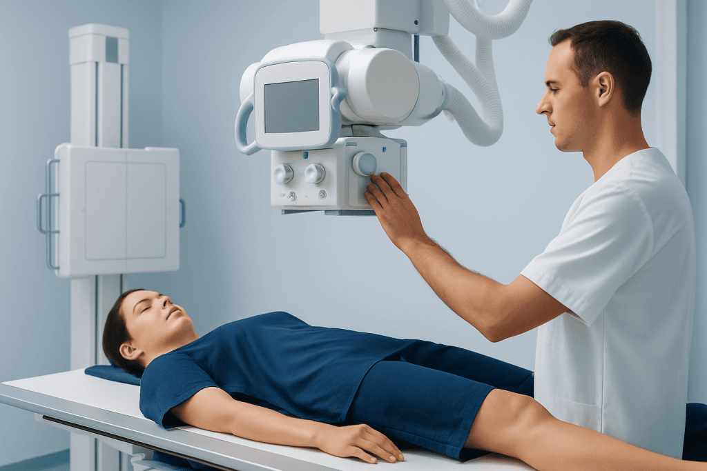
An X-ray machine produces a focused beam of high-energy photons that travels through the body to a detector. Different tissues absorb different amounts of the beam. Dense materials such as bone absorb more radiation and usually appear white or light on an image, while air-filled areas, including the lungs, appear darker. Soft tissues create varying shades of gray.
The examination is usually fast and noninvasive. It can help clinicians assess injuries, joint changes, chest symptoms, dental concerns, digestive problems, and the position of certain medical devices. A plain X-ray is different from computed tomography (CT), which uses multiple X-ray images and usually involves a higher radiation dose than a single standard radiograph.
Not all imaging uses ionizing radiation. Ultrasound uses sound waves, and magnetic resonance imaging (MRI) uses magnetic fields and radiofrequency energy. Depending on the clinical situation, these may be useful alternatives. However, they do not always provide the same information as an X-ray, and the most appropriate test depends on the suspected condition.
Who May Need an X-Ray and How Doctors Decide

Healthcare professionals do not order X-rays simply as a routine step. They first consider symptoms, physical examination findings, medical history, and whether the image is likely to change management. An X-ray may be appropriate after an injury with suspected fracture, for persistent respiratory symptoms, or when a clinician needs to evaluate a specific part of the body.
Children are more sensitive to radiation than adults because their tissues are still developing and they have more years ahead in which a radiation-related effect could theoretically occur. This does not mean children should avoid necessary imaging. Instead, pediatric examinations are carefully adjusted for body size, and clinicians may use ultrasound or other approaches when suitable.
People who are pregnant, may be pregnant, or are trying to conceive should tell the imaging team before the examination. Many X-rays, particularly those performed away from the abdomen and pelvis, may still be possible when clinically necessary. The team can review the urgency, body area being examined, and available alternatives to protect both parent and developing baby.
- Tell the team about possible pregnancy or breastfeeding.
- Share relevant prior imaging results if they are available.
- Ask what question the X-ray is intended to answer.
- Ask whether a non-radiation imaging test would be equally useful in the individual situation.
What Happens During an X-Ray Examination
Before the examination, the radiographer may ask the patient to remove jewelry, metal objects, or clothing items that could block the image. Depending on the body area, the patient may change into a gown. The radiographer explains how to stand, sit, or lie down and positions the body so that the needed structures can be seen clearly.
During image capture, the patient may be asked to remain still or briefly hold their breath. The radiographer usually steps behind a protective barrier or out of the room while activating the machine, but can see and communicate with the patient. The exposure itself lasts a fraction of a second, although positioning and taking several views can make the visit longer.
Protective equipment and careful collimation, which limits the beam to the area of interest, help reduce unnecessary exposure. Lead shielding may be used in selected situations, but current safety practice also emphasizes accurate positioning and limiting the exposed field. The imaging team follows established protocols to obtain a clear image with the minimum appropriate dose.
Images are reviewed by a radiologist or other qualified clinician, then reported to the referring healthcare professional. Results may be available quickly in urgent settings, while non-urgent reports can take longer depending on the service and the complexity of the findings.
Benefits, Risks, and Understanding Cumulative Exposure
The main benefit of an X-ray is timely, useful diagnostic information. A clear image can confirm or rule out important causes of symptoms, support treatment decisions, reduce the need for unnecessary procedures, and help monitor healing. In many situations, this immediate benefit is much more significant than the very small theoretical long-term risk associated with a diagnostic radiation dose.
Ionizing radiation can cause DNA changes. At the low doses used in diagnostic imaging, any cancer risk is estimated from population data and is difficult to measure precisely for an individual examination. Risk is not the same for every person: it can vary with age, the body part exposed, sex, and the total amount of radiation received over a lifetime.
Cumulative exposure matters because radiation doses from multiple examinations add over time. Still, this should not lead patients to decline tests that are clinically needed. Rather, it is a reason to avoid duplicate or low-value imaging, use prior images when possible, and choose the most suitable examination for each medical question.
There is no reliable way to predict whether a particular diagnostic X-ray will cause cancer in one person. A patient should not feel responsible for calculating this alone. The referring clinician and imaging professionals can explain why a study is advised and how its benefits and potential risks apply to the individual.
After the X-Ray: Results and Recovery Timeline
There is generally no recovery period after a standard X-ray. The patient can usually return to work, school, driving, eating, and normal activities immediately. The radiation does not remain in the body after an ordinary X-ray, and the patient does not become radioactive or pose a radiation risk to other people.
The next step depends on the findings and the reason for the examination. A normal image may help the clinician consider other explanations for symptoms. If an injury, infection, or other change is seen, the healthcare team may recommend treatment, follow-up, additional imaging, or referral to an appropriate specialist.
Occasionally, further imaging is needed because a plain X-ray cannot show every type of tissue or problem. For example, a small fracture may not be visible immediately after injury, or a clinician may need a more detailed view of soft tissue. Additional testing does not necessarily mean a serious condition is present; it may simply provide the detail required for an accurate diagnosis.
For international patients, Acibadem International’s multidisciplinary specialists and JCI-accredited hospitals can coordinate diagnostic imaging and follow-up care when clinically appropriate.
Reducing Unnecessary Radiation Exposure
Patients can support safe imaging by sharing previous X-rays, scans, and reports with their healthcare team. Access to earlier studies may prevent unnecessary repeat examinations and can help doctors compare changes over time. It is also helpful to inform the team about recent imaging performed elsewhere.
Before an examination, patients may ask whether the test is necessary now, what information it is expected to provide, and whether another test could be equally effective. These questions are not a reason to delay urgent care; instead, they encourage shared and informed decision-making.
Healthcare professionals reduce exposure through evidence-based referral guidelines, properly maintained equipment, dose-adjusted protocols, precise positioning, and avoiding repeat images whenever possible. For suspected bone and joint injuries, imaging may be part of evaluation alongside a clinical examination and other tools used in orthopedic care.
It is not necessary to avoid all medical imaging. The practical goal is appropriate imaging: the right test, at the right time, for a clear medical reason. When an X-ray is justified, its diagnostic value is usually considered to outweigh the small radiation-related risk.
When to Seek Medical Care
Medical evaluation is important after a significant injury, especially if there is severe pain, visible deformity, inability to bear weight or use a limb, numbness, weakness, or worsening swelling. Urgent assessment is also appropriate for chest pain, severe shortness of breath, coughing up blood, or symptoms that rapidly worsen. A clinician will decide whether an X-ray or another test is needed.
Patients should contact a healthcare professional if symptoms continue despite a normal X-ray, such as persistent bone pain after an injury, ongoing fever and cough, or unexplained worsening discomfort. Some conditions are not visible on a first X-ray, and follow-up may be needed based on symptoms and examination findings.
Anyone with concerns about frequent imaging, possible pregnancy, or a planned scan should discuss them with the referring clinician or radiology team. They can explain the purpose of the test, the expected exposure, and any reasonable alternatives. In an emergency, necessary imaging should not be postponed without medical advice.
Frequently asked questions
Can one X-ray cause cancer?
A single medically necessary standard X-ray is associated with a very small potential cancer risk. The dose is low, and it is not possible to determine that one specific diagnostic X-ray caused cancer in an individual. The benefit of diagnosing or excluding an important health problem usually outweighs this small risk.
Is a CT scan more risky than a standard X-ray?
A CT scan usually uses more radiation than a single plain X-ray because it captures many detailed images from different angles. CT can provide information that a standard X-ray cannot, so it may be the best test in certain circumstances. Clinicians consider the medical need and use dose-optimized protocols.
Does radiation stay in the body after an X-ray?
No. Radiation from a standard diagnostic X-ray passes through the body during the examination and does not remain afterward. The patient does not become radioactive and can safely be around family members, children, and pregnant people.
Should pregnant people avoid all X-rays?
Pregnancy should always be disclosed to the imaging team, but necessary X-rays are not automatically avoided. The team will assess the body area, urgency, and available alternatives, and will use methods to keep exposure as low as possible. Untreated illness or injury can also create risks, so decisions should be individualized.
How can patients lower their exposure to medical radiation?
Patients can keep records of previous imaging, provide earlier reports or images when available, and ask why a test is recommended. They can also ask whether ultrasound or MRI would answer the same clinical question. Necessary imaging should not be skipped without discussing it with a qualified clinician.
Why might a doctor order another X-ray after a normal result?
Some injuries and conditions may not be visible on an initial X-ray, especially early after symptoms begin. A repeat X-ray or a different imaging test may be recommended if pain, swelling, breathing symptoms, or other concerns persist. This helps ensure the diagnosis reflects the patient’s ongoing clinical condition.
References
- World Health Organization
- International Atomic Energy Agency
- U.S. Food and Drug Administration
- Radiological Society of North America
- American College of Radiology
This article is for general information only and is not a substitute for professional medical advice. Please consult a qualified doctor about your individual situation.
Oncology care in Turkey — second opinion and treatment plan
JCI-accredited · board-certified surgeons · reply within 24h
Add us as a Preferred Source to see more of our trusted health content across Google Search, AI Overviews and Discover.