Ear Anatomy — Explained by Medical Evidence, Not Myths
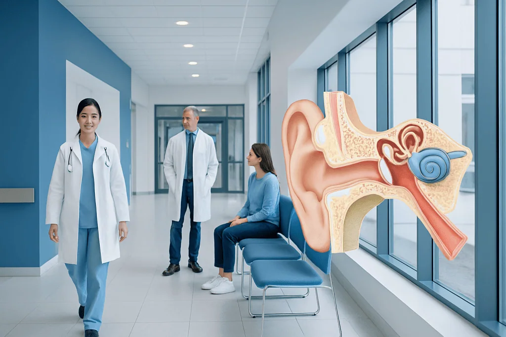
Ear anatomy is divided into the outer ear, middle ear, and inner ear. The ear does more than hear sound; it also helps the body maintain balance.
Key Takeaways
- Ear anatomy is divided into the outer ear, middle ear, and inner ear.
- The ear does more than hear sound; it also helps the body maintain balance.
- Sound travels from the ear canal to the eardrum, through tiny middle-ear bones, and into the inner ear.
- Symptoms such as pain, discharge, sudden hearing loss, or severe dizziness may point to an ear disorder and should be assessed by a doctor.
- Earwax, infections, pressure changes, aging, noise exposure, and injury can affect normal ear function.
Ear anatomy describes how the outer, middle, and inner ear are built and how they work together to support hearing and balance. A clear, evidence-based understanding of ear anatomy can also help explain common symptoms such as ear pain, hearing loss, ringing, dizziness, and pressure.
Overview: what ear anatomy means
Ear anatomy refers to the structure of the ear and the way its parts work together. In medical terms, the ear has three main sections: the outer ear, the middle ear, and the inner ear. These parts do not act separately; they form one connected system that captures sound, transfers vibrations, converts them into nerve signals, and helps the body sense movement and position.
This is why the ear is involved in both hearing and balance. Many myths suggest that the ear is simply a “hearing organ,” but medical evidence shows that the inner ear also contains highly specialized balance structures. When any part of this system is affected by infection, fluid, pressure changes, injury, or age-related changes, a person may notice symptoms such as reduced hearing, ringing, ear fullness, vertigo, or unsteadiness.
Understanding ear anatomy is helpful because it makes common ear problems easier to recognize. For example, pain in the ear canal often points to a different issue than dizziness caused by the inner ear. In clinical practice, doctors use anatomy to narrow down the possible cause of symptoms and to choose the most appropriate examination or treatment.
The outer ear: collecting and directing sound

The outer ear includes the visible ear on the side of the head, called the auricle or pinna, and the external auditory canal, often called the ear canal. The shape of the pinna helps collect sound waves from the environment and direct them into the ear canal. Although it may seem simple, this structure helps the brain detect where sounds are coming from.
The ear canal is lined with skin and tiny hairs, and it contains glands that produce earwax, also called cerumen. Earwax is not dirt; it is a normal protective substance that traps dust, helps reduce infection risk, and keeps the canal from becoming too dry. In many people, earwax naturally moves outward and does not need to be removed.
At the end of the canal is the tympanic membrane, or eardrum. This thin membrane separates the outer ear from the middle ear. When sound waves reach it, the eardrum vibrates. Problems in the outer ear, such as impacted wax, skin irritation, canal infection, or a foreign object, can interfere with the passage of sound and may cause discomfort or temporary hearing reduction.
- Main outer ear structures: pinna, ear canal, eardrum
- Main functions: collect sound, protect deeper structures, channel sound inward
- Common issues: wax blockage, swimmer’s ear, skin inflammation, trauma
The middle ear: turning sound into mechanical movement

The middle ear is a small air-filled space behind the eardrum. Inside it are three tiny bones called the ossicles: the malleus, incus, and stapes. These are sometimes referred to as the hammer, anvil, and stirrup. Their job is to transmit and amplify the eardrum’s vibrations so that sound energy can move efficiently into the inner ear.
The middle ear also connects to the back of the nose through the Eustachian tube. This tube helps equalize air pressure on both sides of the eardrum. When pressure is balanced, the eardrum moves more normally. This is why swallowing, yawning, or chewing may help relieve ear pressure during air travel, altitude changes, or a cold.
If the Eustachian tube does not work well, fluid may build up behind the eardrum and hearing may become muffled. Infections can also develop in the middle ear, especially after upper respiratory infections. These conditions are part of the reason many people search for explanations of ear infection symptoms and middle-ear pressure. Less commonly, changes to the ossicles or eardrum can lead to more persistent conductive hearing loss.
The inner ear: where hearing and balance begin
The inner ear contains delicate, fluid-filled structures that handle two major tasks: hearing and balance. The cochlea is the spiral-shaped hearing organ. Inside it are sensory hair cells that respond to sound vibrations delivered from the middle ear. These vibrations are converted into electrical signals, which then travel along the auditory nerve to the brain.
The balance part of the inner ear includes the semicircular canals, utricle, and saccule. These structures detect head movement, rotation, and changes in position. The brain combines this information with signals from the eyes and muscles to help keep posture stable and vision steady during movement.
Because the hearing and balance organs sit close together, inner-ear conditions can cause more than one symptom at the same time. A person may notice hearing loss together with ringing in the ears or episodes of vertigo. This is often why doctors consider inner-ear disorders when assessing dizziness, tinnitus, or sudden changes in hearing, including conditions related to vertigo.
How the ear works with the brain
Hearing is not completed by the ear alone. The ear captures and converts sound, but the brain interprets what those signals mean. After the cochlea turns vibrations into nerve impulses, the auditory nerve carries them to the brainstem and then to higher hearing centers. This process allows a person to recognize speech, music, warning sounds, and subtle changes in pitch or loudness.
Balance also depends on close communication between the inner ear, the eyes, the brain, and the muscles. When these systems send matching information, movement feels stable and coordinated. If one source sends conflicting signals, a person may feel dizzy, lightheaded, or off balance. That is why dizziness is not always caused by the inner ear, but the ear remains one of the most important places doctors evaluate.
This brain-ear connection also explains why two people with similar ear findings may describe symptoms differently. One may mainly notice muffled hearing, while another may be more aware of ringing or motion sensitivity. Careful clinical assessment helps determine whether the problem lies in sound conduction, sensory function, nerve signaling, or central processing.
Common conditions that affect ear anatomy and function
Different parts of ear anatomy are linked to different types of symptoms. Outer-ear problems may cause itching, pain when the ear is touched, or a feeling of blockage. Middle-ear conditions often cause pressure, temporary hearing reduction, or discomfort during colds and flights. Inner-ear problems are more likely to cause ringing, sensorineural hearing loss, vertigo, or imbalance.
Common causes include earwax impaction, external ear canal infection, middle-ear infection, fluid behind the eardrum, pressure dysfunction, age-related hearing changes, noise-induced hearing loss, and injury to the eardrum. Tinnitus may occur with several of these conditions, especially when hearing pathways are affected. In some cases, dizziness or hearing changes may need further testing to rule out less common disorders.
Evaluation depends on the symptoms and the part of the ear most likely involved. Doctors may examine the ear with an otoscope, test hearing, assess balance, or use imaging when needed. Some patients with structural or chronic ear problems may be referred for cochlear implant evaluation or ear surgery if conservative treatment is not enough, although many ear conditions are managed without surgery.
Protecting ear health and supporting normal function
Good ear care is usually simple and preventive. Avoid inserting cotton swabs, hairpins, or other objects into the ear canal, as these can push wax deeper, irritate the skin, or injure the eardrum. Keeping volume at safer levels, especially with headphones or earbuds, helps protect the cochlea’s delicate hair cells from long-term damage.
It can also help to protect the ears during swimming, noisy work, concerts, or activities with rapid pressure changes. People who fly often or dive may benefit from pressure-equalizing strategies recommended by a doctor. Managing allergies, nasal congestion, or sinus problems may also improve Eustachian tube function in some cases.
Self-care should stay within safe limits. Persistent earwax blockage, recurrent infections, repeated vertigo, or noticeable hearing decline should not be treated only at home. Hearing tests and specialist assessment can identify whether symptoms relate to the outer ear, middle ear, inner ear, or another cause entirely. Near the end of the care pathway, some patients may also need expert review for hearing loss treatment or vestibular assessment.
When to seek medical care
Medical care is appropriate when ear symptoms are severe, persistent, or unusual. A person should arrange an assessment for ongoing ear pain, discharge, repeated infections, hearing difficulty, ringing that does not improve, or dizziness that affects daily activities. Children, older adults, and people with diabetes or weakened immunity may need earlier medical review.
Urgent medical attention is especially important for sudden hearing loss, significant ear injury, severe vertigo, facial weakness, high fever with ear symptoms, or discharge after head trauma. These symptoms do not always mean a serious condition, but they do need prompt evaluation. Treatment is often most effective when the cause is identified early.
Assessment may involve primary care, an ENT specialist, audiology testing, or imaging depending on the findings. For international patients who need specialist evaluation, Acibadem International’s multidisciplinary teams and JCI-accredited hospitals diagnose and treat ear-related conditions using evidence-based ENT, audiology, and surgical care when indicated.
Frequently asked questions
What are the three main parts of ear anatomy?
The ear is divided into the outer ear, middle ear, and inner ear. The outer ear collects sound, the middle ear transfers vibrations, and the inner ear supports both hearing and balance.
What does the inner ear do?
The inner ear contains the cochlea for hearing and the vestibular system for balance. It converts sound vibrations into nerve signals and helps the brain detect motion and position.
Why do the ears pop during flights or altitude changes?
This happens because the Eustachian tube is working to equalize pressure between the middle ear and the outside environment. Swallowing or yawning often helps the pressure balance more comfortably.
Is earwax harmful?
Earwax is usually helpful, not harmful. It protects the ear canal by trapping particles and supporting the skin, but too much wax can sometimes build up and temporarily affect hearing or comfort.
Can ear problems cause dizziness?
Yes, some ear problems can cause dizziness, especially disorders involving the inner ear's balance organs. However, dizziness can also come from many non-ear causes, so proper medical evaluation is important.
When should someone worry about hearing loss?
A doctor should assess hearing loss that is sudden, getting worse, or affecting communication and daily life. Sudden hearing loss in one or both ears needs prompt medical attention because early treatment may matter.
References
- National Institute on Deafness and Other Communication Disorders
- American Academy of Otolaryngology–Head and Neck Surgery
- MedlinePlus
- Merck Manual Consumer Version
- Centers for Disease Control and Prevention
This article is for general information only and is not a substitute for professional medical advice. Please consult a qualified doctor about your individual situation.
Explore treatments in Turkey — costs, top hospitals & a free quote
JCI-accredited · board-certified surgeons · reply within 24h
Add us as a Preferred Source to see more of our trusted health content across Google Search, AI Overviews and Discover.