Echocardiogram Procedure: An Evidence-Based Patient Guide

A standard transthoracic echocardiogram is painless, uses no radiation and usually requires little preparation. The test can assess heart pumping strength, valve function, heart size, fluid around the heart and patterns of blood flow.
Key Takeaways
- A standard transthoracic echocardiogram is painless, uses no radiation and usually requires little preparation.
- The test can assess heart pumping strength, valve function, heart size, fluid around the heart and patterns of blood flow.
- Most people return to their usual activities immediately after a standard echocardiogram.
- Preparation differs for stress and transesophageal echocardiograms, so patients should follow their care team’s specific instructions.
- Echocardiogram findings are interpreted together with symptoms, examination findings and other tests.
An echocardiogram procedure is a noninvasive heart ultrasound that creates moving images of the heart’s chambers, valves and blood flow. It is commonly used to investigate symptoms, assess known heart conditions and help clinicians plan or monitor care.
Echocardiogram procedure explained
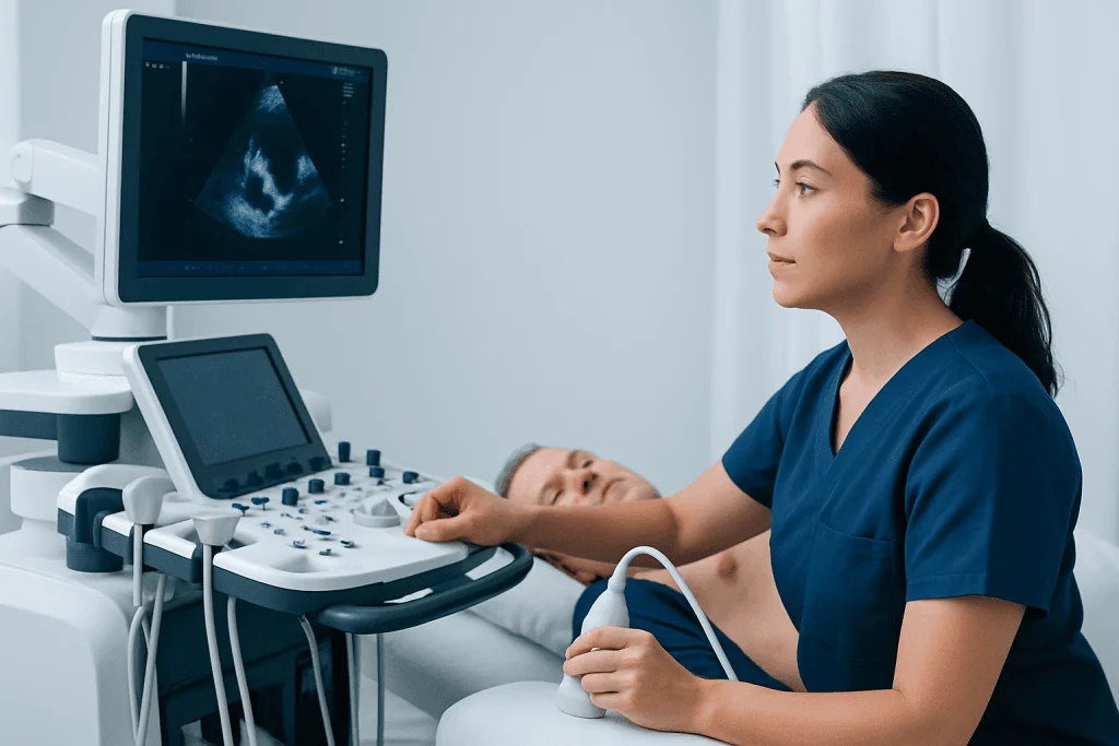
An echocardiogram procedure, often called an echo, is an ultrasound examination of the heart. A trained sonographer places a small device called a transducer on the chest. The device sends harmless sound waves into the body and receives returning echoes, which a computer converts into detailed moving pictures of the heart.
A standard transthoracic echocardiogram (TTE) can show the heart chambers, the heart muscle, the valves and major blood vessels near the heart. Doppler ultrasound may also measure the direction and speed of blood flow. This helps clinicians evaluate how effectively the heart pumps and whether blood flow through a valve appears restricted or leaky.
The examination does not involve ionizing radiation, needles or surgery. It is often ordered when a clinician needs more information about symptoms such as shortness of breath, palpitations, chest discomfort, fainting or ankle swelling, or to follow a known heart condition. An echo may support assessment of conditions such as heart valve disease and heart muscle disorders, but it is only one part of a complete cardiac evaluation.
Who may need an echocardiogram and how it works
A doctor may recommend an echocardiogram when there is a heart murmur, an abnormal electrocardiogram, a family or personal history of heart disease, or symptoms that could be related to heart function. It may also be used before or after certain treatments, after a heart attack, during pregnancy when medically indicated, or to monitor changes in a previously diagnosed condition.
Different echo types answer different clinical questions. A transthoracic echo is performed through the chest wall and is the most common type. A transesophageal echocardiogram (TEE) uses an ultrasound probe passed into the esophagus, allowing closer images of certain heart structures. A stress echocardiogram records heart images before and after exercise or medication that makes the heart work harder. Contrast-enhanced echo may be used when standard images are not clear enough.
Although echocardiogram protocols are designed for safety and consistency, the exact views and measurements vary according to the person’s symptoms and reason for testing. The referring clinician and cardiac imaging team choose the appropriate method, taking account of medical history, mobility, swallowing difficulties, lung disease and any medicines being taken.
What to expect from your first echocardiogram

For a first standard echocardiogram, patients typically check in, review their medical history and change into a gown that opens at the front. The sonographer explains the process and may attach small adhesive electrodes to the chest to record the heart rhythm during imaging. Questions are welcome before the examination begins.
The patient usually lies on the left side on an examination couch, sometimes with the left arm raised or placed behind the head. Warm, water-based gel is applied to areas of the chest. The sonographer moves the transducer over the skin and may ask the patient to hold their breath briefly or change position to obtain clearer views.
Some pressure from the transducer is normal, particularly between the ribs, but a standard TTE should not be painful. The room may be dimmed so the sonographer can see the monitor. Images and measurements are recorded for a cardiologist or appropriately trained physician to review; the sonographer may not be able to discuss final findings during the appointment.
Echocardiogram procedure step by step
A typical transthoracic echocardiogram procedure follows a clear sequence. First, the team confirms the patient’s identity, reason for the test and relevant medical details. The patient then removes clothing from the waist up and wears a gown, while staff maintain privacy and only uncover the area needed for imaging.
Next, electrodes may be placed on the chest and gel is applied. The sonographer obtains images from several standard positions, often along the left side of the breastbone, near the tip of the heart and just below the rib cage. The transducer may be angled in different directions to visualize the chambers, wall movement and valves.
Doppler recordings are then used to assess blood flow. The machine may produce whooshing sounds, which are normal. In some cases, an ultrasound contrast agent is injected through an intravenous line to improve definition of the heart’s inner border; this is not needed for everyone. After imaging is complete, the gel and electrodes are removed and the patient can dress.
The recorded study is reviewed, and a report is sent to the clinician who requested it. Depending on the facility and clinical situation, preliminary or final results may be discussed the same day or at a follow-up appointment. Further evaluation may include other cardiac tests when needed, including cardiology assessment and treatment planning.
What shouldn't I do before an echocardiogram?
For a routine transthoracic echocardiogram, most people can eat, drink and take their regular medicines as usual unless their clinician gives different instructions. It is sensible to wear comfortable two-piece clothing, avoid applying heavy oils or lotions to the chest on the day of the test, and bring a list of medicines and relevant medical information.
Patients should not stop prescribed medicines, including heart medicines, without clear advice from the clinician arranging the test. They should tell the team about pregnancy, implanted cardiac devices, allergies, breathing difficulties and any limitation that could affect positioning during the examination.
Preparation changes for specialized tests. A transesophageal echocardiogram generally requires fasting for a specified period because sedation and an esophageal probe are involved. A stress echocardiogram may require avoiding food, caffeine, tobacco or selected medicines beforehand. These echocardiogram procedure instructions differ by protocol, so the testing center’s directions should always take priority over general advice.
How long does an echogram usually take?
A standard transthoracic echocardiogram commonly takes about 30 to 60 minutes, although the appointment may be longer to allow time for registration, preparation and discussion. Image quality, the number of measurements needed and the person’s ability to change positions can affect the duration.
A stress echocardiogram often takes longer because it includes baseline imaging, monitored exercise or medication-based stress, and repeat imaging. A transesophageal echocardiogram also requires additional time for fasting checks, sedation preparation, recovery monitoring and arranging a responsible adult to take the patient home.
Patients can ask when and how their results will be available before leaving. Urgent findings are generally communicated promptly to the treating clinical team, while nonurgent results are often reviewed in a scheduled consultation so they can be interpreted in the context of the individual’s health.
Will my breasts be exposed during an echocardiogram?
For a transthoracic echocardiogram, the chest needs to be accessible because images are obtained through the skin between the ribs. However, staff should protect privacy throughout the examination. Patients are generally given a gown or drape, and only the small area being scanned should be uncovered at a time.
For many women, the sonographer may need to place the transducer near or beneath the left breast to obtain accurate images of the heart. The breast may be gently moved or supported as necessary, but it does not need to remain exposed beyond what is required for imaging. Patients may request a chaperone, ask for an explanation before repositioning, or state any comfort preferences.
Clear communication is an important part of respectful care. If a patient feels uncomfortable at any stage, they can ask the sonographer to pause, adjust the draping or explain what is needed and why. These requests should not affect the patient’s ability to receive appropriate testing.
Benefits, recovery, risks and when to seek medical care
The main benefit of an echocardiogram is that it provides real-time information about heart structure and function without radiation exposure. It can help identify reduced pumping function, changes in chamber size, valve abnormalities, pressure-related changes and fluid around the heart. It may also guide decisions about monitoring, medicines, procedures or referral to a cardiac specialist.
Recovery after a standard TTE is immediate. Patients can usually eat, drive, work and resume normal activities right away. Mild temporary skin irritation can occur where electrodes were attached. A contrast agent has a small risk of side effects or allergic reaction, and the team monitors patients when it is used. TEE and stress echo have additional, uncommon risks related to sedation, the esophageal probe or exercise, which are explained during consent.
Patients should seek urgent medical care for severe or persistent chest pain, severe shortness of breath, fainting, new weakness on one side of the body, blue lips, or a rapidly worsening heartbeat sensation. These symptoms should not wait for a routine outpatient echocardiogram. For nonurgent symptoms that are new, recurrent or becoming worse, arranging a medical review is appropriate.
Acibadem International’s multidisciplinary specialists and JCI-accredited hospitals evaluate heart concerns and provide diagnostic and treatment planning for international patients. Ongoing care may include assessment for heart valve surgery or other individualized cardiac treatments when clinically appropriate.
Frequently asked questions
Is an echocardiogram painful?
A standard transthoracic echocardiogram is usually painless. The transducer can create mild pressure against the chest, especially between the ribs, but patients should tell the sonographer if it becomes uncomfortable.
Do I need to fast before an echocardiogram?
Fasting is usually not required before a standard transthoracic echocardiogram. It is commonly required before a transesophageal echocardiogram, and preparation may also differ for a stress echo, so patients should follow the instructions from their testing team.
Can an echocardiogram detect blocked arteries?
An echocardiogram does not directly show coronary artery blockages in the same way as coronary CT angiography or cardiac catheterization. However, it may show reduced heart muscle movement that can suggest reduced blood supply, particularly during a stress echocardiogram.
Can I drive after an echocardiogram?
Most people can drive after a standard transthoracic echocardiogram because no sedative is used. Patients should not drive after a transesophageal echocardiogram if they have received sedation and will need a responsible adult to take them home.
What does an abnormal echocardiogram mean?
An abnormal result can refer to many different findings, including changes in heart pumping strength, valve function, chamber size or blood flow. It does not automatically mean a serious problem, and the result should be discussed with the clinician who knows the patient’s symptoms, history and other test results.
How often should an echocardiogram be repeated?
The timing depends on why the test was requested and whether a heart condition is being monitored. Some people need only one examination, while others with valve disease, heart failure or treatment-related monitoring may need periodic studies recommended by their cardiology team.
References
- American Heart Association
- American Society of Echocardiography
- Mayo Clinic
- National Heart, Lung, and Blood Institute
This article is for general information only and is not a substitute for professional medical advice. Please consult a qualified doctor about your individual situation.
Explore treatments in Turkey — costs, top hospitals & a free quote
JCI-accredited · board-certified surgeons · reply within 24h
Add us as a Preferred Source to see more of our trusted health content across Google Search, AI Overviews and Discover.