Elbow Anatomy: What Patients Need to Know
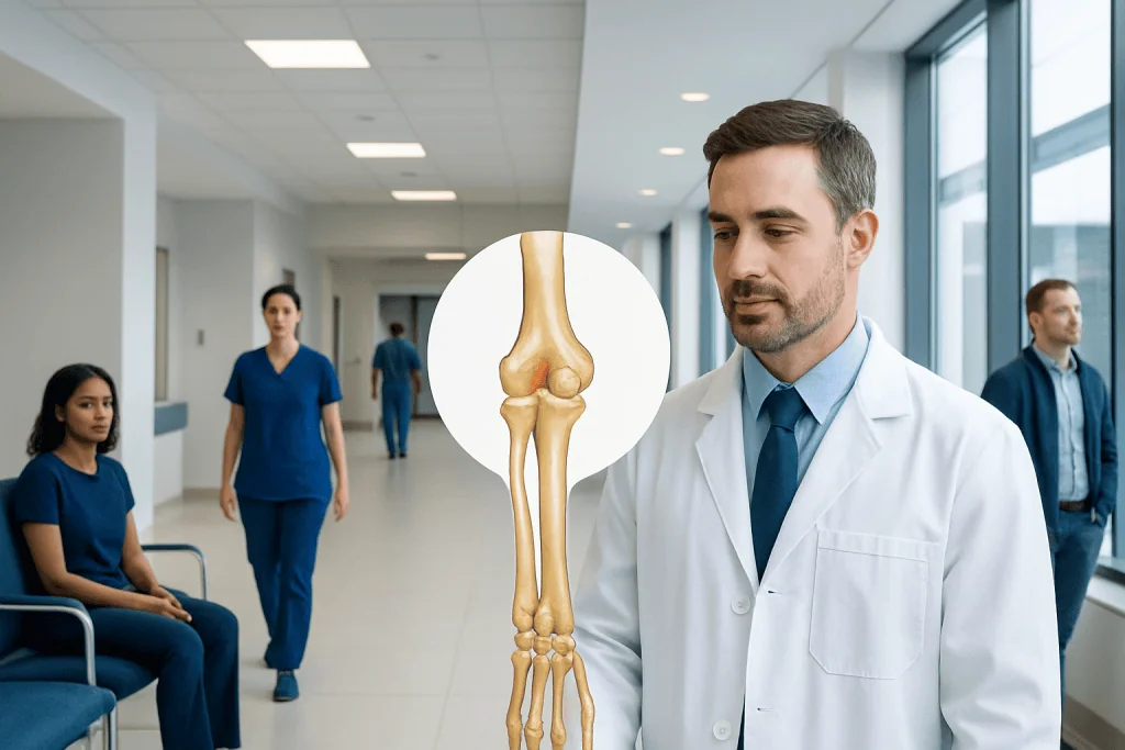
The elbow is made of three bones: the humerus, radius, and ulna. Three linked joints allow bending, straightening, and forearm rotation.
Key Takeaways
- The elbow is made of three bones: the humerus, radius, and ulna.
- Three linked joints allow bending, straightening, and forearm rotation.
- Ligaments provide stability, while muscles and tendons create movement.
- Important nerves pass close to the elbow and can cause tingling or weakness if irritated.
- Pain location and symptom pattern often help doctors identify which elbow structure is affected.
Elbow anatomy refers to the bones, joints, ligaments, muscles, tendons, nerves, and blood vessels that work together to move the arm and hand. Understanding these structures can help patients make sense of common symptoms such as pain, stiffness, weakness, numbness, or reduced motion.
Overview of elbow anatomy
Elbow anatomy includes the structures that allow the arm to bend, straighten, turn the palm up, and turn the palm down. In simple terms, the elbow is not a single hinge but a compact system of bones, cartilage, ligaments, muscles, tendons, nerves, and blood vessels working together. Because the elbow connects the upper arm to the forearm, it plays an important role in daily tasks such as lifting, reaching, dressing, typing, and gripping.
For patients, understanding elbow anatomy can make symptoms easier to describe and less confusing. Pain on the inner elbow, outer elbow, back of the elbow, or front of the elbow may point to different tissues. Numbness in the ring and little fingers, for example, may suggest nerve irritation, while pain with gripping may suggest tendon overload. Learning the basic parts of the elbow helps patients have clearer discussions with their doctor.
The elbow also links shoulder and wrist function. If one part of the arm is weak or stiff, the elbow may take on extra stress. This is why elbow symptoms may develop not only after a direct injury, but also from repeated movement, sports, work tasks, or strain over time.
Bones and joints of the elbow
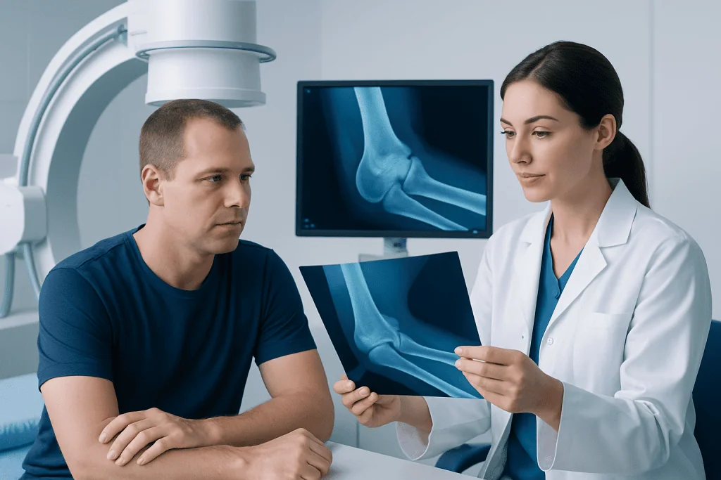
The elbow is formed by three bones: the humerus in the upper arm, and the radius and ulna in the forearm. The lower end of the humerus has shaped surfaces that meet the top ends of the radius and ulna. These surfaces fit together closely, which helps guide motion while maintaining stability.
Patients are often told the elbow is a hinge joint, but it is better understood as a joint complex with three connected joints inside one capsule. The humeroulnar joint mainly allows bending and straightening. The humeroradial joint helps with both flexion-extension and load transfer through the forearm. The proximal radioulnar joint allows the radius to rotate around the ulna, making it possible to turn the palm upward and downward.
Smooth cartilage covers the ends of the bones where they meet. This cartilage reduces friction and helps movement stay comfortable and efficient. When cartilage is damaged by injury, wear, inflammation, or degeneration, the result may be pain, catching, stiffness, or a grinding sensation.
The bony points patients can feel are also clinically important. The tip of the elbow is the olecranon, part of the ulna. The inner bump is the medial epicondyle, and the outer bump is the lateral epicondyle, both part of the humerus. These areas serve as attachment points for tendons and ligaments, which is why they often become tender in overuse conditions.
Ligaments, capsule, and other stabilizing structures

Elbow stability depends on more than bone shape alone. A joint capsule surrounds the elbow, helping hold the joint together while allowing movement. Several ligaments reinforce this capsule and prevent abnormal motion, especially during lifting, pushing, throwing, or falling on an outstretched hand.
The main stabilizers on the inner side are often grouped as the medial or ulnar collateral ligament complex. These ligaments resist forces that push the forearm outward relative to the upper arm, particularly during overhead throwing. On the outer side, the lateral collateral ligament complex helps resist rotational instability. If these ligaments are stretched or torn, the elbow may feel painful, weak, or unstable.
Small fluid-filled sacs called bursae also reduce friction around the joint. The olecranon bursa sits over the tip of the elbow and can become irritated or swollen after repeated pressure, direct impact, or inflammation. This may lead to a soft, localized swelling at the back of the elbow.
When these stabilizing structures are injured, symptoms may differ from muscle pain. Patients may notice a sense of looseness, clicking, pain with pushing up from a chair, or discomfort during sports that involve forceful arm motion. In some cases, doctors may evaluate for tennis elbow or other nearby soft tissue problems if symptoms overlap.
Muscles and tendons that move the elbow
Muscles move the elbow by pulling through tendons attached to bone. The biceps at the front of the upper arm helps bend the elbow and turn the palm upward. The brachialis, located deeper in the front of the arm, is another major elbow flexor. The triceps at the back of the upper arm straightens the elbow. These larger muscles generate much of the force needed for lifting, carrying, pushing, and pulling.
The elbow is also closely connected to the muscles of the forearm. Groups of flexor muscles on the inner side of the forearm help bend the wrist and fingers, while extensor muscles on the outer side help straighten them. Their tendons attach near the medial and lateral epicondyles. Repetitive gripping, lifting, tool use, racket sports, and prolonged computer work can overload these tendon attachments.
Tendon problems around the elbow are common and often develop gradually. Pain on the outer elbow is frequently related to irritation of the wrist extensor tendons, while pain on the inner elbow may involve the wrist flexor-pronator tendons. These conditions are often described as overuse injuries, even though they can affect people who do not play sports.
In some situations, a doctor may recommend targeted rehabilitation such as physical therapy and rehabilitation to improve flexibility, strength, movement patterns, and recovery. Treatment depends on the exact structure involved and whether symptoms are related to strain, inflammation, degeneration, or a tear.
Nerves and blood vessels around the elbow
Several important nerves pass through or near the elbow, which is why elbow problems can sometimes cause symptoms in the hand. The ulnar nerve runs behind the inner side of the elbow through a narrow space often called the cubital tunnel. This is the nerve involved when striking the “funny bone” causes a brief electric-like sensation. If the ulnar nerve becomes irritated or compressed, patients may notice numbness or tingling in the ring and little fingers, hand weakness, or symptoms that worsen when the elbow is bent for long periods.
The median nerve and radial nerve also pass near the elbow. Irritation of these nerves can contribute to pain, altered sensation, or weakness in different parts of the forearm and hand. Because nerve symptoms can mimic tendon or joint problems, doctors usually consider the location of tingling, changes in strength, and whether symptoms are brought on by movement or position.
Arteries and veins also travel through the elbow region. The brachial artery runs in front of the elbow and divides into the radial and ulnar arteries below the joint. Healthy blood flow supports muscle and nerve function and tissue healing after injury. While vascular problems around the elbow are less common than tendon or nerve problems, doctors check circulation when assessing trauma or severe swelling.
A careful physical examination helps separate muscle-tendon pain from nerve compression. In selected cases, imaging or nerve testing may be used if symptoms suggest conditions such as ulnar nerve compression or another nerve-related disorder around the elbow.
How elbow anatomy relates to common symptoms and conditions
Knowing the anatomy of the elbow can help patients understand why symptoms vary so much. Outer elbow pain often involves the common extensor tendon. Inner elbow pain may involve the flexor-pronator tendon group or the ulnar nerve. Pain at the back of the elbow may be linked to the triceps tendon, olecranon bursa, or joint irritation. Pain at the front of the elbow may relate to the biceps tendon or the joint itself.
Stiffness can happen after injury, arthritis, inflammation, or prolonged immobilization. Swelling may suggest bursitis, joint inflammation, bleeding into the joint after trauma, or infection. Clicking, locking, or catching can occur with cartilage injury, loose bodies, or joint surface problems. Weakness may come from pain, tendon injury, nerve compression, or muscle deconditioning.
Some of the more common elbow conditions include tendinopathies such as golfer’s elbow, ligament sprains, bursitis, fractures, dislocations, arthritis, and nerve entrapment. Symptoms can overlap, so it is not always possible for patients to identify the exact cause based on location alone. A structured assessment is often the best way to clarify the diagnosis.
If symptoms persist, worsen, or interfere with work, sleep, sports, or daily activities, medical evaluation can help identify whether the problem is in the joint, tendon, ligament, nerve, or surrounding soft tissue. This is especially important after a fall, direct blow, or sudden loss of strength.
How doctors assess elbow problems
Doctors begin by asking about symptoms, recent injuries, work demands, sports, hand use, and the pattern of pain or numbness. Details matter. Pain that starts after repetitive gripping suggests a different problem from pain that begins after a fall. Tingling in specific fingers may point toward nerve involvement, while swelling and limited motion may suggest joint or bursal inflammation.
During the physical examination, the doctor checks tenderness, swelling, range of motion, strength, joint stability, and nerve function. They may ask the patient to bend and straighten the elbow, rotate the forearm, grip the hand, or resist wrist movement. These simple maneuvers can help identify which structure is irritated.
Imaging is not always needed, but it can be helpful in some situations. X-rays are commonly used to look for fractures, arthritis, dislocation, bone spurs, or alignment problems. Ultrasound can assess tendons, fluid collections, and some nerve issues. MRI may be used when deeper soft tissue injury, ligament damage, cartilage injury, or persistent unexplained symptoms are suspected. When nerve compression is a concern, nerve conduction studies or electromyography may be considered.
If non-surgical measures do not improve symptoms, or if there is significant structural damage, treatment planning may involve orthopedic evaluation and, in selected cases, orthopedic surgery. The goal is to match treatment to the exact anatomy involved rather than treating all elbow pain the same way.
Protecting elbow health and when to seek medical care
Many elbow problems can be reduced by balancing activity and recovery. Helpful measures include using proper technique during sports or repetitive tasks, improving workstation ergonomics, varying movements during long periods of hand use, and building shoulder, forearm, and wrist strength gradually. Warming up before exercise and avoiding sudden increases in training load can also lower strain on tendons and ligaments.
At home, early self-care for mild symptoms may include relative rest, avoiding the movement that triggers pain, using ice for short periods after overuse, and returning to activity gradually. Patients should avoid pushing through sharp pain or ongoing numbness. If a brace, splint, or exercise plan is considered, it is best guided by a qualified clinician so that the underlying problem is not missed.
Medical care is recommended if elbow pain follows trauma, if the joint looks deformed, if swelling is marked, or if the patient cannot move the arm normally. A doctor should also assess symptoms such as persistent pain lasting more than a few weeks, repeated locking or catching, fever with swelling, visible redness, hand weakness, or numbness and tingling that do not settle. These features do not always mean a serious problem, but they do deserve evaluation.
For international patients who need assessment or treatment, Acibadem International’s multidisciplinary specialists and JCI-accredited hospitals diagnose and manage elbow conditions with imaging, rehabilitation, and surgical care when needed, including arthroscopy in selected joint problems.
Frequently asked questions
What are the main parts of the elbow?
The elbow is made up of three bones: the humerus, radius, and ulna. It also includes joints, cartilage, ligaments, muscles, tendons, nerves, bursae, and blood vessels that work together to support movement and stability.
Is the elbow just a hinge joint?
Not exactly. The elbow acts like a hinge for bending and straightening, but it also includes a joint that allows the forearm to rotate so the palm can turn up and down. This is why elbow anatomy is more complex than a simple hinge.
Why does elbow pain happen in different places?
Different parts of the elbow can produce pain in different locations. Outer elbow pain often relates to extensor tendons, inner elbow pain may involve flexor tendons or the ulnar nerve, and back-of-elbow pain may come from the bursa, triceps tendon, or joint.
What does numbness in the ring and little fingers mean?
This symptom can happen when the ulnar nerve is irritated or compressed near the elbow. It may become more noticeable when the elbow stays bent for a long time, such as during sleep, phone use, or desk work.
Can elbow problems improve without surgery?
Yes, many elbow problems improve with non-surgical treatment. Rest, activity modification, rehabilitation, bracing in some cases, and guided exercises are often effective, depending on the cause and severity.
When should someone see a doctor for elbow symptoms?
Medical evaluation is important after an injury, if the elbow is swollen or hard to move, or if symptoms include weakness, numbness, or ongoing pain. A doctor can determine whether the issue involves bone, tendon, ligament, nerve, or the joint itself.
References
- American Academy of Orthopaedic Surgeons
- MedlinePlus
- National Institute of Arthritis and Musculoskeletal and Skin Diseases
- Mayo Clinic
- Cleveland Clinic
This article is for general information only and is not a substitute for professional medical advice. Please consult a qualified doctor about your individual situation.
Joint and spine care in Turkey — expert assessment & treatment
JCI-accredited · board-certified surgeons · reply within 24h
Add us as a Preferred Source to see more of our trusted health content across Google Search, AI Overviews and Discover.