Encephalocele: An Evidence-Based Guide for Patients

Encephalocele is present at birth and happens when the skull does not fully close during early development. Symptoms can range from a visible sac on the head to feeding, breathing, developmental, or neurological problems.
Key Takeaways
- Encephalocele is present at birth and happens when the skull does not fully close during early development.
- Symptoms can range from a visible sac on the head to feeding, breathing, developmental, or neurological problems.
- Diagnosis often begins before birth with ultrasound and is confirmed with imaging such as MRI or CT after delivery.
- Treatment commonly involves coordinated care from pediatric, neurosurgical, and rehabilitation specialists.
- Early evaluation helps guide safe care, monitor complications, and support development over time.
Encephalocele is a rare congenital condition in which brain tissue, membranes, or fluid push through an opening in the skull. Its effects vary widely, so diagnosis and treatment depend on the location, size, and associated brain or developmental changes.
Overview
Encephalocele is a rare neural tube-related birth defect in which part of the brain, its protective coverings, or cerebrospinal fluid protrudes through an opening in the skull. In simple terms, a portion of the skull does not form completely during fetal development, leaving a gap through which tissues may bulge outward. The condition is present at birth, but its impact can differ greatly from one child to another.
The location of the opening matters. An encephalocele may occur in the back of the head, the front of the skull near the forehead or nose, or less commonly in other areas. Some encephaloceles contain mainly fluid and membranes, while others include brain tissue. This helps explain why some children have relatively limited symptoms and others need complex medical support.
Encephalocele is not the same as many other skull or spine conditions, although it belongs to the broader group of congenital central nervous system abnormalities. Families may also hear related terms such as spina bifida, another neural tube defect that affects the spinal column rather than the skull. A careful evaluation helps doctors understand the exact anatomy and what it may mean for growth, brain function, and treatment planning.
Symptoms and Possible Effects
The most noticeable sign of encephalocele is often a sac-like protrusion from the skull. This may be seen on the back of the head or around the forehead and nose, depending on the type. In some babies, the swelling is obvious at birth, while in others the abnormality may be smaller or partly hidden by nearby structures.
Symptoms depend on how much tissue is involved and whether other parts of the brain developed differently. Some children may have no major immediate symptoms beyond the visible opening. Others may develop neurological or physical concerns such as seizures, weakness, problems with coordination, delays in reaching developmental milestones, feeding difficulties, or vision problems.
Possible associated issues can include:
- Hydrocephalus, which is a buildup of fluid in or around the brain
- Differences in facial or skull shape
- Breathing or swallowing difficulties in some front-of-skull cases
- Learning, speech, or developmental challenges
- Increased risk of infection if tissues are exposed or the covering is fragile
Not every child will experience all of these concerns. The range is broad, and outcomes depend on the location of the encephalocele, the amount of brain tissue involved, and whether there are additional congenital abnormalities. For this reason, doctors usually recommend an individualized care plan rather than making assumptions from the diagnosis name alone.
Causes and Risk Factors

Encephalocele develops very early in pregnancy, when the structures that form the brain, skull, and neural tube are still closing and organizing. In many cases, there is no single clear cause. It is generally understood as a congenital developmental abnormality that can result from a combination of genetic and environmental influences.
Researchers have identified several factors that may increase risk, but these do not explain every case. Risk may be higher with certain genetic syndromes or chromosomal conditions, a family history of neural tube defects, and inadequate folate intake before conception and during early pregnancy. Exposure to some harmful substances or poorly controlled maternal medical conditions may also play a role in some pregnancies.
It is important for families to know that encephalocele is not usually caused by anything a parent knowingly did or did not do. Feelings of guilt are common after any congenital diagnosis, but most cases arise from complex developmental processes outside anyone’s control. Genetic counseling can help families understand recurrence risk and discuss future pregnancy planning in a supportive, practical way.
How Encephalocele Is Diagnosed
Many cases of encephalocele are detected before birth. A routine prenatal ultrasound may show a skull defect or a protruding sac, prompting more detailed imaging. Fetal MRI can sometimes provide additional information about the contents of the sac and whether there are related brain abnormalities, helping the medical team plan delivery and newborn care.
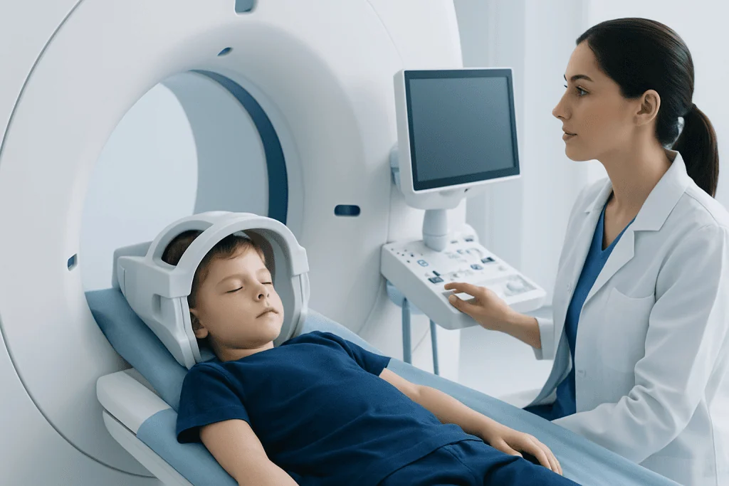
After birth, doctors begin with a physical examination and then use imaging to define the anatomy. MRI is often especially useful because it shows soft tissues, brain structures, and the relationship between the protrusion and the skull opening. CT scanning may also be used to examine the skull bones in detail, while ultrasound may help in selected newborn situations.
Because encephalocele can occur alongside other neurological conditions, the evaluation may also include testing for hydrocephalus, vision or hearing concerns, and developmental needs. In some children, the care team assesses whether the encephalocele is linked to hydrocephalus or other structural findings that may affect treatment timing. This diagnostic process is designed not only to confirm the condition, but also to guide safe, individualized care over time.
Treatment Options and Ongoing Care
Treatment for encephalocele depends on the child’s overall health, the size and location of the defect, and whether brain tissue is involved. In many cases, surgery is the main treatment. The goals are to return or protect tissues as appropriate, close the skull opening, reduce the risk of infection, and support the best possible function and appearance.
Surgical planning may involve pediatric neurosurgeons, craniofacial surgeons, neonatologists, neurologists, anesthesiologists, and rehabilitation specialists. Depending on the anatomy, treatment may include neurosurgery to repair the defect and manage pressure-related complications. If hydrocephalus is present, some children may need additional procedures such as brain shunt surgery to divert excess cerebrospinal fluid.
Not all concerns are resolved by one operation. Some children need staged care, follow-up imaging, seizure management, physical therapy, occupational therapy, speech support, or developmental monitoring. If the encephalocele affects facial structures or the skull shape, reconstructive planning may also be considered as the child grows.
Long-term outcomes vary. Some children do very well with timely treatment and monitoring, while others need ongoing support for neurological or developmental challenges. Families benefit most from regular follow-up with specialists who can watch for changes, coordinate therapies, and adjust the care plan as the child develops.
Daily Care, Prevention, and Family Support
There is no home treatment that can correct encephalocele, but daily care plays an important role in protecting the child and supporting development. Parents and caregivers should follow the medical team’s instructions on positioning, wound care after surgery, feeding, medications, and signs of complications. If there is a visible sac before repair, it should be handled gently and protected from pressure or injury.
For future pregnancies, prevention discussions often focus on folic acid and early prenatal care. Adequate folate intake before conception and in early pregnancy is known to lower the risk of some neural tube defects. While this cannot prevent every case of encephalocele, it is an important evidence-based step that healthcare professionals commonly recommend.
Family support is also part of treatment. A diagnosis involving the brain or skull can be emotionally overwhelming, especially when it is made during pregnancy or soon after birth. Clear communication with the medical team, developmental follow-up, and access to counseling or parent support resources can help families feel more prepared and less isolated.
In selected cases, children may also need rehabilitation or supportive therapies as they grow. The aim is to help with movement, communication, feeding, learning, and independence according to each child’s needs rather than a fixed expectation.
When to Seek Medical Care
Medical attention is needed promptly whenever encephalocele is suspected during pregnancy or after birth. A visible swelling on a newborn’s head, an unusual mass near the nose or forehead, poor feeding, breathing difficulty, repeated vomiting, seizures, or unusual sleepiness should all be assessed by a qualified doctor without delay. Early evaluation helps reduce risks and allows specialists to plan imaging and treatment safely.
Families should also seek medical review if a child with known encephalocele develops fever, redness or leakage around a surgical site, increasing head size, irritability, worsening headaches, new weakness, or developmental regression. These symptoms may suggest infection, pressure changes, or another complication that needs timely care.
Because management often requires several specialties, many families are referred to centers with pediatric neurosurgery and advanced imaging expertise. Acibadem International’s multidisciplinary specialists and JCI-accredited hospitals diagnose and treat complex conditions such as encephalocele for international patients, including coordinated assessment and pediatric neurosurgery when appropriate.
Frequently asked questions
Is encephalocele the same as spina bifida?
No. Both are congenital neural tube-related defects, but encephalocele affects the skull and tissues connected to the brain, while spina bifida affects the spine. They are related in developmental origin but involve different parts of the nervous system.
Can encephalocele be seen before birth?
Yes, many cases are identified during pregnancy on prenatal ultrasound. If doctors need more detail, fetal MRI may help clarify the size, location, and contents of the protrusion.
Does every baby with encephalocele need surgery?
Many babies do need surgery, but the timing and exact approach depend on the anatomy and the child's overall condition. Specialists decide this after imaging and examination, with the goals of protecting the brain, closing the defect, and reducing complications.
What problems can happen along with encephalocele?
Some children may have hydrocephalus, seizures, developmental delay, feeding issues, or vision and coordination problems. Others may have fewer associated concerns, especially if the defect is smaller or involves less brain tissue.
Can encephalocele be prevented?
Not all cases can be prevented. However, adequate folic acid intake before pregnancy and in early pregnancy, along with regular prenatal care, may help reduce the risk of some neural tube defects.
What is the outlook for a child with encephalocele?
The outlook varies widely from child to child. It depends on the location and size of the defect, whether brain tissue is involved, and whether there are other brain or developmental conditions. Early specialist care can help families understand the expected course and available support.
This article is for general information only and is not a substitute for professional medical advice. Please consult a qualified doctor about your individual situation.
Explore treatments in Turkey — costs, top hospitals & a free quote
JCI-accredited · board-certified surgeons · reply within 24h
Add us as a Preferred Source to see more of our trusted health content across Google Search, AI Overviews and Discover.

More from the Health Library

Herbal Tea: An Evidence-Based Guide for Patients

Agitation: A Complete Medical Overview

Menorrhagia: An Evidence-Based Guide for Patients

Cathisophobia: What Patients Need to Know

Sugar Free Gummy Bears: A Complete Medical Overview