Endovascular Neurosurgery for Stroke: Who May Benefit and How Fast It Must Happen

This treatment is mainly used for ischemic stroke caused by a blocked large brain artery. Mechanical thrombectomy can improve outcomes when performed quickly in carefully selected patients.
Key Takeaways
- This treatment is mainly used for ischemic stroke caused by a blocked large brain artery.
- Mechanical thrombectomy can improve outcomes when performed quickly in carefully selected patients.
- Not every stroke is treated this way, so urgent brain imaging is needed to decide.
- Stroke symptoms are a medical emergency and require immediate emergency care.
- Treatment decisions depend on timing, brain scans, clot location, and the patient’s overall condition.
Endovascular neurosurgery for stroke is a minimally invasive treatment used to remove or bypass a clot in certain types of ischemic stroke. It can be highly effective for selected patients, but fast recognition, emergency transport, and urgent brain imaging are essential.
Overview
Endovascular neurosurgery for stroke refers to minimally invasive procedures performed inside the blood vessels to treat certain kinds of stroke. In practice, this most often means mechanical thrombectomy, a procedure in which a specialist guides a thin tube called a catheter through an artery to the blocked blood vessel in the brain and removes the clot. The goal is to restore blood flow before more brain tissue is permanently injured.
This treatment is used mainly for ischemic stroke, which happens when a blood clot blocks blood flow to part of the brain. It is not the standard treatment for every stroke. Some patients are treated with clot-dissolving medicine, some with thrombectomy, and some with both, depending on scan results, symptom timing, and the location of the blockage.
Time matters because brain cells can be damaged quickly when oxygen supply is reduced. Even so, decisions are not based on the clock alone. Modern emergency stroke care also uses brain imaging to identify whether salvageable brain tissue remains and whether a large artery blockage is present. This helps doctors decide who may benefit most from endovascular treatment.
Who May Benefit Most
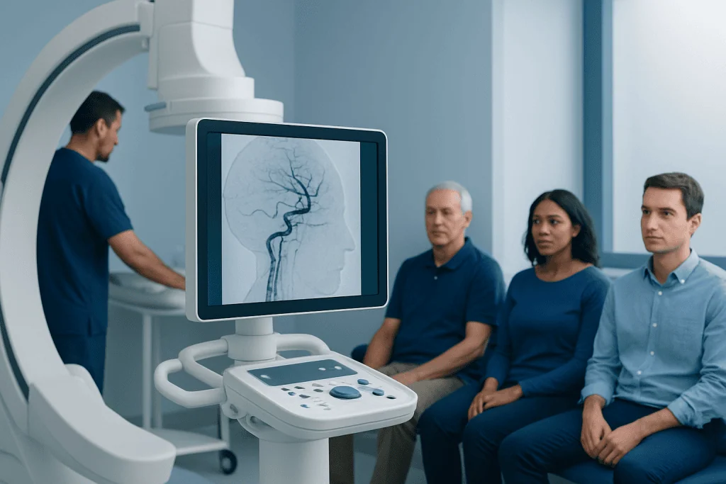
The people most likely to benefit are those with an acute ischemic stroke caused by a large vessel occlusion. This means a major artery in the brain, such as the middle cerebral artery or internal carotid artery, is blocked. These blockages can cause severe symptoms and carry a high risk of long-term disability if blood flow is not restored quickly.
Doctors look at several factors when deciding whether thrombectomy is appropriate. These include the time symptoms began or the last time the person was known to be well, the severity of symptoms, the patient’s level of function before the stroke, and findings on CT, CT angiography, or perfusion imaging. A patient may still be considered even if the exact time of stroke is uncertain, especially if advanced imaging suggests that some brain tissue may still be saved.
Patients with disabling stroke symptoms may be considered for this treatment if imaging shows a suitable blockage and the benefits are expected to outweigh the risks. In some cases, thrombectomy is performed after clot-busting medication, while in others it is used when that medication is not suitable. Emergency teams often assess related conditions such as stroke subtype and blood vessel anatomy before making a final plan.
- Sudden weakness or numbness, especially on one side of the body
- Difficulty speaking or understanding speech
- Sudden vision loss or double vision
- Severe balance problems or trouble walking
- Facial drooping
How Fast It Must Happen

Endovascular treatment for stroke is time-sensitive. In general, the earlier blood flow is restored, the better the chance of preserving brain function. For many patients, thrombectomy is considered within the first several hours after symptoms begin. In selected cases, especially when advanced imaging shows brain tissue at risk but not yet permanently damaged, treatment may still be possible later.
This does not mean people should wait to see whether symptoms improve. Stroke symptoms can come and go, and even temporary improvement may be followed by worsening. Calling emergency services immediately is the safest step because paramedics can begin early assessment and transport the patient to a stroke-ready hospital without delay.
Hospitals aim to move rapidly from arrival to brain imaging and then to treatment when appropriate. The exact timeline varies by the patient’s condition and the hospital pathway, but every stage is designed to shorten delays. Quick action is especially important in large vessel occlusion, where each minute of reduced blood flow can affect recovery.
How Doctors Diagnose and Decide
Diagnosis begins with recognizing that stroke is a medical emergency. In the emergency department, doctors perform a neurological examination and ask when symptoms started or when the person was last seen well. Blood tests, heart monitoring, and assessment of breathing, blood pressure, and blood sugar help guide immediate care.
Brain imaging is central to the decision. A non-contrast CT scan is commonly used first to rule out bleeding. CT angiography can show whether a major artery is blocked, and perfusion imaging may help estimate how much brain tissue is already injured and how much might still be rescued. In some hospitals, MRI may also be used in carefully selected cases.
The treatment team then weighs the expected benefit against the risks. These risks can include bleeding in the brain, injury to the blood vessel, contrast-related complications, or failure to fully reopen the artery. Multidisciplinary stroke care may involve neurologists, interventional specialists, neuroradiologists, anesthesiologists, and intensive care staff, especially when planning stroke treatment tailored to the individual patient.
What Happens During Endovascular Treatment
Mechanical thrombectomy is usually performed in a specialized angiography suite. After the skin is cleaned and numbed, a catheter is inserted into an artery, often in the groin or sometimes the wrist. Using live X-ray guidance, the specialist advances the catheter through the blood vessels toward the blocked artery in the brain.
Devices such as stent retrievers or aspiration catheters are then used to capture or suction out the clot. The aim is to reopen the artery and restore circulation as safely and completely as possible. Depending on the situation, the patient may receive local anesthesia with sedation or, in some cases, general anesthesia.
After the procedure, the patient is monitored closely in a stroke unit or intensive care setting. Follow-up neurological checks and repeat imaging may be needed. Doctors also investigate why the stroke happened so they can plan prevention, which may include medicines, control of vascular risk factors, and occasionally other procedures such as carotid artery treatment when neck artery narrowing is involved.
Benefits, Risks, and Recovery
For the right patient, thrombectomy can significantly improve the chance of a better functional recovery. Reopening a blocked large artery may reduce the extent of brain injury, lower disability, and support a return to daily activities. The amount of recovery varies widely and depends on factors such as the size and location of the stroke, how quickly treatment occurred, age, other health conditions, and whether the artery was successfully reopened.
As with any invasive procedure, there are risks. Possible complications include bleeding in the brain, damage to the artery, movement of clot fragments, reaction to contrast material, and swelling in the brain after the stroke. Even when the procedure is technically successful, some patients may still have lasting symptoms because a portion of brain tissue was already severely injured before blood flow could be restored.
Recovery often continues for weeks to months. Many patients benefit from rehabilitation, including physical, occupational, and speech therapy. Ongoing care may also address swallowing, mood changes, fatigue, and cognitive difficulties. Follow-up after stroke rehabilitation is an important part of improving independence and quality of life.
Prevention and Self-Care After Stroke
Preventing another stroke becomes a major priority after emergency treatment. Doctors try to identify the cause, which may include atrial fibrillation, carotid artery disease, small vessel disease, high blood pressure, diabetes, high cholesterol, or smoking. Treatment plans are individualized and may include antiplatelet or anticoagulant medicines, cholesterol-lowering therapy, and careful blood pressure management.
Daily habits also matter. Stopping smoking, staying physically active within medical guidance, eating a balanced diet, limiting alcohol, sleeping well, and taking prescribed medicines consistently can all support long-term vascular health. Keeping follow-up appointments is important because risk factors often need ongoing adjustment over time.
Family members and caregivers can help by learning the warning signs of recurrent stroke and supporting rehabilitation goals at home. For international patients who need advanced evaluation and coordinated recovery planning, Acibadem International’s multidisciplinary specialists and JCI-accredited hospitals diagnose and treat complex stroke conditions, including brain aneurysm and other neurovascular disorders when relevant to the patient’s care.
When to Seek Emergency Help
Anyone with possible stroke symptoms should receive emergency care immediately. Common warning signs include face drooping, arm weakness, speech difficulty, sudden confusion, sudden trouble seeing, severe dizziness, or a sudden severe headache. A simple way to remember the most common signs is FAST: Face, Arm, Speech, Time.
People should not drive themselves if a stroke is suspected unless there is absolutely no alternative. Emergency medical services can alert the hospital in advance and direct the patient to a center equipped for urgent stroke imaging and intervention. This can save valuable time.
Even if symptoms improve within minutes, evaluation is still essential. A transient ischemic attack, or “mini-stroke,” can be a warning sign of a more serious event. Prompt assessment helps doctors reduce the risk of future stroke and decide whether further testing or treatment is needed.
Frequently asked questions
Is endovascular neurosurgery used for every stroke?
No. It is mainly used for selected cases of ischemic stroke caused by blockage in a large brain artery. The decision depends on urgent brain imaging, symptom timing, and the patient’s overall medical condition.
What is the difference between thrombectomy and clot-busting medicine?
Clot-busting medicine is given through a vein to help dissolve certain clots. Mechanical thrombectomy is a catheter-based procedure that physically removes a clot from a blocked brain artery. Some patients receive both treatments, while others may be eligible for only one of them.
How quickly should a person get to the hospital if stroke is suspected?
Immediately. Stroke is a medical emergency, and early treatment offers the best chance of protecting brain tissue and improving recovery. Calling emergency services right away is safer than waiting to see if symptoms go away.
Can someone still be treated if symptoms started several hours ago?
Sometimes, yes. Although earlier treatment is usually better, some patients may still benefit later if advanced imaging shows that part of the brain can still be saved. This is why emergency evaluation remains important even if some time has passed.
What are the main risks of mechanical thrombectomy?
Possible risks include bleeding in the brain, injury to a blood vessel, movement of clot fragments, and complications related to contrast dye or anesthesia. Doctors recommend the procedure only when the expected benefits are likely to outweigh these risks.
What happens after the procedure?
After thrombectomy, the patient is monitored closely in a stroke unit or intensive care setting. Follow-up care usually includes repeat assessments, prevention planning, and rehabilitation such as physical, speech, or occupational therapy if needed.
References
- World Stroke Organization
- American Heart Association
- National Institute of Neurological Disorders and Stroke
- European Stroke Organisation
- Centers for Disease Control and Prevention
This article is for general information only and is not a substitute for professional medical advice. Please consult a qualified doctor about your individual situation.
Neurosurgery in Turkey — costs, top hospitals & a free quote
JCI-accredited · board-certified surgeons · reply within 24h
Add us as a Preferred Source to see more of our trusted health content across Google Search, AI Overviews and Discover.
Check your numbers in seconds
BMI, calories, due date, blood pressure and 30+ more clinical calculators — free, instant, doctor-reviewed ranges.