Exstrophy Bladder Surgery: Procedure, Recovery and Results

Bladder exstrophy is a rare congenital condition in which the bladder develops outside the body through an opening in the lower abdomen. Surgery is usually planned in stages and may begin soon after birth, although timing depends on anatomy, overall health and specialist assessment.
Key Takeaways
- Bladder exstrophy is a rare congenital condition in which the bladder develops outside the body through an opening in the lower abdomen.
- Surgery is usually planned in stages and may begin soon after birth, although timing depends on anatomy, overall health and specialist assessment.
- Continence may require later procedures, catheterization, medicines or bladder augmentation in some patients.
- Lifelong follow-up with pediatric and adult urology teams is important to monitor kidney function, urinary infections and continence.
- Most people with bladder exstrophy can attend school, work, form relationships and lead active lives with appropriate medical care.
Exstrophy bladder surgery is a series of reconstructive operations used to close and rebuild the bladder, abdominal wall, pelvic structures and urinary outlet in people born with bladder exstrophy. Treatment is highly individualized and aims to preserve kidney health, achieve urinary continence where possible, and support comfort, mobility and quality of life over time.
Overview: What Is Exstrophy Bladder Surgery?
Exstrophy bladder surgery is reconstructive treatment for bladder exstrophy, a congenital condition in which the bladder is open and visible on the lower abdominal wall at birth. The urinary opening, pelvic bones, lower abdominal muscles and genital structures may also be affected. Surgery closes and protects the bladder and helps reconstruct the urinary tract so urine can be stored and passed more normally.
There is no single operation that is right for every child or adult. Care is usually provided by a multidisciplinary team that may include pediatric urologists, reconstructive urologists, orthopedic surgeons, anesthesiologists, nephrologists, continence nurses and psychologists. The overall plan is designed around bladder size and function, kidney health, pelvic anatomy, continence goals and the person’s age.
Many patients need more than one procedure during childhood or later in life. The long-term priorities are protecting the kidneys, reducing skin exposure to urine, improving urinary control when possible, and helping the person participate fully in everyday activities.
How the Surgery Works and Who May Be a Candidate
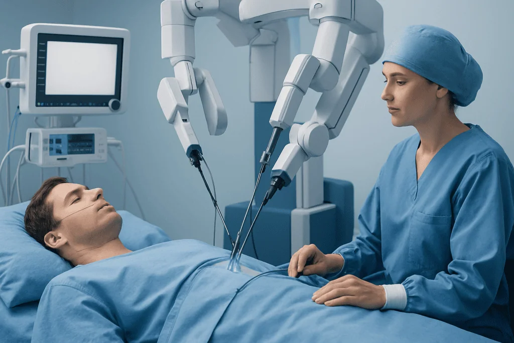
Bladder exstrophy repair generally involves placing the bladder back inside the pelvis, closing the bladder and abdominal wall, and reconstructing the urinary outlet. In newborns, this initial repair may also include bringing together the pelvic bones when they are widely separated. Temporary tubes or catheters are used after surgery to drain urine and allow tissues to heal.
Most children with classic bladder exstrophy are evaluated for repair soon after birth. The exact timing depends on the baby’s stability, bladder tissue, pelvic anatomy, associated conditions and the experience of the specialist center. Some people first receive care in infancy and later need further reconstruction for continence, reflux, small bladder capacity or genital reconstruction.
Later reconstructive surgery may be considered for children, adolescents or adults who have persistent urine leakage, recurrent infections, difficulty emptying the bladder, bladder stones, kidney concerns or complications from earlier repairs. Before recommending surgery, the team considers bladder capacity, continence potential, kidney drainage, bowel health, mobility, family support and the person’s own treatment goals.
Evaluation commonly includes physical examination, kidney and bladder ultrasound, urine testing, blood tests to assess kidney function, and imaging or urodynamic testing when needed. Urodynamics measures how the bladder stores and releases urine, helping the team plan safe and appropriate treatment.
Step by Step: What Happens During Bladder Exstrophy Repair?

The child or adult receives general anesthesia and is asleep throughout the procedure. The surgical team first assesses the bladder plate and nearby structures. The bladder is carefully mobilized, closed to create a reservoir for urine, and positioned inside the pelvis. The lower abdominal wall is then reconstructed to cover and protect the repair.
Depending on the anatomy, surgeons may repair the bladder neck and urethra to improve future urinary control. If the pelvic bones are separated, an orthopedic surgeon may perform pelvic osteotomy, which involves carefully repositioning the pelvic bones to support closure. This is not required in every case, but it may improve the stability of the reconstruction in selected patients.
Drainage tubes are usually left in place in the bladder and, in some cases, the ureters. These prevent pressure from building up while the tissues heal. The surgical approach, number of incisions and need for later operations vary considerably, so the treating team should explain the planned technique and expected milestones for the individual patient.
Some patients later need procedures to increase bladder capacity, improve continence, address vesicoureteral reflux or create a catheterizable channel. When the bladder cannot safely store urine or cannot be reconstructed adequately, urinary diversion may be discussed. These decisions are individualized and made after detailed counseling about benefits, limitations and long-term follow-up.
Recovery Timeline, Pain Control and Follow-Up
Recovery begins in the hospital, where clinicians monitor urine output, wound healing, bowel function, hydration and pain. Hospital stay length varies with the type of repair, whether pelvic osteotomy was performed, and the person’s overall recovery. Babies and children may need positioning or immobilization measures for a period after pelvic reconstruction to protect the repair.
Pain after bladder surgery is expected, but it is actively treated. The care team uses age-appropriate pain medicines and may combine different methods to improve comfort while limiting side effects. Parents and caregivers are taught how to recognize pain, protect catheters and dressings, and contact the surgical team if there are concerns.
At home, activity restrictions, catheter care and follow-up appointments are important. Tubes are removed only when the surgeon confirms that healing is adequate. Families should follow the team’s instructions about bathing, lifting, school attendance, constipation prevention and fluid intake. Sudden fever, worsening pain, vomiting, reduced urine drainage, new swelling or a wound that opens should be reported promptly.
Long-term follow-up continues beyond the early recovery period. Regular assessments may include blood pressure checks, kidney and bladder imaging, urine tests and discussion of continence, catheterization, sexual health and emotional wellbeing. As children grow, planned transitions to adult urology care help maintain continuity.
Benefits, Risks and Expected Results
The main benefits of exstrophy bladder surgery are protection of the exposed bladder, reconstruction of the abdominal wall and urinary tract, and the opportunity for improved bladder storage and urinary continence. Successful reconstruction can also support physical comfort, participation in school and work, social confidence and long-term kidney protection.
As with any major operation, risks include bleeding, infection, anesthesia-related complications, wound problems and blood clots. Urinary-specific risks include urine leakage from the repair, blockage of drainage tubes, urinary tract infections, reflux of urine toward the kidneys, bladder stones, reduced bladder capacity and ongoing incontinence. Pelvic osteotomy has additional bone-healing and mobility considerations.
It is important to understand that an anatomically successful closure does not always mean immediate or complete continence. The bladder must grow and develop adequate capacity, and some people need later bladder-neck procedures, medicines, intermittent catheterization or augmentation. The need for additional surgery does not necessarily mean the initial repair failed; staged care is common in this condition.
Experienced reconstructive urology teams review these potential outcomes before treatment and tailor monitoring to the person’s needs. Decisions should balance the likely benefit of surgery with kidney safety, bladder function and the practical impact of lifelong urinary care.
What Is the Success Rate of Bladder Exstrophy Surgery?
There is no single success rate for bladder exstrophy surgery because “success” can mean different things: initial bladder closure, preservation of kidney function, voluntary urinary continence, the ability to void without catheterization, or avoidance of further operations. Results also vary according to the severity of the exstrophy, bladder development, associated abnormalities, surgical technique and the experience of the treating center.
Initial closure is often achievable in specialized centers, but many patients require additional procedures as they grow. Achieving urinary continence is a longer-term goal and may depend on bladder capacity, bladder neck function and the ability to empty safely. Some patients become continent through the urethra, while others achieve social continence using clean intermittent catheterization or a surgically created catheterizable channel.
Families may find it helpful to ask their surgical team which outcomes are most realistic for their child, what additional procedures may be expected, and how kidney function will be monitored. Individualized expectations are more meaningful than a single percentage because anatomy and treatment plans differ substantially.
Is Bladder Surgery Recovery Painful?
Recovery from bladder exstrophy surgery can be uncomfortable, particularly in the first days after a major reconstruction, but pain should be anticipated and treated. General anesthesia is used during the procedure, and the hospital team provides regular pain relief afterward. In infants and young children, clinicians also watch for nonverbal signs of discomfort, such as irritability, poor feeding or changes in sleep.
Pain often improves gradually as the incision heals and drainage tubes are removed. Movement may be limited after pelvic reconstruction, and some discomfort may occur with coughing, position changes or bowel movements. Constipation can worsen discomfort, so the care plan may include measures to support regular bowel function.
Caregivers should not wait until pain becomes severe before asking for help. Persistent or increasing pain, pain with fever, a hard or swollen abdomen, poor urine drainage or unusual lethargy needs prompt assessment by the surgical team. Pain plans should always be individualized by qualified clinicians.
What Is Life Like After Bladder Removal?
Bladder removal, also called cystectomy, is not the usual first approach for classic bladder exstrophy. It may be considered only in selected, complex situations when the bladder cannot safely function or be reconstructed. If the bladder is removed, surgeons create another route for urine to leave the body, known as urinary diversion.
Options can include an incontinent diversion, where urine drains continuously into a collection pouch through a stoma, or a continent reservoir that is emptied at intervals using a catheter. The most appropriate option depends on kidney function, bowel health, hand function, lifestyle, prior surgery and the person’s preferences. Each method requires education and ongoing medical follow-up.
People can live active, fulfilling lives after bladder removal, including studying, working, traveling, exercising and maintaining relationships. Adjustment takes time, and support from continence nurses, urologists, dietitians and mental health professionals can be valuable. Regular monitoring remains essential because urinary diversions can have long-term effects on kidney function, infection risk and metabolic balance.
Frequently asked questions
What is bladder exstrophy?
Bladder exstrophy is a rare condition present at birth in which the bladder forms outside the lower abdominal wall. It can affect the bladder, urethra, abdominal muscles, pelvic bones and genital structures. Specialist reconstructive care is needed from infancy through adulthood.
When is bladder exstrophy surgery performed?
Many babies are assessed for initial repair soon after birth, but the timing is individualized. The decision depends on the baby’s health, the bladder and pelvic anatomy, and whether other medical conditions are present. Further procedures may be planned later as the child grows.
Will a child be able to control urine after bladder exstrophy surgery?
Urinary control is possible for many patients, but it may take time and sometimes requires additional treatment. Bladder growth, bladder-neck function and the ability to empty safely all influence the outcome. Some people use intermittent catheterization or other continence strategies as part of long-term care.
What is the life expectancy of someone with bladder exstrophy?
With appropriate surgery, kidney monitoring and lifelong urologic follow-up, many people with bladder exstrophy can have a typical life expectancy. Individual health outlook depends mainly on kidney function, urinary tract complications, infections and associated conditions. Regular specialist care helps identify and manage concerns early.
Can bladder exstrophy cause kidney problems?
It can increase the risk of kidney problems if urine does not drain properly, if there is significant reflux toward the kidneys, or if recurrent infections occur. Kidney function is therefore monitored throughout life using blood tests, urine tests and imaging. Early treatment of urinary complications is important.
When should someone seek medical care after bladder exstrophy surgery?
Urgent medical advice is needed for fever, chills, worsening pain, vomiting, little or no urine draining from a catheter, new swelling, heavy bleeding or an opening in the wound. A clinician should also assess recurrent urinary tract symptoms, new leakage, changes in continence or concerns about catheterization. Families should use the contact instructions provided by their surgical team.
References
- European Association of Urology
- American Urological Association
- National Institute of Diabetes and Digestive and Kidney Diseases
- Children's Hospital of Philadelphia
- National Organization for Rare Disorders
This article is for general information only and is not a substitute for professional medical advice. Please consult a qualified doctor about your individual situation.
Explore treatments in Turkey — costs, top hospitals & a free quote
JCI-accredited · board-certified surgeons · reply within 24h
Add us as a Preferred Source to see more of our trusted health content across Google Search, AI Overviews and Discover.