Flow Cytometry: A Complete Medical Overview

Flow cytometry is a diagnostic lab technique that examines the physical and chemical features of individual cells. It is often used on blood, bone marrow, lymph node, or other body fluid samples.
Key Takeaways
- Flow cytometry is a diagnostic lab technique that examines the physical and chemical features of individual cells.
- It is often used on blood, bone marrow, lymph node, or other body fluid samples.
- The test can help classify leukemias, lymphomas, immune deficiencies, and other blood or immune system problems.
- Results usually need to be interpreted together with symptoms, blood counts, imaging, and sometimes biopsy findings.
- Flow cytometry itself is not a treatment; it is a tool that helps guide diagnosis, treatment planning, and follow-up.
Flow cytometry is a laboratory method that analyzes cells one by one as they pass through a laser, allowing doctors to identify cell types and abnormal patterns. It is commonly used to help diagnose and monitor blood disorders, immune conditions, infections, and some cancers.
Overview: what flow cytometry is and why it matters
Flow cytometry is a laboratory technique that measures the characteristics of cells or particles suspended in fluid. As the sample moves through the machine in a narrow stream, cells pass one at a time through a laser. Sensors then detect how the cells scatter light and whether they carry special fluorescent markers attached to proteins on or inside the cell.
In practical terms, flow cytometry helps doctors answer important questions such as: What kinds of cells are present? Are they mature or immature? Do they look like normal immune cells, or do they show a pattern linked to leukemia, lymphoma, or another disorder? Because it examines many cells quickly and in detail, it can reveal patterns that are not obvious under a standard microscope alone.
Rather than being a disease itself, flow cytometry is a diagnostic tool. It is often part of a larger medical evaluation that may also include a physical examination, complete blood count, blood smear, imaging, genetic testing, or tissue biopsy. This combined approach helps doctors make a more accurate diagnosis and choose the most appropriate next steps.
How flow cytometry works
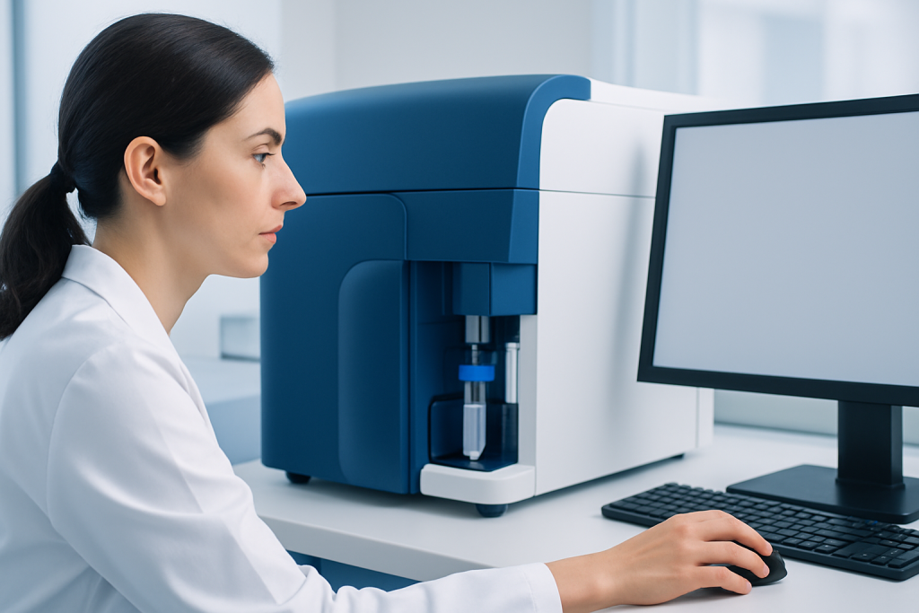
The process starts with a sample that contains cells in liquid form. This may be blood, bone marrow, lymph node material, spinal fluid, or another body fluid, depending on the medical question. The laboratory prepares the sample and often adds antibodies tagged with fluorescent dyes. These antibodies bind to specific markers on cells, such as proteins found on B cells, T cells, plasma cells, or immature blood cells.
When the cells pass through the instrument, the laser detects several features at once. Forward scatter gives information related to cell size, while side scatter helps estimate internal complexity or granularity. Fluorescent signals show whether the cell carries the markers being tested. A computer then displays these findings as plots or graphs, allowing specialists to identify cell populations and abnormal cell groups.
One advantage of flow cytometry is that it can analyze thousands of cells in a short time. This speed and precision make it especially useful when doctors need to distinguish between similar-looking conditions or track very small numbers of abnormal cells after treatment. In some laboratories, related techniques can also sort selected cells for further study, although that use depends on the clinical setting.
When doctors use flow cytometry

Flow cytometry has a wide range of medical uses, but it is especially important in blood and immune system disorders. Doctors commonly request it when a person has abnormal blood counts, enlarged lymph nodes, unexplained fevers, recurrent infections, unusual bruising, or findings that suggest a possible bone marrow problem. It may help classify diseases such as leukemia or support the workup of lymphoma.
It is also used to evaluate immune function. For example, flow cytometry can measure different immune cell populations, including T cells, B cells, and natural killer cells. This may help in the assessment of suspected immune deficiencies, autoimmune conditions, certain infections, or the effects of immunosuppressive medicines.
Common clinical uses include:
- Classifying leukemias and lymphomas
- Evaluating unexplained abnormal blood cell counts
- Assessing plasma cell disorders and some bone marrow diseases
- Monitoring response to treatment and checking for residual abnormal cells
- Investigating some inherited or acquired immune system disorders
- Analyzing cells in body fluids, such as cerebrospinal fluid or pleural fluid, when clinically appropriate
Because the test identifies cell markers in detail, it can provide information that helps distinguish one condition from another. This is especially useful when symptoms overlap or when routine blood tests raise concern but do not yet point to a single diagnosis.
What to expect before, during, and after the test
What a patient experiences depends on the type of sample needed. If flow cytometry is done on a blood sample, the process is similar to a standard blood draw. A healthcare professional places a needle into a vein, collects the sample, and sends it to the laboratory. In that situation, no special preparation is often needed unless the doctor gives specific instructions.
In other cases, the sample may come from bone marrow, a lymph node, or another body fluid. A bone marrow procedure can involve local anesthesia and sometimes additional support for comfort. If a tissue sample or fluid aspiration is required, the medical team explains the purpose, how to prepare, and what temporary after-effects such as soreness or bruising might occur.
Results are usually not interpreted in isolation. Patients may receive a report with technical terms describing cell markers, percentages, or populations, but the meaning depends on the overall clinical context. A normal or abnormal pattern does not replace a full medical assessment, and many reports require interpretation by a hematologist, pathologist, or other specialist familiar with the patient’s symptoms and other test results.
Understanding flow cytometry results
Flow cytometry results describe the types of cells found and whether they express particular markers. Doctors often look for an “immunophenotype,” which is the pattern of proteins present on a group of cells. Some patterns suggest normal immune cells, while others may indicate a clonal or abnormal population that requires further investigation.
For example, in suspected blood cancers, flow cytometry can help determine whether abnormal cells are of B-cell, T-cell, myeloid, or plasma cell origin. This distinction matters because treatment choices and prognosis often differ by cell type. It may also help detect minimal residual disease in some settings, meaning a very small number of abnormal cells that remain after treatment and could influence follow-up planning.
Even so, results are not always straightforward. An abnormal pattern does not automatically confirm cancer, and a normal result does not rule out every condition. Sample quality, the number of cells present, recent treatment, and the exact panel of markers used can all affect interpretation. Doctors may recommend repeat testing, biopsy, imaging, or bone marrow biopsy when more information is needed.
Benefits, limits, and related tests
The main strengths of flow cytometry are speed, sensitivity, and detail. It can analyze many cells rapidly and identify subtle differences between them. This makes it valuable in situations where doctors need to classify a disease accurately, assess immune status, or monitor treatment over time.
However, like any medical test, it has limits. Flow cytometry does not evaluate the tissue architecture of a lymph node or bone marrow in the same way a biopsy does. It also depends on having a good-quality sample with enough viable cells. In some disorders, abnormal cells may be patchy or scarce, so results need to be interpreted carefully alongside other findings.
Related tests may include complete blood count, peripheral smear, cytogenetics, molecular testing, imaging, and pathology review. In cancer care, flow cytometry may be part of a broader evaluation that can also involve hematology assessment and bone marrow transplantation planning or medical oncology care, depending on the diagnosis. This stepwise approach helps ensure that a test result is translated into a clear and appropriate care plan.
When to seek medical care
Flow cytometry is not a test people usually request on their own. It is ordered when a doctor believes it may help explain symptoms, abnormal blood counts, or suspicious findings from other tests. A person should seek medical care if they have ongoing fatigue, repeated infections, unexplained bruising or bleeding, swollen lymph nodes, persistent fevers, night sweats, or unintentional weight loss.
Medical attention is also important when routine blood tests show unusual white blood cell, red blood cell, or platelet levels. These results do not always mean a serious illness, but they do deserve proper evaluation. Early review by a qualified clinician can help determine whether the cause is temporary, benign, immune-related, infectious, or linked to a blood disorder that needs further testing.
If flow cytometry is recommended, patients may wish to ask why the test is being ordered, what sample is needed, how long results may take, and whether additional tests are likely. In complex cases, multidisciplinary specialists can help coordinate diagnosis and follow-up. Acibadem International’s multidisciplinary teams in JCI-accredited hospitals evaluate and treat international patients who need advanced diagnostic workups for blood, immune, and cancer-related conditions.
Frequently asked questions
What is flow cytometry used for?
Flow cytometry is used to identify and measure different types of cells in a sample. Doctors often use it to help diagnose blood cancers, immune disorders, and other conditions that affect blood or body fluids.
Is flow cytometry a blood test?
It can be. Flow cytometry is often performed on a blood sample, but it can also be done on bone marrow, lymph node tissue, spinal fluid, or other body fluids depending on the clinical situation.
Does an abnormal flow cytometry result mean cancer?
Not always. An abnormal result means the cells show a pattern that needs interpretation, but it must be considered together with symptoms, examination findings, and other tests. Some non-cancerous immune or inflammatory conditions can also affect results.
How long does it take to get flow cytometry results?
Timing varies by laboratory, sample type, and the complexity of the test panel. Some results may be available relatively quickly, while others take longer if additional specialist review or related testing is needed.
Do people need to prepare for a flow cytometry test?
For a routine blood sample, special preparation is often not needed unless a doctor gives instructions. If the sample comes from bone marrow or another procedure, the healthcare team will explain how to prepare and what to expect.
Can flow cytometry monitor treatment response?
Yes, in some conditions it helps doctors monitor how well treatment is working. It may also detect very small numbers of remaining abnormal cells in selected blood cancers, which can support follow-up decisions.
References
- World Health Organization
- National Cancer Institute
- MedlinePlus
- Centers for Disease Control and Prevention
- American Society of Hematology
This article is for general information only and is not a substitute for professional medical advice. Please consult a qualified doctor about your individual situation.
Explore treatments in Turkey — costs, top hospitals & a free quote
JCI-accredited · board-certified surgeons · reply within 24h
Add us as a Preferred Source to see more of our trusted health content across Google Search, AI Overviews and Discover.

More from the Health Library



Related Specialists

Dr. Özgür Özer
Gynecology & Obstetrics
Dr. Burcu Uprak
Internal Medicine
Prof. Dr. Nesliar Eser Kutsal
Gastroenterology
Prof. Dr. Bünyamin Kaplan
Radiation Oncology