Flow Volume Loop: An Evidence-Based Guide for Patients

A flow volume loop displays airflow against lung volume during a forced breathing maneuver. The shape of the loop may suggest obstructive, restrictive, or upper-airway patterns.
Key Takeaways
- A flow volume loop displays airflow against lung volume during a forced breathing maneuver.
- The shape of the loop may suggest obstructive, restrictive, or upper-airway patterns.
- Results must be interpreted alongside symptoms, medical history, examination findings, and other lung-function measurements.
- Test quality matters: an incomplete effort, coughing, or poor seal around the mouthpiece can affect the graph.
- Spirometry is generally safe and noninvasive, although some people may briefly feel light-headed after forceful breathing.
A flow volume loop is a graph created during spirometry, a breathing test that measures the speed and amount of air a person can breathe out and in. It can help clinicians recognize patterns associated with airway narrowing, restriction, upper-airway obstruction, or poor test technique, but it does not diagnose a condition on its own.
What Is a Flow Volume Loop?
A flow volume loop is a visual record produced during spirometry, a common pulmonary function test. It plots airflow on the vertical axis and the volume of air moved on the horizontal axis. The graph shows what happens when a person takes a full breath in, then blows out as hard and fast as possible until the lungs feel empty, followed by a full breath back in.
The upper part of the graph usually represents forceful exhalation, while the lower part represents inhalation. Clinicians look at the loop’s overall size, peak flow, and contour rather than relying on one feature alone. It is one part of a wider spirometry report that may also include measurements such as forced vital capacity (FVC) and forced expiratory volume in one second (FEV1).
A flow volume loop is best understood as a pattern-recognition tool. It can provide useful clues about how air moves through the airways, but it cannot by itself confirm asthma, chronic obstructive pulmonary disease (COPD), vocal cord problems, or another diagnosis. A qualified clinician interprets it in the context of the person’s age, height, sex, symptoms, smoking or exposure history, and the quality of the test.
How the Test Is Performed
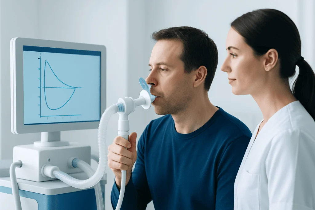
Spirometry is usually performed in a clinic, pulmonary function laboratory, or hospital outpatient setting. The person sits upright, wears a nose clip, and seals their lips around a disposable mouthpiece. After breathing in fully, they are asked to exhale as forcefully and completely as possible, then breathe in quickly and deeply again.
Several attempts are usually needed because spirometry depends on effort and technique. The healthcare professional coaches the person to start promptly, blow hard, and continue exhaling long enough. Results are compared for consistency; this helps ensure that the displayed loop reflects lung function rather than an isolated weak or interrupted effort.
In some cases, spirometry is repeated after an inhaled bronchodilator medicine that relaxes airway muscles. Comparing pre- and post-bronchodilator values can help clinicians assess whether airflow limitation improves after medication. A clinician may also request additional tests, such as lung-volume measurements, gas-transfer testing, imaging, or exercise testing, when needed.
Most people tolerate the test well. Forceful breathing can briefly cause coughing, chest tightness, or light-headedness. The test team can pause if symptoms occur, and people should inform them about recent surgery, a respiratory infection, chest pain, coughing up blood, or any condition that may make forceful exhalation unsuitable.
What Clinicians Look For on the Graph

In a typical loop, exhaled airflow rises rapidly to a high point called the peak expiratory flow and then gradually falls as lung volume decreases. The inhalation portion forms a smooth curve below the horizontal line. Normal shape and size vary between individuals, so predicted reference values based on personal characteristics are important.
An obstructive pattern may show a reduced ratio of FEV1 to FVC and a concave, or “scooped,” appearance on the exhalation side of the loop. This can occur when smaller airways narrow or collapse earlier during exhalation. Asthma and COPD are common examples, although the loop alone cannot distinguish between them.
A restrictive pattern may produce a loop that looks relatively narrow or small because the total volume of air exhaled is lower. The basic contour may remain fairly preserved. Spirometry can suggest restriction, but measuring total lung capacity is generally needed to confirm it, since a low FVC may also result from other factors.
Flattening of part of the loop can sometimes suggest obstruction involving larger or upper airways. For example, flattening mainly during inhalation, mainly during exhalation, or during both phases may point to different possible locations or behaviors of airway narrowing. These findings require careful review, because poor technique may produce similar-looking changes.
Why a Flow Volume Loop May Be Ordered
A clinician may request spirometry with a flow volume loop when a person has ongoing shortness of breath, wheezing, chronic cough, chest tightness, reduced exercise tolerance, or recurrent respiratory symptoms. It may also be used to assess occupational or environmental exposures, monitor an established lung condition, or check response to treatment.
The test is often part of an evaluation for suspected asthma or COPD. It can be especially helpful when symptoms fluctuate, occur with exercise, follow exposure to smoke or irritants, or persist after a respiratory infection. However, normal spirometry between episodes does not always rule out asthma, and further assessment may be appropriate when symptoms remain suggestive.
Flow volume loops may also contribute to the assessment of unexplained noisy breathing, voice-related breathing symptoms, possible central airway narrowing, or breathing concerns before certain operations. In these situations, clinicians may combine results with an ear, nose, and throat examination, chest imaging, laryngoscopy, or other specialized testing.
For people already diagnosed with a respiratory condition, repeated spirometry can show whether lung function is stable, improving, or changing over time. A single result is only a snapshot; trends, symptoms, medication use, and daily functioning are often more informative than one graph viewed in isolation.
Understanding Results and Their Limits
It is understandable to look at a spirometry graph and wonder whether it is “normal.” However, interpreting a flow volume loop requires more than recognizing a shape. Clinicians first confirm that the test meets quality standards, then review numerical values, predicted ranges, repeatability, and the person’s clinical situation.
A loop that appears abnormal does not necessarily mean serious disease. Submaximal effort, a delayed start, coughing, air leakage around the mouthpiece, early stopping, or variable inhalation can alter the graph. Conversely, a loop that looks broadly normal does not always exclude early disease or a condition that occurs only at certain times, such as during exercise or allergen exposure.
Some people have more than one factor affecting their breathing. For example, airway obstruction and reduced lung volume can coexist, and obesity, chest-wall conditions, neuromuscular weakness, or prior lung surgery may influence results. This is why a clinician may arrange complete pulmonary function testing rather than drawing conclusions from basic spirometry alone.
Patients can ask their clinician what the loop suggests, whether the test quality was acceptable, how the FEV1 and FVC compare with expected values, and whether additional testing is needed. Bringing a list of symptoms, triggers, medications, and relevant exposure history can make the discussion more useful.
Preparing for Spirometry and Supporting Lung Health
Preparation instructions vary depending on why the test is being performed and which medicines a person uses. The testing center may advise avoiding smoking, vaping, alcohol, strenuous exercise, or a heavy meal for a period before the appointment. They may also give specific directions about whether to temporarily withhold inhalers; people should not stop prescribed medicine unless the testing team or their clinician has advised them to do so.
Wearing comfortable clothing can make deep breathing easier. It is helpful to tell the staff about recent illness, pregnancy, surgery, heart conditions, or a history of fainting with medical tests. Clear communication allows the team to decide whether the test should proceed, be adapted, or be postponed.
Long-term lung health is supported by avoiding tobacco smoke and vaping, reducing exposure to workplace dusts and chemical fumes when possible, staying physically active within personal limits, and keeping recommended vaccinations up to date. People with a diagnosed respiratory condition should follow their personalized treatment plan and review inhaler technique regularly.
Acibadem International’s multidisciplinary specialists and JCI-accredited hospitals evaluate breathing symptoms and lung-function findings for international patients when further assessment or treatment is needed.
When to Seek Medical Care
People should arrange a medical appointment for persistent cough, repeated wheezing, breathlessness that affects ordinary activities, unexplained reduced exercise tolerance, or symptoms that repeatedly interrupt sleep. An assessment is also sensible when respiratory symptoms follow workplace exposure, smoking or vaping, a significant infection, or a change in prescribed inhaler needs.
Urgent medical care is important for severe or rapidly worsening shortness of breath, chest pain or pressure, bluish or gray lips or face, confusion, fainting, coughing up blood, or difficulty speaking in full sentences because of breathlessness. These symptoms may have causes beyond lung disease and should not be assessed using a flow volume loop or home monitoring alone.
For people with known asthma, COPD, or another respiratory condition, a worsening pattern of symptoms, increasing use of quick-relief medicine, or a noticeable decline in usual activity should prompt contact with their healthcare professional. Early review can help clarify the cause and support an appropriate care plan.
Frequently asked questions
Is a flow volume loop the same as spirometry?
A flow volume loop is one graph produced during spirometry. Spirometry also provides numerical measurements, including FEV1 and FVC, which clinicians use alongside the graph. Together, these results help assess how air moves in and out of the lungs.
Can a flow volume loop diagnose asthma?
A flow volume loop can show airflow patterns that may be consistent with asthma, particularly if obstruction improves after a bronchodilator. It cannot diagnose asthma by itself, because symptoms and spirometry findings may vary over time. A clinician considers the full history and may recommend additional testing.
What does a scooped-out flow volume loop mean?
A scooped or concave exhalation curve can suggest airflow obstruction, often involving smaller airways. It may be seen in conditions such as asthma or COPD, but it can also be affected by test quality. The pattern should be interpreted with the spirometry values and clinical assessment.
Can anxiety affect spirometry results?
Anxiety can make it harder to perform the breathing maneuver consistently, especially if it leads to rapid, interrupted, or hesitant breathing. The test staff can provide coaching, allow rest between attempts, and repeat the maneuver when appropriate. Anxiety itself does not create a specific diagnostic loop pattern.
Do abnormal spirometry results always mean lung disease?
No. An abnormal result may reflect technique, temporary respiratory illness, incomplete effort, or factors outside the lungs. When results are unexpected, clinicians may repeat spirometry or request more complete pulmonary function testing before reaching conclusions.
How long does a spirometry test take?
The breathing maneuvers themselves are brief, but the full appointment commonly takes longer because several acceptable and repeatable attempts are needed. If testing is repeated after a bronchodilator, additional waiting time is required. The testing center can provide more specific timing instructions.
References
- American Thoracic Society
- European Respiratory Society
- National Heart, Lung, and Blood Institute
- Global Initiative for Asthma
- Global Initiative for Chronic Obstructive Lung Disease
This article is for general information only and is not a substitute for professional medical advice. Please consult a qualified doctor about your individual situation.
Explore treatments in Turkey — costs, top hospitals & a free quote
JCI-accredited · board-certified surgeons · reply within 24h
Add us as a Preferred Source to see more of our trusted health content across Google Search, AI Overviews and Discover.