Foci: Symptoms, Causes, and Treatment — Complete Patient Guide

Foci means one or more small, localized areas that look different from surrounding tissue. The term does not name a disease by itself; it describes a finding that needs clinical context.
Key Takeaways
- Foci means one or more small, localized areas that look different from surrounding tissue.
- The term does not name a disease by itself; it describes a finding that needs clinical context.
- Some foci are harmless, while others may reflect infection, inflammation, bleeding, reduced blood flow, or tumors.
- Symptoms vary by organ and many foci cause no symptoms at all.
- Evaluation may include imaging, blood tests, follow-up scans, or biopsy depending on the finding.
- Treatment focuses on the underlying cause, not the word foci itself.
Foci are localized areas of change in tissue that may be found on imaging scans, laboratory tests, or physical examination. The term itself is descriptive rather than a diagnosis, so symptoms, causes, and treatment depend on where the foci are located and what is causing them.
What does foci mean?
Foci is the plural of focus. In medicine, it refers to one or more localized areas that appear different from surrounding tissue. A radiology report, pathology result, or doctor’s note may use this word to describe a spot, patch, lesion, or area of change that needs interpretation in the full clinical picture.
This is why the word can feel confusing for patients. Foci is not a final diagnosis by itself. It may describe something temporary and mild, such as a small inflammatory change, or something that needs more attention, such as an infection, reduced blood supply, scar tissue, or a growth.
The meaning depends on several factors: the organ involved, the person’s symptoms, the size and appearance of the finding, and whether it changes over time. For example, foci in the brain, liver, thyroid, breast, lungs, or bones are evaluated differently because each organ has its own common patterns and causes.
In patient-friendly terms, foci means “small areas doctors have noticed.” The next step is understanding what kind of change they represent and whether any treatment is needed.
How foci may be found
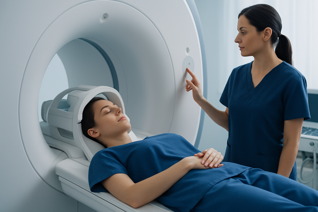
Foci are often discovered during tests done for another reason. A person may have an ultrasound, CT, MRI, mammogram, X-ray, or blood test because of pain, headache, cough, fatigue, or a routine checkup. The report may mention single or multiple foci, focal changes, or focal lesions.
Sometimes a doctor can identify a focus during a physical exam, such as a small skin change, swollen lymph node, or breast lump. In other cases, the finding is microscopic and only appears on pathology after a biopsy or surgery.
Common examples include:
- Small white matter foci on a brain MRI
- Focal nodules in the thyroid or liver on ultrasound
- Foci of infection or inflammation in the lungs on CT
- Focal calcifications in the breast on mammography
- Focal changes in the digestive tract seen during endoscopy
Because the term is broad, patients benefit from asking where the foci are located, how large they are, whether they look benign or suspicious, and what follow-up is recommended.
Symptoms linked to foci

Foci do not cause a single standard set of symptoms. Many produce no symptoms at all and are found incidentally. When symptoms do occur, they usually come from the underlying condition and the body part involved.
For example, brain foci may be associated with headache, dizziness, numbness, weakness, balance problems, seizures, or changes in vision or memory. Lung foci may be linked to cough, fever, shortness of breath, or chest discomfort. Liver or gallbladder foci may be associated with abdominal pain, nausea, or abnormal liver tests.
In the breast, thyroid, skin, or lymph nodes, a person may notice a lump, thickening, tenderness, or visible skin changes. In bones or joints, focal abnormalities may lead to pain, swelling, reduced movement, or fractures if the area is weakened.
It is also common for a report to mention chronic or nonspecific foci that do not explain a person’s symptoms. In that situation, the clinician determines whether the finding is clinically important or simply an unrelated observation.
Causes and risk factors
The causes of foci vary widely. Some are benign and stable over time, while others require timely treatment. Broad categories include infection, inflammation, vascular changes, trauma, degenerative change, benign cysts or nodules, autoimmune disease, congenital differences, and cancer.
In the brain, small foci may reflect migraine-related changes, aging, prior tiny vessel injury, inflammation, demyelinating disease, or less commonly an active lesion. If the finding raises concern for disorders such as multiple sclerosis, doctors look closely at the pattern, location, and symptoms rather than relying on one report term alone.
In other organs, focal findings may be caused by cysts, adenomas, calcifications, abscesses, scarring, or primary and secondary tumors. Some foci represent healing tissue after past infection or injury. Others may relate to chronic conditions such as high blood pressure, diabetes, smoking-related lung disease, or immune disorders.
Risk factors depend on the site, but may include age, tobacco use, heavy alcohol use, obesity, chronic inflammation, family history, exposure to infections, vascular risk factors, or a personal history of cancer. This is why medical history matters so much when interpreting any focal finding.
How doctors diagnose and interpret foci
Diagnosis starts with context. A doctor considers symptoms, medical history, medications, age, family history, and the exact wording of the report. Important details include whether the foci are single or multiple, small or large, well-defined or irregular, active or chronic-appearing, and whether they enhance with contrast or show restricted diffusion on imaging.
Further evaluation may include repeat imaging, targeted ultrasound, blood tests, infection testing, inflammatory markers, hormone tests, or referral to a specialist such as a neurologist, pulmonologist, endocrinologist, or oncologist. In some cases, advanced studies such as MRI, CT imaging, or PET-CT help clarify the nature of the finding.
If imaging cannot determine the cause with confidence, a biopsy may be recommended. Tissue sampling can show whether a focus is inflammatory, infectious, benign, precancerous, or malignant. Doctors balance the benefit of certainty against the risks of invasive testing, especially when a lesion is very small or likely benign.
Many patients worry when they read radiology terms before speaking with their doctor. That concern is understandable, but it is important to remember that report language is often intentionally broad. A finding described as foci usually means “areas that need interpretation,” not “proof of serious disease.”
Treatment options for foci
There is no single treatment called “treatment for foci” because the word describes a finding, not a disease. Care is directed at the underlying cause. Some people need no immediate treatment and only periodic monitoring, especially when the foci look stable and benign.
If infection is responsible, treatment may involve antimicrobial medicines and follow-up imaging. If inflammation or autoimmune disease is the cause, doctors may recommend anti-inflammatory or immune-modifying therapy. Vascular causes are often managed by treating blood pressure, cholesterol, diabetes, smoking, or other stroke and heart risk factors.
When a focal lesion is suspicious for cancer or clearly malignant, treatment may include surgery, systemic therapy, or radiation depending on the organ and stage. For selected cases, doctors may use biopsy first to guide decision-making. If the finding is linked to a known condition such as brain tumor, treatment planning is tailored carefully to the individual.
Supportive care is also important. Pain control, rehabilitation, nutrition, and management of related symptoms can improve quality of life while the underlying cause is being clarified or treated.
Monitoring, self-care, and prevention
Not all foci can be prevented, but overall health measures may reduce the risk of some underlying causes. Controlling blood pressure, cholesterol, and blood sugar supports brain and vascular health. Avoiding tobacco, limiting alcohol, staying physically active, and keeping up with recommended vaccinations can also lower the risk of certain infections, lung changes, and chronic disease.
Self-care after a focal finding mainly means following the recommended plan. That may include repeat imaging at a set interval, taking prescribed medicines, attending specialist visits, and watching for new symptoms. Skipping follow-up can make it harder to know whether a finding is stable, improving, or changing.
Patients can also help by keeping copies of imaging reports, bringing prior scans for comparison, and sharing complete medical history with their care team. Comparison with earlier studies is often one of the most useful tools for deciding whether a focus is old and harmless or newly significant.
For international patients who need coordinated evaluation, Acibadem International’s multidisciplinary specialists in JCI-accredited hospitals diagnose and treat a wide range of focal findings using imaging, pathology, and organ-specific expertise.
When to seek medical care
A person should contact a doctor if a test report mentions foci and the next steps are unclear, especially if there are persistent symptoms or a personal history of cancer, autoimmune disease, or significant infection. Prompt review helps place the finding in context and determine whether monitoring or additional testing is needed.
Urgent medical care is appropriate for warning signs such as sudden weakness, facial drooping, trouble speaking, severe new headache, seizures, chest pain, coughing up blood, severe shortness of breath, high fever, confusion, or rapid worsening of symptoms. These symptoms may relate to conditions that need immediate evaluation rather than the report wording itself.
If a doctor recommends repeat imaging or biopsy, it is wise not to delay without discussing the reason. Many focal findings are benign, but timely follow-up is the safest way to confirm that and to identify the smaller number that need treatment.
Reassurance and caution can coexist. Most focal findings are best understood step by step with a qualified clinician who can explain what the result means for that individual person.
Frequently asked questions
Is foci a disease?
No. Foci is a descriptive medical term that means localized areas of change. It does not identify the exact cause, so doctors use symptoms, imaging details, and sometimes biopsy to determine what it represents.
Are foci always serious?
No, many foci are benign, old, or incidental findings that do not cause harm. However, some can reflect infection, inflammation, vascular problems, or tumors, so proper interpretation and follow-up are important.
Can foci go away on their own?
Some can. Foci caused by temporary inflammation, infection, or minor injury may improve or resolve over time, while others remain stable for years. Follow-up imaging may be used to check whether a finding is changing.
What tests are used to investigate foci?
The most appropriate tests depend on the organ involved. Doctors may use ultrasound, CT, MRI, blood tests, endoscopy, or biopsy to better understand the nature of the focal finding.
Do multiple foci mean cancer?
Not necessarily. Multiple foci can occur with many noncancerous conditions, including inflammation, infection, vascular change, or benign nodules. The pattern, location, and accompanying symptoms are more informative than the number alone.
Should a person worry about foci on an MRI or CT report?
It is understandable to feel concerned, but the term itself should not be taken as a diagnosis. Many imaging reports use broad language first, and a doctor then explains whether the finding appears harmless, needs monitoring, or requires further testing.
This article is for general information only and is not a substitute for professional medical advice. Please consult a qualified doctor about your individual situation.
Explore treatments in Turkey — costs, top hospitals & a free quote
JCI-accredited · board-certified surgeons · reply within 24h
Add us as a Preferred Source to see more of our trusted health content across Google Search, AI Overviews and Discover.
Check your numbers in seconds
BMI, calories, due date, blood pressure and 30+ more clinical calculators — free, instant, doctor-reviewed ranges.

More from the Health Library

Oxidative Damage Stress — Explained by Medical Evidence, Not Myths

Januvia: What Patients Need to Know

Cardiac Sphincter: An Evidence-Based Guide for Patients

Ridges in Nails: A Complete Medical Overview

Nasal Cannula: A Complete Medical Overview