

Frontal Bone — Explained by Medical Evidence, Not Myths
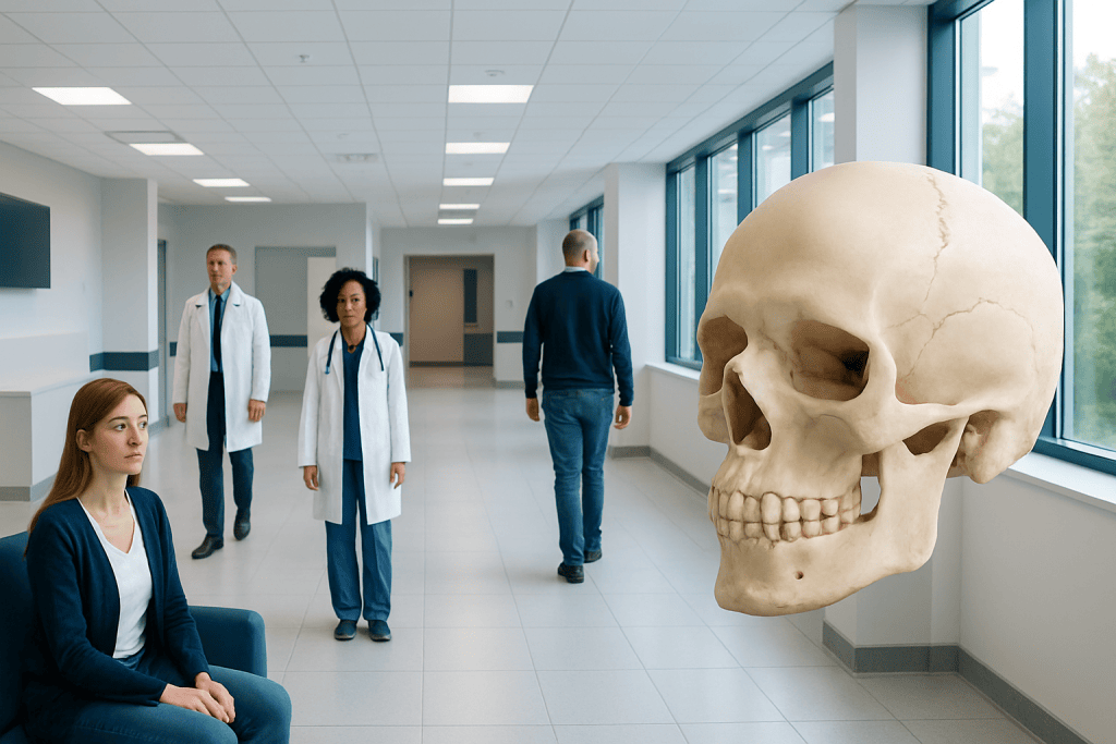
The frontal bone forms the forehead and part of the skull and eye sockets. It protects the frontal lobes of the brain and supports facial shape.
Key Takeaways
- The frontal bone forms the forehead and part of the skull and eye sockets.
- It protects the frontal lobes of the brain and supports facial shape.
- The frontal bone also contains the frontal sinuses, which connect to the nasal cavity.
- Injuries, congenital differences, infection, and tumors can affect this bone.
- Persistent pain, swelling, deformity, or symptoms after head trauma should be medically assessed.
The frontal bone is the large bone that forms the forehead, part of the roof of the eye sockets, and the front of the skull. It helps protect the brain, shapes the upper face, and contains the frontal sinuses, making it important in both structure and function.
Overview: what the frontal bone is
The frontal bone is the broad, curved bone at the front of the skull. In simple terms, it is the bone of the forehead, but its role goes beyond appearance. It also forms part of the bony roof of the eye sockets and contributes to the front portion of the cranial vault that protects the brain.
From a medical point of view, the frontal bone is important because it combines protection, support, and internal anatomy in one structure. It helps shield the frontal lobes of the brain from injury, gives shape to the upper face, and houses the frontal sinuses in most adults. These air-filled spaces are part of the sinus system connected to the nose.
The frontal bone begins as separate parts in early life and normally fuses during childhood. This normal development matters because doctors assess skull growth, facial symmetry, and suture closure when examining infants and children. In adults, the bone is fully formed and usually discussed in relation to anatomy, trauma, sinus disease, or imaging findings.
Where it is located and what structures it forms
The frontal bone sits at the very front of the skull. Its most visible part is the forehead, but it extends backward over the eyes and upward to meet other skull bones. It connects with the parietal bones near the top of the head and with bones of the nose, cheek area, and upper eye socket region below.
Anatomically, the frontal bone has several parts. The squamous part forms the forehead. The orbital part forms much of the roof of each eye socket. The nasal part helps form the upper bridge of the nose and supports structures around the sinus drainage pathway. These relationships explain why injuries or disease in this area can affect the head, eyes, and nose at the same time.
Doctors may describe features such as the brow ridges, the glabella between the eyebrows, and the supraorbital margins above the eyes. Small openings or notches in this region allow nerves and blood vessels to pass through. Because the bone is so closely related to nearby soft tissues, symptoms can sometimes involve numbness, bruising, vision changes, or sinus pressure depending on the underlying problem.
What the frontal bone does

The frontal bone has several essential functions. Its most important role is protection. It forms a hard barrier over the frontal portion of the brain, helping reduce the risk of direct injury from everyday impacts and trauma. No skull bone can prevent all harm, but this protective function is central to why the frontal bone matters clinically.
It also provides structure and support. The frontal bone helps create the upper contours of the face, supports the eyebrows and forehead, and contributes to the architecture of the eye sockets. This support is not only cosmetic; it helps maintain the normal position of the eyes and nearby tissues.
Another key function involves the frontal sinuses. These air-filled cavities within the frontal bone are part of the paranasal sinus system. Their exact roles are still discussed in medical science, but they are thought to help reduce skull weight, influence voice resonance, and participate in conditioning inhaled air together with the rest of the sinus and nasal system. Problems affecting the frontal sinuses may overlap with sinusitis symptoms such as pressure, congestion, and headache.
Development, normal variation, and common misconceptions
The frontal bone develops early in life from paired centers of ossification, which gradually unite. In infants and young children, sutures between skull bones remain open to allow brain growth. Over time, these sutures normally fuse in a predictable pattern. This process is one reason clinicians pay attention to head shape and growth milestones during childhood.
There is also normal variation in the frontal bone from person to person. Forehead contour, brow prominence, and frontal sinus size can differ widely without indicating disease. Some people have large frontal sinuses, while others have small ones or, rarely, little sinus development in that area. Imaging can show these variations clearly.
A common myth is that the frontal bone is simply a solid plate with no internal complexity. In reality, it contains sinus spaces in many adults and has close relationships with nerves, blood vessels, the eyes, and the brain. Another misconception is that any forehead bump means a skull problem. Many bumps are due to normal anatomy or minor soft-tissue changes rather than a bone disorder. Even so, a new, persistent, or painful lump should be assessed by a qualified clinician.
Conditions and injuries that can affect the frontal bone
The frontal bone can be affected by trauma, infection, developmental conditions, and, less commonly, tumors. A frontal bone fracture may happen after a fall, sports injury, vehicle accident, or other strong impact. Because this area lies near the brain and eye sockets, doctors evaluate these injuries carefully. Some fractures are minor and stable, while others may involve the frontal sinus, orbit, or nearby intracranial structures.
Frontal sinus disease can also involve the frontal bone. Inflammation or infection in the sinus may cause pressure above the eyes, headache, tenderness, or swelling. Although many sinus infections improve with routine care, complicated cases may need specialist review, imaging, or procedural treatment. Depending on the cause, management may overlap with endoscopic sinus surgery when medical therapy alone is not enough.
In children, a clinician may assess the skull if there are concerns about unusual head shape or early closure of skull sutures. One example is craniosynostosis, in which one or more sutures close too early and can affect skull growth. Other less common issues include benign bony growths, congenital defects, or lesions involving the frontal bone that are found on physical examination or imaging.
Because the frontal bone is part of the skull, doctors may also investigate related symptoms such as persistent headaches, neurological changes, or deformity after injury. Evaluation sometimes involves a multidisciplinary team including emergency physicians, ENT specialists, neurosurgeons, maxillofacial surgeons, or radiologists depending on the suspected problem.
How doctors evaluate frontal bone problems
Evaluation begins with a medical history and physical examination. A doctor asks about recent injury, pain, sinus symptoms, headaches, changes in vision, swelling, fever, or neurological symptoms such as confusion or vomiting. The forehead, scalp, eyes, and nose are examined for bruising, tenderness, step-off deformity, lacerations, fluid leakage, or signs of infection.
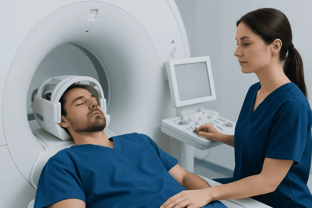
Imaging is often important. A CT scan is commonly used when a fracture or sinus complication is suspected because it shows bone detail clearly. MRI may be used when soft tissue, brain, or orbital involvement needs closer assessment. In non-urgent settings, imaging findings are interpreted together with symptoms because normal anatomical variation is common.
Doctors may also assess eye movement, vision, facial sensation, and nasal function. If sinus disease is suspected, an ENT specialist may perform nasal endoscopy. When trauma is significant or symptoms suggest deeper injury, related imaging or treatment may overlap with brain surgery planning, although many patients do not need surgery and are managed conservatively.
Treatment options and self-care
Treatment depends entirely on the cause. A normal frontal bone seen on imaging needs no treatment. Minor soft-tissue forehead injuries may improve with rest, ice, and observation if there are no warning signs. Fractures, however, range from simple injuries to complex cases that require specialist care. Treatment may include monitoring, pain control, wound care, antibiotics in selected situations, or surgery when there is deformity, sinus obstruction, cerebrospinal fluid leakage, or involvement of nearby structures.
When frontal sinus disease is the main issue, treatment often starts with medical care directed by a clinician. This may include measures to reduce inflammation, improve drainage, and treat infection when appropriate. If symptoms are persistent, recurrent, or complicated, doctors may consider procedural options. In selected facial trauma cases, reconstructive treatment can overlap with maxillofacial surgery depending on the location and extent of injury.
Self-care should stay within safe limits. People should avoid pressing repeatedly on a painful or swollen forehead lump, return to sports too early after head injury, or self-diagnose sinus pain if symptoms are severe or prolonged. Protecting the head during sports, using seat belts, and seeking assessment after significant facial or head trauma are practical ways to reduce complications.
Near the end of the care journey, some patients may need coordinated follow-up across specialties. Acibadem International’s multidisciplinary specialists and JCI-accredited hospitals evaluate and treat frontal bone conditions for international patients when advanced imaging, ENT care, neurosurgical input, or facial trauma management is needed.
When to seek medical care
Medical care is recommended after a significant blow to the forehead or face, especially if there is loss of consciousness, severe headache, vomiting, confusion, drowsiness, vision changes, nosebleeding that is difficult to control, or clear fluid leaking from the nose. These symptoms do not always mean a serious injury, but they need prompt professional assessment.
A doctor should also evaluate persistent forehead pain, a new hard lump, swelling that does not improve, fever with sinus pain, or tenderness around the brow area lasting more than a few days. In children, unusual head shape, developmental concerns, or forehead asymmetry should be discussed with a pediatrician or specialist.
If symptoms are sudden or severe, emergency evaluation is the safest choice. For non-urgent concerns, a primary care doctor, ENT specialist, neurologist, or surgeon may guide the next steps based on the likely cause.
Frequently asked questions
What is the frontal bone?
The frontal bone is the bone that forms the forehead and part of the front of the skull. It also helps form the roof of the eye sockets and contains the frontal sinuses in many adults.
Is the frontal bone the same as the forehead?
Not exactly. The forehead is the visible external region, while the frontal bone is the underlying skull bone that gives the forehead its shape and also extends deeper into the skull and orbit area.
Can the frontal bone be broken?
Yes. A strong impact can cause a frontal bone fracture, especially in falls, sports injuries, or vehicle accidents. Because the bone is near the brain and eyes, doctors usually assess these injuries carefully with examination and often imaging.
Why does the frontal bone matter in sinus problems?
The frontal sinuses are located inside the frontal bone above the eyes. When these sinuses become inflamed or blocked, they can cause pressure, headache, tenderness, and sometimes swelling in the forehead area.
Are forehead shape differences usually normal?
Many differences in forehead contour are part of normal anatomy. Brow shape, forehead slope, and sinus size vary between people, but a new change, marked asymmetry, or symptoms such as pain should be medically reviewed.
When should someone worry about pain over the frontal bone?
Pain should be assessed if it follows head trauma, becomes severe, lasts more than a few days, or comes with fever, swelling, vision changes, vomiting, or confusion. These features may suggest injury, sinus disease, or another condition that needs evaluation.
This article is for general information only and is not a substitute for professional medical advice. Please consult a qualified doctor about your individual situation.
Explore treatments in Turkey — costs, top hospitals & a free quote
JCI-accredited · board-certified surgeons · reply within 24h
Add us as a Preferred Source to see more of our trusted health content across Google Search, AI Overviews and Discover.

More from the Health Library

39.5c to F: A Complete Medical Overview

Crabs STD: An Evidence-Based Guide for Patients

Acth Hormone: What Patients Need to Know

White Gourd Melon: An Evidence-Based Guide for Patients

Tips Procedure: A Complete Medical Overview

Normal Heart Rate: A Complete Medical Overview
Orthopedic Surgery & Traumatology Specialists at Acibadem

Prof. Dr. Arel Gereli
Orthopedic Surgery & Traumatology
Fzt. Asım Sakallı
Physical Medicine & Rehabilitation
Fzt. Aslı Hacıoğlu
Physical Medicine & Rehabilitation
Assoc. Prof. Dr. Ata Can
Orthopedic Surgery & Traumatology