Fuchs Disease Surgery: Procedure, Recovery and Results

Fuchs disease surgery most often involves endothelial keratoplasty, which replaces the cornea’s damaged inner cell layer. DMEK and DSAEK are common procedures; the best choice depends on the eye, lens status, surgeon assessment, and individual needs.
Key Takeaways
- Fuchs disease surgery most often involves endothelial keratoplasty, which replaces the cornea’s damaged inner cell layer.
- DMEK and DSAEK are common procedures; the best choice depends on the eye, lens status, surgeon assessment, and individual needs.
- Vision commonly improves gradually over weeks to months, and follow-up visits are essential during recovery.
- Cataract surgery may be performed alone or combined with corneal transplantation when Fuchs dystrophy is advanced.
- Surgery has important benefits but also risks, including graft detachment, rejection, infection, pressure changes, and the need for further treatment.
Fuchs disease surgery is usually considered when corneal swelling from Fuchs endothelial corneal dystrophy causes persistent blurred vision, glare, or daily-life difficulties. Modern endothelial keratoplasty replaces the damaged inner corneal cell layer and can restore clearer vision for many suitable patients, although healing and visual improvement take time.
Overview: How Fuchs Disease Surgery Helps
Fuchs disease surgery treats vision loss caused by Fuchs endothelial corneal dystrophy, an inherited or age-related condition in which the cornea’s innermost cells gradually stop working well. These endothelial cells normally pump fluid out of the cornea. When too many are damaged, fluid builds up, the cornea swells, and vision may become hazy, distorted, glare-sensitive, or worse on waking.
The most common surgical approach is endothelial keratoplasty, a form of corneal transplant that replaces only the unhealthy inner corneal layers rather than the full thickness of the cornea. This targeted approach is designed to restore the cornea’s fluid balance and transparency while preserving as much of the person’s own cornea as possible. Early Fuchs disease may be monitored without surgery, particularly when symptoms remain mild.
Fuchs endothelial corneal dystrophy can affect both eyes, although symptoms and progression may differ between them. A cornea specialist can determine whether reduced vision is mainly due to corneal swelling, cataract, another eye condition, or a combination of causes. Readers can also learn more about Fuchs dystrophy and its effects on the cornea.
How the Procedure Works and Who May Be a Candidate
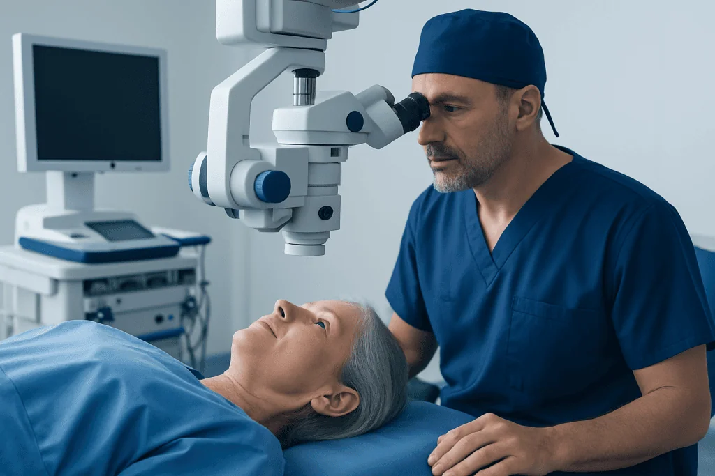
The endothelial layer does not reliably regenerate once substantial cell loss has occurred. Endothelial keratoplasty therefore uses donor corneal tissue containing healthy endothelial cells to take over the pumping function. The two main techniques are Descemet membrane endothelial keratoplasty (DMEK), which transplants an exceptionally thin layer, and Descemet stripping automated endothelial keratoplasty (DSAEK), which includes a thin layer of supporting corneal tissue.
DMEK may offer excellent visual clarity in appropriate eyes, while DSAEK can be a suitable option in certain complex eyes or when the anatomy makes handling an ultra-thin graft less practical. The surgeon discusses the expected advantages, limitations, and recovery considerations of each technique. In some cases, a different procedure, such as Descemet stripping only, may be discussed for carefully selected patients with healthy peripheral endothelial cells.
A person may be considered for surgery when corneal edema causes troublesome blur, glare, painful surface blisters, reduced function, or a meaningful decline in quality of life. The decision is individualized. Examination may include vision testing, corneal thickness measurement, slit-lamp examination, endothelial cell assessment, and corneal imaging. Cataract severity, glaucoma, retinal disease, previous eye surgery, and overall eye health all influence planning.
When a cataract is also contributing to poor vision, a surgeon may recommend cataract surgery first, endothelial keratoplasty first, or a combined operation. The appropriate plan depends on the degree of corneal swelling and the likelihood that cataract surgery could stress the remaining endothelial cells. Corneal transplant evaluation and treatment can help clarify whether endothelial keratoplasty is appropriate.
Fuchs Disease Surgery: Step by Step
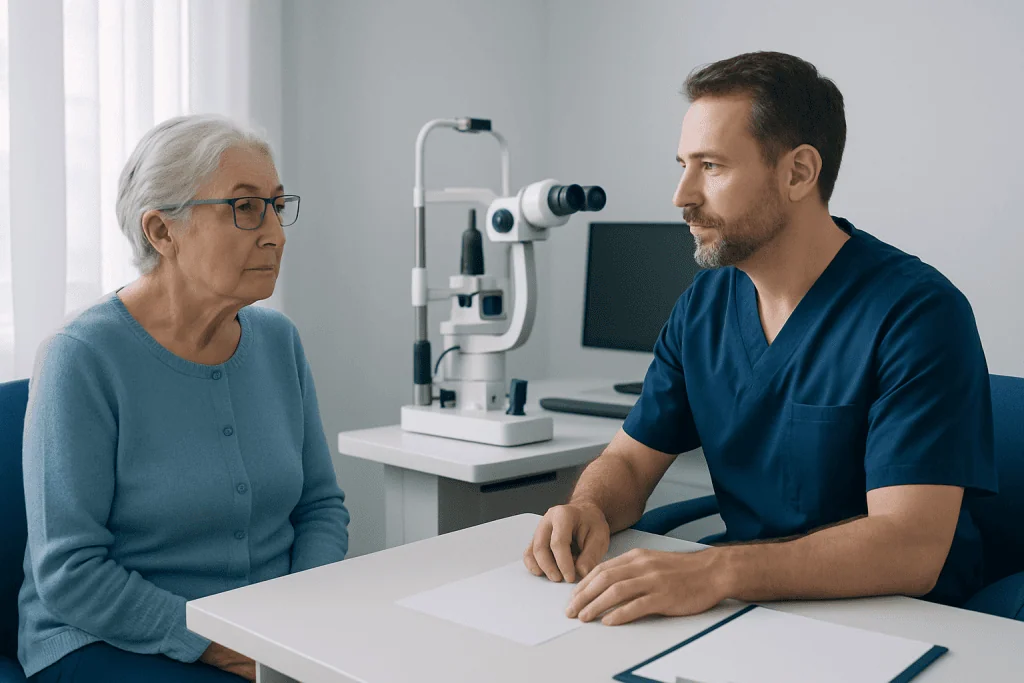
Endothelial keratoplasty is typically performed as an outpatient procedure under local anesthesia with sedation, although the anesthetic plan is tailored to the individual. The eye is numbed, cleaned, and held open with a small instrument. Patients should expect to be awake but comfortable if local anesthesia and sedation are used.
The surgeon makes very small incisions in the cornea and removes the diseased Descemet membrane and endothelial cells from the central back surface of the cornea. Prepared donor tissue is inserted through a tiny opening and carefully unfolded or positioned inside the eye. A temporary air or gas bubble is placed beneath the graft to hold it against the back of the cornea while it adheres.
For the first hours or days, the surgeon may ask the patient to lie flat on their back for specified periods so the bubble can support graft attachment. Instructions differ according to the procedure, bubble type, and surgeon’s protocol. The air or gas bubble gradually absorbs on its own, and vision may be quite blurred while it remains.
If cataract surgery is performed at the same time, the cloudy natural lens is removed and replaced with an artificial intraocular lens before or during the corneal procedure. This combined approach is sometimes called a triple procedure. It can avoid a second operation but may make prediction of the final glasses prescription less precise than in some staged approaches.
Benefits, Risks and What Results to Expect
The principal benefit of Fuchs disease surgery is reduction of corneal swelling, which can improve clarity, contrast, glare, and day-to-day visual function. Many people notice vision becoming clearer as the graft begins pumping fluid effectively and the cornea returns toward a more normal thickness. Final visual quality also depends on cataract, refractive error, macular or optic nerve conditions, and any longstanding corneal scarring.
As with all transplant surgery, complications are possible. These include graft detachment, incomplete clearing of the cornea, raised or lowered eye pressure, bleeding, infection, inflammation, irregular astigmatism, and graft rejection. A detached graft may require an additional air or gas injection, often called rebubbling. In some cases, the graft may fail and another transplant may be needed.
Rejection can occur even years after surgery, though it is often treatable when recognized promptly. New redness, light sensitivity, pain, or a sudden decline in vision should be reported urgently to the eye care team. Prescribed steroid eye drops and regular monitoring are important for lowering inflammation and identifying concerns early.
A thorough discussion before surgery should cover realistic goals. Surgery can improve corneal clarity but cannot reverse visual loss caused by unrelated retinal, optic nerve, or neurological conditions. The ophthalmologist can explain what outcome is reasonably expected for the individual eye.
What Is the Success Rate of Fuchs Dystrophy Surgery?
Fuchs dystrophy surgery has generally favorable outcomes when performed for appropriate patients by an experienced corneal surgeon, particularly with modern endothelial keratoplasty techniques. However, there is no single success rate that applies to every person because studies define success differently and outcomes vary according to the procedure used, the health of the eye, donor tissue, surgical factors, and length of follow-up.
In practical terms, a successful result means that the graft remains attached and clear, the corneal swelling improves, and vision reaches the best level allowed by the rest of the eye. DMEK and DSAEK can both provide durable clearing of the cornea, but some patients need a rebubbling procedure for graft attachment or, less commonly, repeat transplantation.
People should ask their surgeon about their personal prognosis rather than relying on a general fuchs dystrophy surgery success rate. Factors such as glaucoma, prior glaucoma surgery, severe pre-existing swelling or scarring, previous transplants, and retinal disease may affect both risks and final vision. Continued long-term checkups remain important even after vision has stabilized.
Fuchs Surgery Recovery Time and Aftercare
Fuchs surgery recovery time varies considerably. The first days often involve blurred vision, light sensitivity, a foreign-body sensation, and limited activity while the graft settles. DMEK may clear more quickly than DSAEK for some people, but neither procedure produces an immediate final result. Vision commonly improves progressively over several weeks, and fuchs dystrophy surgery recovery time may extend for several months before the eye reaches a stable outcome.
Patients are usually prescribed antibiotic drops for a short period and anti-inflammatory steroid drops for a longer period. They should use medications exactly as directed, avoid rubbing or pressing on the eye, protect the eye from injury, and attend scheduled follow-up visits. Temporary positioning requirements after surgery are especially important because they help the graft remain attached.
Driving, work, exercise, swimming, eye makeup, heavy lifting, and air travel may need to be paused or modified initially. The care team gives individualized advice based on healing, visual function, and whether an air or gas bubble remains in the eye. If a gas bubble is present, air travel and high-altitude travel may be unsafe until the surgeon confirms that it has fully resolved.
Acibadem International’s multidisciplinary specialists and JCI-accredited hospitals can assess and treat corneal conditions for international patients, with follow-up plans tailored to the procedure and the patient’s travel needs. Consultation is particularly useful when cataract, glaucoma, or retinal conditions may also influence surgical planning.
How Painful Is Fuchs Dystrophy?
Fuchs dystrophy is often not painful in its early and moderate stages. More commonly, it causes painless morning blur, fluctuating vision, glare, halos around lights, and difficulty seeing in low light. These symptoms result from fluid in the cornea and may become more persistent as the condition progresses.
Pain can develop if marked swelling leads to small blisters on the corneal surface, known as bullous keratopathy. When blisters break, they can cause sharp discomfort, tearing, light sensitivity, and a gritty sensation. This deserves prompt assessment because the surface of the eye may need protective treatment while the underlying corneal problem is addressed.
After endothelial keratoplasty, discomfort is usually mild to moderate and short-lived, but symptoms vary. Severe or increasing pain is not something to manage alone, especially when combined with redness, nausea, headache, or worsening vision. The surgical team should be contacted promptly for advice.
How Long Does It Take to Recover From Cataract Surgery if I Have Fuchs Dystrophy?
Recovery from cataract surgery in someone with Fuchs dystrophy can be slower and less predictable than routine cataract recovery because the remaining endothelial cells may have limited capacity to clear fluid from the cornea. If the cornea remains relatively healthy before surgery, visual recovery may still be smooth. If endothelial function is significantly reduced, corneal swelling can persist for weeks or longer after cataract removal.
Before surgery, the ophthalmologist evaluates corneal thickness, the appearance of endothelial changes, the presence of morning blur, and the amount of edema. These findings help estimate whether cataract surgery alone is reasonable or whether combining it with endothelial keratoplasty may better address both problems. There is no universal approach because each eye has different risks and visual priorities.
Some patients have staged treatment, with cataract surgery and corneal transplantation performed separately. Others have both in one operation. Clear communication about expected recovery, postoperative drops, and follow-up is essential. Cataract surgery assessment can be coordinated with corneal evaluation when both conditions are present.
What Worsens Fuchs Dystrophy and When to Seek Medical Care
Fuchs dystrophy progresses because endothelial cells are gradually lost over time. Aging and inherited susceptibility are important factors, and it commonly affects both eyes. Eye surgery, especially cataract surgery in an already vulnerable cornea, can temporarily or sometimes persistently increase swelling because it places stress on endothelial cells. This does not mean surgery should be avoided when needed; it means careful preoperative planning is important.
Anything that causes additional injury or inflammation in the eye may complicate corneal health. People should avoid eye rubbing and should tell eye specialists about their Fuchs diagnosis before any eye procedure. They should not stop prescribed treatments or use unprescribed eye drops to treat persistent blur, as some products may be unsuitable for their particular eye condition.
Medical care should be sought promptly for sudden worsening of vision, new significant eye pain, marked redness, light sensitivity, discharge, flashes or a curtain-like shadow in vision, or symptoms after eye surgery that are getting worse rather than better. Routine review with an ophthalmologist is also appropriate when morning blur lasts longer, glare increases, or daily activities become more difficult.
For associated visual concerns, patients may also benefit from assessment for glaucoma treatment or other eye conditions when indicated. The safest next step is an examination by a qualified ophthalmologist, ideally a cornea specialist when Fuchs dystrophy is suspected or confirmed.
Frequently asked questions
Is Fuchs disease surgery the same as a full corneal transplant?
Usually, no. Most modern surgery for Fuchs dystrophy is endothelial keratoplasty, which replaces only the damaged inner corneal layers. A full-thickness corneal transplant is generally reserved for selected situations, such as significant corneal scarring or when another transplant approach is not suitable.
Can Fuchs dystrophy be treated without surgery?
Yes, mild symptoms may be managed with observation and measures recommended by an ophthalmologist. However, these approaches do not replace lost endothelial cells. Surgery may become appropriate when corneal swelling and vision problems meaningfully affect daily life.
Will I need glasses after Fuchs dystrophy surgery?
Many people still need glasses after surgery for distance, reading, or astigmatism. If cataract surgery is also performed, the intraocular lens plan can influence spectacle needs. The final prescription is usually determined only after the eye has healed and vision has stabilized.
Can Fuchs dystrophy return after endothelial keratoplasty?
The transplanted endothelial cells can function for many years, but no corneal graft lasts forever in every person. Graft cell loss, rejection, or failure can occur over time and may sometimes require additional treatment. Ongoing follow-up helps detect changes early.
What are warning signs of corneal graft rejection?
Possible warning signs include increasing redness, light sensitivity, pain, and a new drop in vision. These symptoms can have several causes, but they should be reported urgently after corneal transplant surgery. Prompt treatment can often help protect graft function.
Can I have cataract surgery if I have Fuchs dystrophy?
Yes, many people with Fuchs dystrophy can have cataract surgery, but the cornea should be assessed carefully beforehand. Depending on endothelial health and corneal swelling, the surgeon may recommend cataract surgery alone, combined surgery, or staged procedures. The plan should be individualized by an ophthalmologist.
References
- American Academy of Ophthalmology
- National Eye Institute
- Eye Bank Association of America
- Mayo Clinic
- National Health Service
This article is for general information only and is not a substitute for professional medical advice. Please consult a qualified doctor about your individual situation.
Explore treatments in Turkey — costs, top hospitals & a free quote
JCI-accredited · board-certified surgeons · reply within 24h
Add us as a Preferred Source to see more of our trusted health content across Google Search, AI Overviews and Discover.