Glenoid Fracture: A Complete Medical Overview

The glenoid is the shallow socket of the shoulder joint and is part of the scapula, or shoulder blade. Glenoid fractures commonly occur after significant trauma, including falls, sports injuries, or road traffic collisions.
Key Takeaways
- The glenoid is the shallow socket of the shoulder joint and is part of the scapula, or shoulder blade.
- Glenoid fractures commonly occur after significant trauma, including falls, sports injuries, or road traffic collisions.
- X-rays and CT scans help clinicians identify fracture patterns, displacement, and shoulder-joint stability.
- Some stable fractures can heal with a sling and guided rehabilitation, while displaced or unstable injuries may require surgery.
- Early assessment is important because untreated joint instability or poor alignment can affect shoulder movement and long-term comfort.
A glenoid fracture is a break involving the socket portion of the shoulder blade, where the upper arm bone meets the shoulder. Treatment depends on whether the joint surface remains aligned and stable, with careful rehabilitation playing an important role in recovery.
What Is a Glenoid Fracture?
A glenoid fracture is a break in the glenoid, the cup-shaped part of the shoulder blade that forms the socket of the ball-and-socket shoulder joint. The head of the upper arm bone, called the humerus, sits in this socket. Although the glenoid is relatively small, it has an important role in keeping the shoulder stable while allowing a wide range of movement.
These fractures are less common than fractures of the collarbone or upper arm. They may involve the edge, or rim, of the socket; the joint surface itself; or the upper part of the shoulder blade near the socket. A fracture may occur on its own or alongside shoulder dislocation, ligament damage, or other injuries around the chest, arm, or shoulder.
The main clinical concern is whether the fracture changes the smoothness of the joint surface or affects the stability of the shoulder. Prompt assessment helps the care team choose the safest treatment approach and plan rehabilitation around the person’s injury, activity needs, and overall health.
Symptoms and How It May Feel
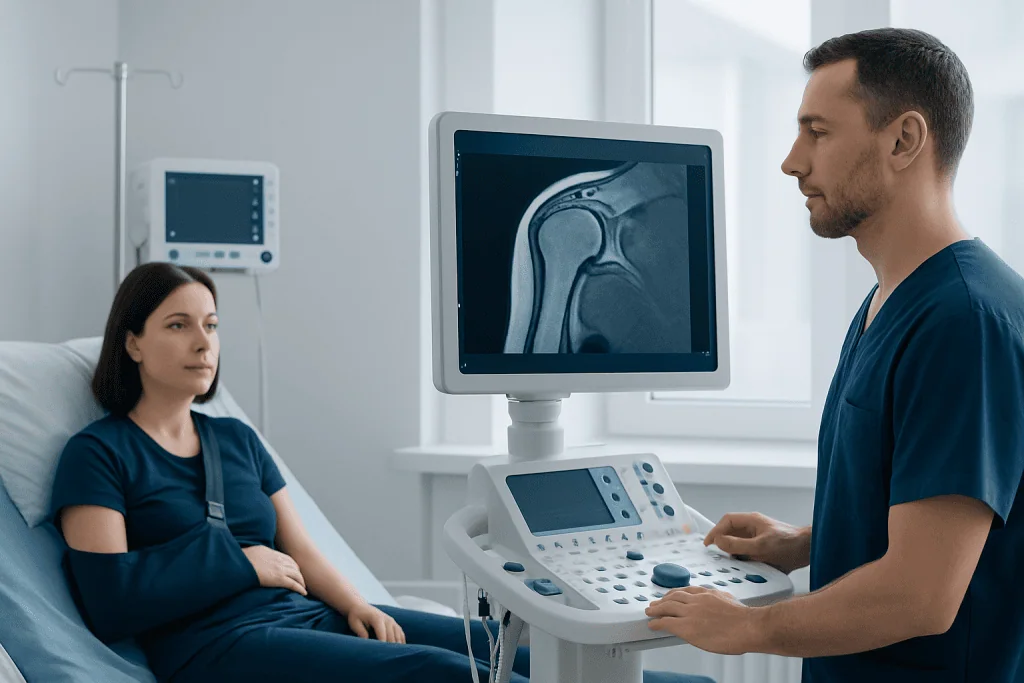
Most people develop sudden shoulder pain after an injury. Pain may be severe, especially when trying to lift, rotate, or bear weight through the arm. The shoulder may look swollen or bruised, and holding the arm close to the body can feel more comfortable.
Movement is often limited because of pain, swelling, and muscle guarding. Some people notice clicking, catching, or a feeling that the shoulder may slip out of place. These symptoms can occur when the glenoid rim is affected or when a fracture occurs with a shoulder dislocation.
A medical assessment is important because symptoms alone cannot show the exact fracture type. Numbness, tingling, weakness in the hand or arm, a cool or pale hand, or difficulty moving the fingers may indicate associated nerve or blood-vessel concerns and should be assessed urgently.
Causes and Risk Factors
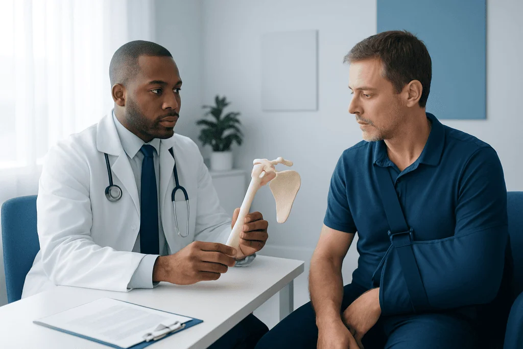
Glenoid fractures usually result from forceful trauma to the shoulder. Common causes include a fall directly onto the shoulder or an outstretched arm, a road traffic collision, contact sports, cycling accidents, and workplace injuries. The force may drive the humeral head into the glenoid, fracture the socket rim, or cause a dislocation that pulls or compresses the bone.
In younger adults, these injuries are often related to high-energy trauma or athletic injury. In older adults, a lower-energy fall may cause a fracture when bone strength is reduced. Conditions that weaken bone, such as osteoporosis, can increase fracture risk, although a clinician will consider the full injury history rather than assume a cause.
Repeated shoulder dislocations can also damage the rim of the glenoid. This may produce a bony injury associated with instability, sometimes described as a bony Bankart lesion. Related shoulder instability problems require individualized evaluation because the amount of bone involved can influence treatment decisions.
How a Glenoid Fracture Is Diagnosed
Diagnosis begins with a history of the injury and a physical examination. A clinician checks shoulder shape, swelling, tenderness, range of movement, and signs of instability. They also assess circulation, sensation, and muscle function in the arm and hand, particularly after a dislocation or high-impact injury.
Standard shoulder X-rays are usually the first imaging test. They can show many fractures and identify whether the shoulder is dislocated. However, the glenoid is a complex three-dimensional structure, so a CT scan is often used to define the fracture more clearly. CT imaging can show the size of fragments, the amount of displacement, and whether the joint surface is affected.
MRI may be considered when clinicians need more information about soft tissues, such as the rotator cuff, labrum, cartilage, or ligaments. It is not always required for the initial fracture diagnosis. The imaging findings are considered alongside symptoms, the person’s functional goals, and any associated injuries.
Treatment Options and Recovery Planning
Treatment aims to restore a stable, comfortable shoulder with the best possible joint alignment and movement. A stable fracture with little or no displacement may be treated without surgery. This commonly includes a sling for comfort and protection, pain management recommended by a clinician, and follow-up imaging to confirm that healing is progressing appropriately.
Gentle movement is introduced at a time chosen by the treating team. Starting rehabilitation too early can stress a healing fracture, while immobilization for too long can contribute to stiffness. A physiotherapist may guide exercises that gradually restore mobility, shoulder-blade control, strength, and confidence in daily activities.
Surgery may be advised if the fracture is significantly displaced, involves an important portion of the joint surface, causes instability, or occurs with injuries that need repair. The procedure may involve realigning the fragments and securing them with plates, screws, or other fixation devices. In selected cases, arthroscopic techniques may be used. Shoulder surgery is planned according to the fracture pattern and the patient’s individual needs.
Recovery time varies. Bone healing commonly takes several weeks, but regaining strength, movement, and sport- or work-specific function can take longer. Regular follow-up helps identify stiffness, ongoing instability, nerve symptoms, or delayed healing early.
Daily Care, Rehabilitation and Prevention
During the early phase of recovery, people should follow their clinician’s instructions about sling use, sleeping position, wound care after surgery, and permitted arm movements. It is usually important to avoid lifting, pushing, pulling, overhead activity, or returning to sport before the shoulder has been cleared for these activities.
Rehabilitation is not only about increasing range of motion. It also focuses on the muscles that support the shoulder blade and rotator cuff, helping the shoulder work in a coordinated way. Exercises should be progressed gradually and should not be copied from another person’s plan, as restrictions can differ substantially between fracture patterns and surgical repairs.
Not every accident can be prevented, but risk may be reduced by using appropriate protective equipment for cycling and contact sports, improving balance and home safety to reduce fall risk, and supporting bone health through adequate nutrition, weight-bearing activity where suitable, and evaluation of osteoporosis risk when indicated. For people with repeated dislocations, assessment for shoulder dislocation and instability can help guide prevention strategies.
When to Seek Medical Care
Urgent medical care is appropriate after a significant shoulder injury, particularly when there is severe pain, visible deformity, inability to move the arm, rapid swelling, or a sensation that the shoulder has come out of place. A shoulder injury should not be forced back into position outside a medical setting.
Emergency assessment is especially important if the hand or arm becomes numb, weak, pale, blue, cool, or increasingly painful. These signs may suggest pressure on nerves or blood vessels and need prompt evaluation. New shortness of breath, chest pain, neck pain, or symptoms after major trauma also require urgent care.
People already receiving treatment should contact their care team if pain or swelling is worsening rather than improving, a surgical wound becomes increasingly red or drains fluid, fever develops, or new loss of sensation or movement occurs. Acibadem International’s multidisciplinary specialists and JCI-accredited hospitals assess and treat shoulder injuries for international patients.
Frequently asked questions
Is a glenoid fracture serious?
A glenoid fracture can be significant because it involves the socket of the shoulder joint. Its seriousness depends on the location, amount of displacement, joint stability, and whether there are associated injuries. Accurate imaging and orthopedic assessment help determine the appropriate treatment.
Can a glenoid fracture heal without surgery?
Yes, some glenoid fractures can heal without surgery when the fracture is stable and the joint surface remains suitably aligned. Treatment may include temporary immobilization, follow-up appointments, and a structured rehabilitation program. Surgery may be recommended when alignment or stability cannot be maintained non-surgically.
How long does recovery from a glenoid fracture take?
Initial bone healing often takes several weeks, but full recovery can take months. The timeline depends on the fracture pattern, whether surgery was needed, associated soft-tissue injuries, and participation in rehabilitation. A treating clinician can provide the most relevant timeline for the individual injury.
Will shoulder movement return after a glenoid fracture?
Many people regain useful shoulder movement with appropriate treatment and rehabilitation. Some stiffness or weakness is common early in recovery, especially after a period of sling use. Outcomes may be affected by fracture severity, cartilage injury, instability, and adherence to the rehabilitation plan.
What is the difference between a glenoid fracture and a shoulder dislocation?
A glenoid fracture is a break in the shoulder socket bone, while a shoulder dislocation occurs when the upper arm bone comes out of the socket. The two injuries can happen together, particularly when a dislocation damages the rim of the glenoid. Both need medical evaluation after an acute injury.
Can a glenoid fracture cause arthritis later in life?
A fracture involving the joint surface can increase the long-term risk of post-traumatic shoulder arthritis, especially if the surface heals unevenly. This does not mean arthritis will definitely develop. Restoring alignment where possible, following rehabilitation advice, and attending follow-up visits can support joint health.
References
- American Academy of Orthopaedic Surgeons
- Orthopaedic Trauma Association
- American Shoulder and Elbow Surgeons
- National Institute of Arthritis and Musculoskeletal and Skin Diseases
This article is for general information only and is not a substitute for professional medical advice. Please consult a qualified doctor about your individual situation.
Joint and spine care in Turkey — expert assessment & treatment
JCI-accredited · board-certified surgeons · reply within 24h
Add us as a Preferred Source to see more of our trusted health content across Google Search, AI Overviews and Discover.