Hemangioma: Diagnosis, Outlook, and Modern Treatment Approaches

A hemangioma is a non-cancerous blood-vessel growth that can appear on the skin or inside the body. Many infantile hemangiomas grow for a period, then slowly shrink over time.
Key Takeaways
- A hemangioma is a non-cancerous blood-vessel growth that can appear on the skin or inside the body.
- Many infantile hemangiomas grow for a period, then slowly shrink over time.
- Treatment is not always necessary, but it may be recommended if a hemangioma affects vision, breathing, feeding, hearing, skin integrity, or comfort.
- Diagnosis is often based on a physical exam, though ultrasound or MRI may be used in selected cases.
- Modern treatment options can include observation, medicines, laser therapy, or procedures for carefully chosen patients.
Hemangioma is a benign growth made of extra blood vessels. Many hemangiomas, especially in infants, improve over time without treatment, but some need closer follow-up or medical care depending on their size, location, symptoms, and effect on function or appearance.
Overview: what a hemangioma is and what to expect
A hemangioma is a benign, meaning non-cancerous, growth formed by extra blood vessels. It may be present at birth or appear in the first weeks of life, and it can develop on the skin, under the skin, or less commonly in internal organs such as the liver. The most common type seen in babies is infantile hemangioma, which often follows a predictable pattern of growth and gradual improvement.
In many cases, a hemangioma does not cause serious health problems and can be safely monitored. However, some hemangiomas need closer attention because of where they are located, how quickly they grow, or whether they interfere with normal activities such as seeing, feeding, breathing, hearing, or moving comfortably. Ulceration, bleeding, or pain may also prompt treatment.
Although the word “tumor” may be used medically for any growth, a hemangioma is not a cancer. This distinction is important for patients and families, because the outlook is usually reassuring. The main questions are whether the lesion is truly a hemangioma, whether it is likely to improve on its own, and whether treatment would help protect function, skin health, or long-term appearance.
How hemangiomas appear and change over time
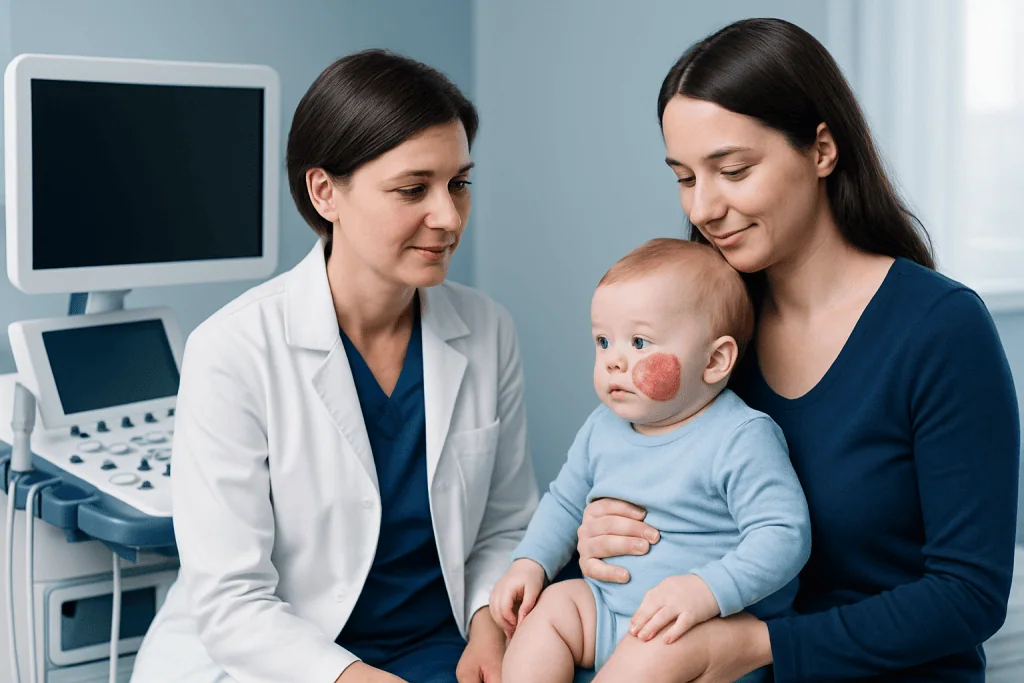
Hemangiomas can look different depending on how deep they are in the tissue. Superficial hemangiomas often appear bright red and raised, sometimes described as “strawberry” marks. Deeper hemangiomas may look bluish, skin-colored, or swollen rather than bright red. Mixed hemangiomas contain both superficial and deep components.
Infantile hemangiomas usually have a growth phase during early infancy, followed by a plateau and then a slow shrinking phase called involution. This natural history helps guide care. A lesion that is small, stable, and not causing problems may only need periodic review, while one in a high-risk area may need early treatment even if the child seems otherwise well.
Not every vascular mark is a hemangioma. Some birthmarks are vascular malformations, which behave differently and do not usually follow the same grow-then-shrink pattern. Distinguishing between these conditions matters because treatment planning and long-term outlook are different. In some situations, doctors may also consider related vascular conditions such as vascular malformations when evaluating a birthmark or soft tissue mass.
- Common locations include the face, scalp, neck, chest, and back.
- Some hemangiomas occur near the eye, nose, lips, airway, diaper area, or liver and may need closer follow-up.
- Color, size, and texture can change over time, especially in the first year of life.
Causes and risk factors

The exact cause of hemangioma is not fully understood. It develops when blood vessels grow in an unusual way, but this is not usually linked to anything a parent did or did not do during pregnancy. In most cases, hemangiomas occur sporadically, meaning they happen without a clear inherited pattern.
Some factors are associated with a higher chance of infantile hemangioma. These include female sex, premature birth, low birth weight, and multiple pregnancy. Even so, many children with these risk factors never develop a hemangioma, while others with no known risk factors do.
Hemangiomas can also occur in adults, though the patterns and common locations may differ. A doctor may need to distinguish an adult vascular lesion from other benign or more concerning skin or soft tissue conditions. Because several vascular lesions can look similar, expert assessment is helpful when a growth is enlarging, painful, deep, or atypical in appearance.
Diagnosis: how doctors confirm hemangioma
Diagnosis often begins with a careful history and physical examination. Doctors look at when the lesion appeared, how fast it has grown, whether it is changing color or texture, and whether symptoms such as ulceration, pain, feeding difficulty, noisy breathing, or visual obstruction are present. In straightforward cases, especially superficial infantile hemangiomas, examination alone may be enough to make the diagnosis.
Imaging tests may be used when the diagnosis is uncertain or when the hemangioma appears deep, extensive, or close to important structures. Ultrasound can help show blood flow and the depth of the lesion. MRI may be recommended if doctors need a more detailed view of the surrounding tissues or if the lesion involves the head and neck, spine, airway, or internal organs. In selected cases, a child may also be assessed for syndromes associated with large segmental hemangiomas.
Doctors also evaluate whether a hemangioma is low risk or high risk. A small lesion on the trunk may simply need observation, while a lesion near the eye, on the lip, in the beard area, or in skin folds may need earlier intervention. When a vascular lesion is unusual or raises questions, assessment by specialists in dermatology, pediatrics, radiology, surgery, or vascular anomalies can help refine the diagnosis and care plan.
Treatment options and when they are used
Treatment depends on the hemangioma’s type, stage, location, symptoms, and likely impact over time. Observation is appropriate for many uncomplicated hemangiomas, especially when they are small and not affecting function. Families may be advised to monitor the lesion with regular photographs and follow-up visits so growth patterns can be tracked.
When treatment is needed, medication is often the first approach for problematic infantile hemangiomas. Beta-blocker therapy is commonly used under medical supervision because it can slow growth and help the lesion shrink. Some patients may also be considered for laser therapy, especially for superficial redness or residual skin changes, or for surgery when the lesion causes persistent problems, leaves significant tissue changes, or does not respond adequately to other options. Depending on the case, doctors may discuss laser treatment or pediatric surgery as part of a broader care plan.
Internal hemangiomas, including some liver hemangiomas, are managed according to symptoms, size, and complications rather than appearance. Most still do not require urgent treatment, but they do require proper evaluation. In more complex cases, a multidisciplinary team may coordinate dermatology, pediatrics, radiology, surgery, ENT, ophthalmology, or other specialties to choose the least invasive and most effective option.
After a hemangioma shrinks, some residual changes may remain, such as loose skin, fine blood vessels, discoloration, or a fibrofatty bump. These changes are not dangerous, but they can affect comfort or appearance. In selected patients, doctors may consider follow-up care through services such as dermatology or reconstructive procedures if residual tissue changes are significant.
Daily care, prevention, and self-care tips
There is no known way to prevent most hemangiomas from developing. The most helpful step is early recognition and appropriate follow-up, especially if the lesion is in a sensitive area or growing quickly. Parents and patients can support care by documenting changes over time and sharing clear photographs with the care team when requested.
Skin care matters most when a hemangioma is raised, irritated, or prone to ulceration. Keeping the area clean and protected from friction can reduce discomfort. If the lesion is in the diaper area, skin folds, or around the lips, gentle cleansing and barrier protection may be recommended. Any home treatment should be discussed with a clinician, especially for infants.
Sun protection can help reduce irritation and protect surrounding skin, although it does not remove a hemangioma. Patients should avoid picking, scratching, or using unapproved topical products on the lesion. Regular review is important because a hemangioma that was once low risk can occasionally become more troublesome if it enlarges, breaks down, or starts affecting nearby structures.
- Take serial photos in similar lighting to document change.
- Watch for pain, open sores, crusting, bleeding, or sudden swelling.
- Follow the treatment plan exactly if medicine has been prescribed.
- Ask a doctor before trying over-the-counter creams or alternative remedies.
When to seek medical care
Medical review is advisable for any new or enlarging vascular lesion when the diagnosis is uncertain. Early assessment is especially important in babies younger than a few months, because some hemangiomas grow rapidly during this stage. Prompt evaluation can help determine whether observation is enough or whether treatment should begin early to prevent complications.
Patients should seek medical care sooner if a hemangioma is near the eye, nose, lips, beard area, ear, airway, genital area, hands, or feet, or if it interferes with feeding, vision, hearing, breathing, or movement. Care is also important if the skin breaks down, the lesion bleeds repeatedly, becomes painful, develops signs of infection, or seems to be growing unusually fast. Deep lesions, multiple skin hemangiomas, or suspected internal hemangiomas may also need imaging and specialist review.
For complex cases, coordinated care can be useful. Acibadem International’s multidisciplinary specialists in JCI-accredited hospitals diagnose and treat hemangioma for international patients, including cases that may need advanced imaging, interventional radiology, or input from teams experienced in pediatric vascular anomalies.
Outlook and long-term considerations
The outlook for hemangioma is generally very good. Many infantile hemangiomas become less noticeable with time, and some nearly disappear. Even when treatment is needed, the goal is usually to protect function, reduce complications, and improve long-term skin or tissue outcome rather than to respond to an emergency.
Long-term follow-up depends on the lesion’s location and severity. A child with a small trunk hemangioma may need only occasional review, while one with a facial or airway lesion may need coordinated follow-up across several specialties. Residual cosmetic changes can be addressed later if they remain bothersome after the lesion has completed most of its natural involution.
Because each hemangioma behaves a little differently, families benefit from individualized guidance. Clear diagnosis, timely monitoring, and treatment when indicated help most patients do well. When questions arise about growth, symptoms, or treatment choices, a qualified doctor can explain the expected course and the most appropriate next steps.
Frequently asked questions
Is a hemangioma dangerous?
Most hemangiomas are not dangerous and do not become cancer. They are benign blood-vessel growths, but some can cause problems if they are large, ulcerated, or located near the eye, airway, lips, or other sensitive areas.
Do all hemangiomas need treatment?
No. Many hemangiomas can be safely observed because they eventually shrink over time. Treatment is usually considered when the lesion affects function, causes pain or skin breakdown, grows rapidly, or is likely to leave more significant long-term changes.
At what age do infantile hemangiomas usually improve?
Infantile hemangiomas often grow most in early infancy and then gradually begin to shrink. Improvement usually happens over several years, not all at once, so regular follow-up helps track the expected pattern.
How is a hemangioma diagnosed?
Doctors often diagnose hemangioma with a medical history and physical examination. If the lesion is deep, unusually large, or in a sensitive area, ultrasound or MRI may be used to confirm the diagnosis and guide treatment planning.
Can a hemangioma bleed or become infected?
Yes, especially if the surface breaks down and forms an ulcer. Repeated bleeding, increasing pain, drainage, or signs of infection should be assessed by a doctor promptly so the area can be treated and protected.
What is the difference between a hemangioma and a vascular malformation?
A hemangioma usually has a typical pattern of growth followed by slow shrinking, especially in infancy. A vascular malformation is a different type of blood-vessel abnormality that usually grows with the person and often needs a different evaluation and treatment plan.
References
- American Academy of Pediatrics
- American Academy of Dermatology
- National Organization for Rare Disorders
- National Health Service
- Society for Pediatric Dermatology
This article is for general information only and is not a substitute for professional medical advice. Please consult a qualified doctor about your individual situation.
Explore treatments in Turkey — costs, top hospitals & a free quote
JCI-accredited · board-certified surgeons · reply within 24h
Add us as a Preferred Source to see more of our trusted health content across Google Search, AI Overviews and Discover.