Hemangioma Treatment: How It Works, Results and What to Expect

Most infantile hemangiomas shrink naturally over time and do not require a procedure. Treatment is considered when a hemangioma threatens vision, breathing, feeding, skin integrity or long-term appearance.
Key Takeaways
- Most infantile hemangiomas shrink naturally over time and do not require a procedure.
- Treatment is considered when a hemangioma threatens vision, breathing, feeding, skin integrity or long-term appearance.
- Beta-blocker medicines are commonly used for problematic infantile hemangiomas; laser therapy and surgery are selected for specific situations.
- Recovery after hemangioma surgery varies with the lesion’s size, depth and location, but initial wound healing often takes about one to two weeks.
- Hemangiomas are benign and do not turn into melanoma, although a changing skin lesion should still be assessed by a clinician.
Hemangioma treatment is tailored to the type, location, size and behavior of the blood-vessel growth. Many hemangiomas need observation only, while those affecting function, ulcerating, bleeding or likely to leave significant changes may benefit from medication, laser treatment or surgery.
Hemangioma treatment: how it works and who may need it
Hemangioma treatment aims to protect normal function, prevent complications and improve healing or appearance when needed. A hemangioma is a non-cancerous growth made up of blood vessels. The term is often used for infantile hemangiomas, which usually appear in the first weeks of life, grow most rapidly during early infancy and then gradually fade over years. Hemangiomas can also occur in adults, including as vascular growths on the skin or within internal organs.
Many hemangiomas are safely monitored rather than actively treated. A clinician may recommend treatment if the lesion is growing quickly, painful, ulcerated, repeatedly bleeding, infected, or located near the eye, nose, mouth, airway, ear, genital area or another site where it can affect function. A visible lesion may also be treated when it has a high likelihood of leaving stretched skin, excess tissue or a noticeable scar after natural involution.
Care is individualized. Dermatologists, pediatricians, plastic surgeons, vascular specialists, ophthalmologists and other clinicians may work together depending on the hemangioma’s location. The best option is based on the person’s age, general health, whether the lesion is superficial or deep, and whether it is actively changing.
How hemangioma treatment works
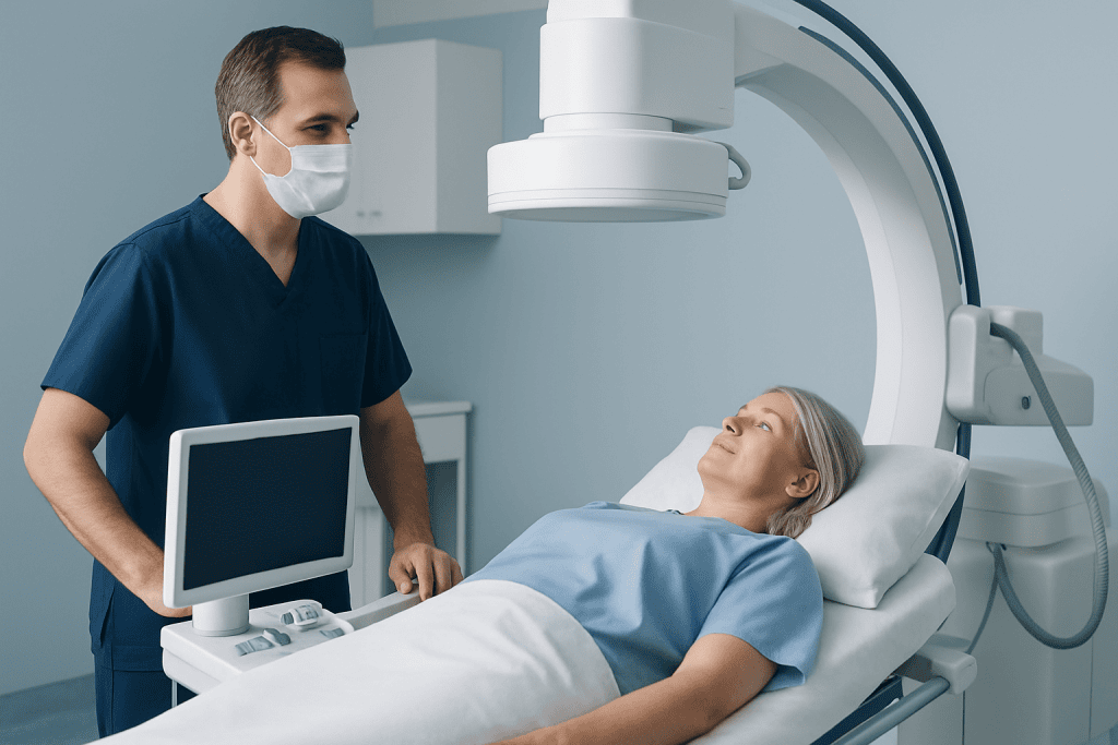
Observation is appropriate for many uncomplicated lesions. During active monitoring, families may be asked to take regular photographs and attend follow-up visits so growth, color, ulceration and functional effects can be reviewed. This approach avoids unnecessary treatment while allowing timely action if the hemangioma begins to cause problems.
For higher-risk infantile hemangiomas, a doctor may prescribe an oral beta-blocker medicine, commonly propranolol, after assessing whether it is suitable. These medicines can reduce blood flow within the lesion and influence the signals that support its growth. The hemangioma often becomes softer and lighter in color after treatment begins, although the timing and degree of response vary. Regular clinical review is important because these medicines are not appropriate for everyone.
Laser treatment may be used for selected superficial residual redness, small vascular marks, or certain complications such as ulceration. It targets blood vessels with focused light while seeking to limit injury to surrounding skin. Laser treatment is not necessary for every hemangioma and is usually considered after specialist assessment.
Surgery removes all or part of a hemangioma or corrects tissue left behind after it has shrunk. It may be considered for a well-defined lesion that is causing functional impairment, has not responded adequately to other management, continues to bleed or ulcerate, or leaves excess skin or contour changes. Surgical planning aims to remove the lesion as safely as possible while minimizing scars.
Candidacy and assessment before treatment
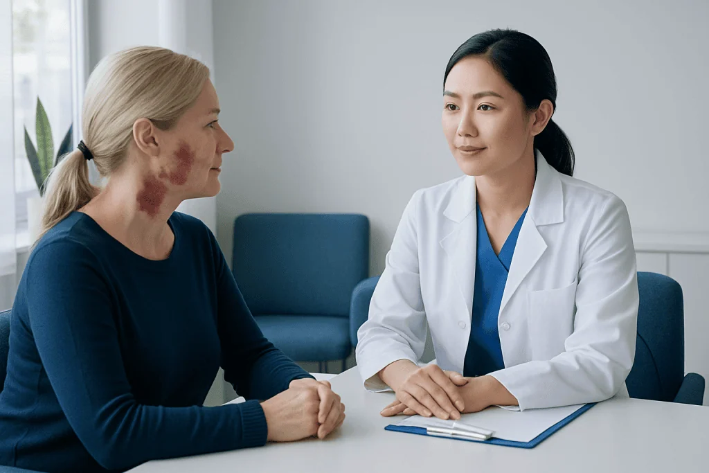
A careful assessment helps distinguish a hemangioma from other vascular birthmarks and skin lesions. The clinician will ask when the lesion appeared, how fast it has changed, whether it has bled or broken down, and whether it causes pain or affects daily activities. For infants, feeding, breathing, vision and hearing are particularly important considerations.
Most skin hemangiomas can be diagnosed from their appearance and clinical history. Ultrasound may be used when the lesion is deep, when its extent is unclear, or when blood flow needs to be assessed. Magnetic resonance imaging may be recommended for some large, deep or anatomically complex hemangiomas. Blood tests and other evaluations are only used when there is a specific clinical reason.
Before medication or a procedure, clinicians review medical history, current medicines, allergies and relevant heart, breathing or blood-sugar concerns. For surgery, the team also discusses anesthesia, expected scar placement, wound care and the possibility that more than one treatment approach may be needed. A specialist can also assess whether a lesion may represent a different vascular condition, such as vascular malformations, which are managed differently from infantile hemangiomas.
What happens during hemangioma surgery or laser treatment
The steps depend on the treatment selected. For medication-based care, the first visit generally includes an examination, discussion of expected benefits and side effects, and a plan for follow-up. Families should follow the prescribing clinician’s instructions carefully and should not stop or adjust medication without medical advice.
Laser treatment is often performed in an outpatient setting. Protective eye shields are used when appropriate, and the skin may be cooled or numbed. The clinician applies brief pulses of laser energy to the target area. Some people require more than one session, usually spaced apart to allow the skin to recover and the response to be evaluated.
Hemangioma surgery is usually planned after the surgeon has assessed the lesion’s boundaries and nearby structures. Depending on its location and the patient’s age, it may be performed with local anesthesia, sedation or general anesthesia. The surgeon makes an incision designed, where possible, to follow natural skin lines, removes or reshapes the targeted tissue, controls bleeding and closes the wound with stitches, skin adhesive or dressings.
After either procedure, the care team provides individualized instructions on pain control, cleansing, dressing changes, activity limits and follow-up. In selected cases, plastic surgery techniques may help address residual skin, contour differences or scarring after the vascular component has improved.
Benefits, risks and expected results
The potential benefits of hemangioma treatment include slowing growth, lightening color, reducing bulk, supporting normal vision or feeding, relieving ulcer-related discomfort and limiting permanent skin changes. With successful treatment, many lesions become flatter, softer and less noticeable. However, results are gradual for many people, and the final appearance can continue to evolve as a child grows or the skin heals.
All treatments have possible risks. Beta-blocker medicines can cause side effects in some patients and require clinician supervision. Laser treatment may temporarily cause redness, swelling, bruising, blistering or pigment changes; scarring is uncommon but possible. Surgery can result in bleeding, infection, wound separation, changes in sensation, an unfavorable scar, recurrence of visible vascular tissue, or a need for further treatment.
Not every hemangioma can be removed completely without trade-offs, especially if it is large, deep or close to important structures. A balanced discussion should compare the likely course without treatment against the potential benefits and limitations of intervention. The treating team can explain what outcome is realistic for the individual lesion.
For international patients needing coordinated assessment, Acibadem International’s multidisciplinary specialists and JCI-accredited hospitals diagnose and treat hemangiomas using individualized medical, laser and surgical approaches.
How long does it take to recover from hemangioma surgery?
Initial recovery from hemangioma surgery commonly involves mild swelling, bruising, tenderness and temporary redness around the incision. The wound surface often heals in approximately one to two weeks, although the exact timing depends on the size and location of the surgery, the method of closure, and the individual’s health. Young children may need help avoiding rubbing, scratching or pulling at dressings.
Most people can gradually return to routine activities according to the surgeon’s advice. Activities that could stretch the incision, cause impact or increase bleeding risk may need to be postponed. If stitches require removal, the timing is decided by the surgeon based on the body area and wound healing.
Scar maturation takes much longer than surface healing. A scar may remain pink, firm or slightly raised for several months before softening and fading. Protecting healing skin from sun exposure and attending follow-up appointments can support the best possible outcome. New or worsening pain, spreading redness, fever, pus-like drainage or wound opening should be reported promptly.
Is hemangioma a serious problem?
Most hemangiomas are not serious and do not require treatment. Infantile hemangiomas are benign, meaning they are not cancer, and many become smaller and less visible over time. Nevertheless, the location and behavior of a hemangioma matter more than its appearance alone.
A hemangioma needs urgent specialist attention when it interferes with vision, breathing, feeding, hearing or movement; breaks down into an ulcer; bleeds persistently; appears infected; or grows rapidly in a sensitive area. Large facial hemangiomas or multiple skin hemangiomas may sometimes require additional assessment because they can be associated with other health concerns in a small number of cases.
Prompt review does not mean a serious outcome is expected. It allows clinicians to identify lesions that may benefit from early treatment, when preventing complications is often easier. Parents and adults should seek personalized advice rather than relying only on photos or online descriptions.
What foods should you avoid with a hemangioma?
There are no specific foods proven to shrink a hemangioma or prevent it from growing. For most people, a balanced diet with adequate fluids, protein, fruits, vegetables and whole grains supports general health and normal skin healing. Food restrictions are not routinely needed solely because someone has a hemangioma.
If a person is taking medication for a hemangioma, the prescriber may give specific instructions about taking it with food or what to do during illness or poor intake. These instructions are important, particularly for infants and young children, because some medicines require careful timing around feeds. Families should ask the prescribing team before introducing supplements, herbal remedies or restrictive diets.
After surgery, nutrition that supports recovery is generally more useful than avoidance diets. A clinician may suggest individualized advice if there are allergies, swallowing concerns, diabetes, poor appetite, or another medical condition affecting nutrition.
Can hemangiomas turn into melanoma? When to seek medical care
Hemangiomas do not turn into melanoma. A hemangioma is a benign blood-vessel growth, while melanoma is a cancer that develops from pigment-producing skin cells. They are different conditions with different causes and patterns of change.
Any new or changing skin lesion still deserves medical assessment if it becomes irregular in shape, develops varied or very dark color, grows unexpectedly, crusts repeatedly, bleeds without an obvious reason, or does not heal. These changes do not automatically indicate melanoma, but an in-person examination is the safest way to clarify the cause. For information about suspicious pigmented lesions, see melanoma.
Medical care should also be sought promptly for a known hemangioma that ulcerates, becomes increasingly painful, shows signs of infection, bleeds and does not stop with gentle direct pressure, or affects eyesight, breathing, feeding or other normal function. Early review can provide reassurance and help determine whether observation, medication, laser treatment or surgery is appropriate.
Frequently asked questions
What is the best treatment for a hemangioma?
The best hemangioma treatment depends on the lesion’s type, size, location, growth pattern and symptoms. Observation is appropriate for many uncomplicated hemangiomas, while medication, laser therapy or surgery may be recommended for higher-risk or troublesome lesions. A qualified clinician should assess the individual case.
At what age should a hemangioma be treated?
Treatment may be started during infancy when a hemangioma is growing quickly or threatens function, skin health or long-term appearance. Early assessment is especially important for lesions near the eye, nose, lips, airway or diaper area. Some stable lesions can be safely monitored instead.
How long does it take to recover from hemangioma surgery?
Surface wound healing often takes around one to two weeks, but recovery varies according to the surgical site, depth of the lesion and the type of closure used. Bruising and swelling usually improve gradually, while scar maturation may take several months. The surgeon’s aftercare plan should guide return to activities.
Is hemangioma a serious problem?
Most hemangiomas are benign and are not serious. However, lesions that affect vision, breathing, feeding, hearing, skin integrity or normal movement need timely medical evaluation. A doctor can determine whether treatment is needed.
What foods should you avoid with a hemangioma?
No foods are known to worsen or improve hemangiomas in most people, so a special diet is usually unnecessary. If medication is being used, the clinician may give instructions about doses and meals or feeds. Supplements and restrictive diets should be discussed with a healthcare professional first.
Can hemangiomas turn into melanoma?
No. Hemangiomas are benign blood-vessel growths and do not transform into melanoma, which is a cancer of pigment-producing skin cells. A new or changing dark skin lesion should still be examined by a clinician to establish the correct diagnosis.
References
- American Academy of Pediatrics
- American Academy of Dermatology
- National Institute of Arthritis and Musculoskeletal and Skin Diseases
- Mayo Clinic
- International Society for the Study of Vascular Anomalies
This article is for general information only and is not a substitute for professional medical advice. Please consult a qualified doctor about your individual situation.
Persistent digestive symptoms? Get evaluated in Turkey
JCI-accredited · board-certified surgeons · reply within 24h
Add us as a Preferred Source to see more of our trusted health content across Google Search, AI Overviews and Discover.

More from the Health Library


Related Specialists
Prof. Dr. Celaletdin Camcı
Medical Oncology
Embriyolog Seda Özkan
Vitro Fertilization and Reproductive Medicine Center
Dr. Ayşe Dişli Gürler
Gynecology & Obstetrics
Dr. Sema Tekin Koşargelir
Cosmetic Dermatology