Hemispherectomy: Candidacy, Procedure Steps, and Recovery Timeline

Hemispherectomy is considered for carefully selected people with severe, drug-resistant seizures arising predominantly from one hemisphere of the brain. Modern procedures usually disconnect the affected hemisphere rather than completely removing it; this is often called hemispherotomy or functional hemispherectomy.
Key Takeaways
- Hemispherectomy is considered for carefully selected people with severe, drug-resistant seizures arising predominantly from one hemisphere of the brain.
- Modern procedures usually disconnect the affected hemisphere rather than completely removing it; this is often called hemispherotomy or functional hemispherectomy.
- Detailed presurgical testing is essential to confirm where seizures start and to assess expected effects on movement, vision, language, and thinking.
- Recovery involves intensive monitoring, rehabilitation, seizure follow-up, and ongoing support for the patient and family.
- The procedure has significant risks, but it may substantially reduce seizures and improve quality of life for appropriate candidates.
Hemispherectomy is a highly specialized epilepsy surgery used when severe seizures arise mainly from one damaged side of the brain and do not respond adequately to medication. It aims to stop abnormal electrical activity from spreading, while rehabilitation helps the person adapt after surgery.
Overview: what is hemispherectomy?
Hemispherectomy is a major form of epilepsy surgery that treats seizures arising primarily from one side, or hemisphere, of the brain. It is reserved for people whose seizures remain disabling despite appropriate trials of antiseizure medicines and whose evaluation shows that one hemisphere is the main source of seizure activity. The central goal is to prevent abnormal electrical signals from the affected hemisphere from spreading to the healthier side of the brain.
Despite its name, modern hemispherectomy usually does not mean removing an entire half of the brain. In many cases, surgeons perform a functional hemispherectomy or hemispherotomy, creating surgical disconnections within and around the affected hemisphere while removing only limited tissue when needed. This approach is designed to achieve seizure control while reducing some complications associated with older anatomical techniques.
The operation is performed most often in children, because certain severe epilepsy syndromes begin early in life and the developing brain has considerable capacity to adapt. However, selected adolescents and adults may also be assessed. Hemispherectomy is not appropriate for every person with epilepsy; it requires evaluation in a comprehensive epilepsy surgery program with expertise in neurology, neurosurgery, neuropsychology, imaging, and rehabilitation.
Who may be a candidate?
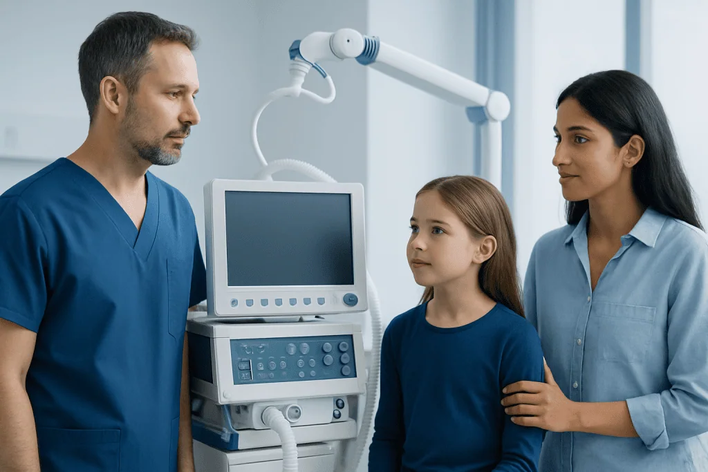
Potential candidates generally have drug-resistant epilepsy, meaning seizures continue despite appropriate treatment with at least two suitable antiseizure medications. The seizures are typically frequent, severe, and harmful to daily life, learning, development, safety, or independence. Examples include repeated focal seizures, seizure clusters, or drop attacks that lead to falls and injuries.
A key requirement is evidence that seizures begin mainly in one hemisphere and that this side is substantially diseased or not functioning normally. Conditions that may lead to consideration of hemispherectomy include extensive injury from a perinatal stroke, hemimegalencephaly (abnormal enlargement and development of one hemisphere), Sturge-Weber syndrome, Rasmussen encephalitis, severe cortical malformations, and selected cases of damage after infection or trauma.
Many candidates already have weakness, reduced hand function, visual-field loss, or developmental effects related to the underlying brain condition. The team carefully considers whether surgery may worsen these difficulties and whether the likely benefit in seizure control outweighs that risk. Surgery is generally less suitable when seizures clearly arise from both hemispheres, when the healthier hemisphere cannot support vital functions, or when testing does not identify a sufficiently localized seizure source.
Families and adult patients should understand that candidacy is not decided from one test alone. It is a structured, individualized decision based on the person’s seizure burden, brain development, abilities, goals, support needs, and the findings of a multidisciplinary epilepsy team.
How the presurgical evaluation works

Before recommending hemispherectomy, clinicians complete a detailed epilepsy assessment. This commonly includes a full seizure history, neurological examination, review of medication response, and prolonged video-electroencephalography (video-EEG). Video-EEG records brain-wave activity alongside video of seizures, helping the team determine where seizures begin and how they spread.
Brain imaging is central to planning. High-resolution MRI can show structural abnormalities, old injuries, or developmental differences. Depending on the situation, the team may also use PET, SPECT, magnetoencephalography, or advanced MRI techniques to clarify brain function and seizure networks. In some complex cases, invasive EEG monitoring with electrodes placed on or in the brain may be discussed, although it is not necessary for everyone.
Neuropsychological testing assesses memory, language, attention, learning, behavior, and daily functioning at a level appropriate to the patient’s age and abilities. Physical, occupational, speech, and visual assessments may also be completed. These evaluations establish a baseline and help anticipate rehabilitation needs after surgery.
The team then reviews the findings together with the patient and family. Discussions cover expected seizure outcomes, existing neurological differences, possible new or worsened deficits, alternatives to surgery, and the practical recovery plan. Seeking a second opinion from an experienced epilepsy surgery center can be reasonable when such a life-changing procedure is being considered.
Hemispherectomy procedure: step by step
Hemispherectomy is performed under general anesthesia by a specialist neurosurgical team. The exact method depends on the individual’s anatomy, underlying condition, prior surgery, and surgical plan. The procedure may be described as anatomical hemispherectomy, functional hemispherectomy, or hemispherotomy; functional disconnection approaches are now commonly used.
First, the surgeon makes an incision in the scalp and temporarily removes a section of skull, called a craniotomy, to reach the brain. Using operating microscopes, image guidance, and careful neuroanatomical landmarks, the surgeon disconnects pathways that allow seizure activity to travel from the affected hemisphere to the rest of the brain. These pathways may include connections through the corpus callosum and deeper white-matter structures.
Some abnormal brain tissue may be removed, especially if it is contributing to seizures or is necessary to safely complete the disconnection. In a functional procedure, much of the affected hemisphere may remain physically in place but no longer communicates effectively with the other side. The surgeon controls bleeding, closes the protective tissue layers, replaces or secures the bone, and closes the scalp.
After surgery, the patient is transferred to intensive care or a high-dependency unit for close observation. The operation is complex and can last many hours, but timing varies considerably. Families should ask the surgical team how the planned approach applies to their specific situation rather than relying on a standard timeline.
Benefits, limitations, and possible risks
The principal potential benefit of hemispherectomy is a major reduction in seizures, and some appropriately selected patients become seizure-free. Better seizure control may reduce emergency visits, injuries, medication burden, and the disruption seizures cause to sleep, education, behavior, and family life. In young children, controlling severe ongoing seizures may also support developmental progress by reducing the impact of persistent epileptic activity on the brain.
Results vary, and surgery cannot guarantee seizure freedom. Some people continue to have seizures after the operation and may still need antiseizure medication. Medication changes should only be made with guidance from the epilepsy specialist, often after a period of postoperative monitoring. Ongoing neurological, developmental, and psychological support remains important regardless of seizure outcome.
All brain surgery carries risks. These include bleeding, infection, stroke, swelling, fluid buildup around the brain (hydrocephalus), blood clots, anesthesia complications, and, rarely, life-threatening complications. Hydrocephalus can occur months or even years after surgery and may require treatment such as a shunt to drain excess fluid.
Because one hemisphere is disconnected, weakness on the opposite side of the body, loss of fine hand function, and loss of part of the visual field may occur or worsen. Effects on language, thinking, behavior, swallowing, or movement depend on which hemisphere is affected, existing abilities, age, and how the brain has reorganized. The surgical and rehabilitation teams explain these risks in personal terms before any decision is made.
Recovery timeline and rehabilitation
The first days after hemispherectomy are spent in hospital with frequent neurological checks, pain management, fluid monitoring, and observation for complications. The patient may initially be sleepy, uncomfortable, or temporarily less able to communicate or move. Seizures may still occur early after surgery, and the care team monitors them closely rather than judging the final outcome from the first few days.
Hospital stay length varies based on age, recovery speed, complications, and rehabilitation needs. Physical therapy often begins in hospital to support safe movement, positioning, balance, and strength. Occupational therapy can help with dressing, feeding, hand use, and other daily activities, while speech and language therapy may be recommended for communication, swallowing, or cognitive needs.
During the following weeks and months, rehabilitation is usually the most active part of recovery. Children may need support returning to school, learning new skills, or accessing educational accommodations. Adults may need assistance with mobility, self-care, work planning, and emotional adjustment. Improvements can continue over a long period as the brain and body adapt, particularly with consistent therapy.
Follow-up appointments review wound healing, seizure control, medication plans, imaging when needed, and rehabilitation progress. Families should keep a seizure record and report new symptoms promptly. Acibadem International’s multidisciplinary specialists and JCI-accredited hospitals can assess and treat complex epilepsy surgery cases for international patients, with coordinated postoperative rehabilitation and follow-up planning.
When to seek medical care
People with ongoing seizures should seek assessment by a neurologist, particularly if seizures continue after trials of appropriate medication, cause injuries, interfere with learning or daily activities, or are associated with developmental regression. A referral to a comprehensive epilepsy center can help determine whether advanced treatments, including surgery, may be appropriate.
Urgent medical care is needed if a seizure lasts longer than five minutes, repeated seizures occur without the person returning to their usual level of awareness, breathing is difficult, a significant injury occurs, or this is a first seizure. Emergency services should also be contacted when a person is pregnant, has diabetes, is in water, or has other circumstances that increase immediate risk.
After brain surgery, patients and caregivers should contact the surgical team urgently for fever, increasing redness or drainage from the incision, severe or worsening headache, repeated vomiting, unusual drowsiness, new weakness, new speech difficulties, confusion, or a major change in seizures. These symptoms do not always indicate a serious problem, but prompt review is important.
Frequently asked questions
What is the difference between hemispherectomy and hemispherotomy?
Hemispherectomy is a broad term for surgery that prevents one brain hemisphere from driving seizures. In an anatomical hemispherectomy, most of the affected hemisphere is removed. A functional hemispherectomy or hemispherotomy primarily disconnects seizure pathways and usually removes less tissue, which is why these approaches are commonly used today.
Can an adult have a hemispherectomy?
Yes, although hemispherectomy is performed more commonly in children. Adults may be considered when they have severe drug-resistant seizures arising from one extensively damaged hemisphere and detailed testing supports surgery. The expected benefits and functional risks need individualized assessment.
Will a person be seizure-free after hemispherectomy?
Many carefully selected patients have a substantial reduction in seizures, and some become seizure-free. However, no operation can promise this outcome, and some people continue to have seizures or need antiseizure medication afterward. Follow-up with an epilepsy specialist remains necessary.
How long does recovery from hemispherectomy take?
Initial hospital recovery commonly takes days to weeks, depending on the person’s condition and rehabilitation needs. Functional recovery continues over months and may extend longer, especially when physical, occupational, speech, or educational therapies are required. Progress is individual and often occurs gradually.
Does hemispherectomy affect intelligence or personality?
The effects depend on the underlying brain condition, the hemisphere involved, the person’s age, pre-existing abilities, and seizure control after surgery. Some people gain developmental or cognitive benefits when severe seizures are controlled, while others may have challenges involving language, attention, mood, or behavior. Neuropsychological evaluation and follow-up help identify and support these needs.
Why is rehabilitation important after hemispherectomy?
Rehabilitation helps patients adapt to movement, balance, visual, communication, and daily-living changes after surgery. Therapy can also support school participation, independence, and family routines. Starting rehabilitation early and continuing it according to individual needs can make recovery more manageable.
References
- International League Against Epilepsy
- National Institute of Neurological Disorders and Stroke
- Epilepsy Foundation
- Johns Hopkins Medicine
- Cleveland Clinic
This article is for general information only and is not a substitute for professional medical advice. Please consult a qualified doctor about your individual situation.
Explore treatments in Turkey — costs, top hospitals & a free quote
JCI-accredited · board-certified surgeons · reply within 24h
Add us as a Preferred Source to see more of our trusted health content across Google Search, AI Overviews and Discover.

More from the Health Library



Related Specialists

Dr. Gülhan İpek Deniz
Medical Oncology
Prof. Dr. Recep Kurnaz
Orthopedic Surgery & Traumatology
Assoc. Prof. Dr. Tuğçe Apaydın
Endocrinology
Dr. Sevim Buzkan Ertuğrul
Acibadem Life Clinical Service