Histology: An Evidence-Based Guide for Patients

Histology examines tissue structure under a microscope to help diagnose disease. It is commonly performed on biopsy or surgical samples by a pathologist.
Key Takeaways
- Histology examines tissue structure under a microscope to help diagnose disease.
- It is commonly performed on biopsy or surgical samples by a pathologist.
- Histology can identify inflammation, infection, benign changes, and cancer.
- Results are usually reported in a pathology report that must be interpreted in clinical context.
- Not every abnormal histology result means cancer, and many findings are treatable.
- Patients should discuss histology results with their doctor to understand what they mean for care.
Histology is the study of body tissues under a microscope, usually after a biopsy or surgery. For patients, it matters because histology helps confirm a diagnosis, explain symptoms, and guide the most appropriate treatment plan.
Overview: what histology means for patients
Histology is the microscopic study of tissues. In everyday medical care, the term usually refers to the laboratory examination of a tissue sample taken during a biopsy, endoscopy, or surgery. This process helps doctors see how cells are arranged, whether tissue looks healthy or abnormal, and what kind of disease may be present.
For patients, histology is important because it often provides the clearest answer when scans, blood tests, or symptoms alone are not enough. A histology result can help confirm inflammation, infection, autoimmune disease, benign growths, precancerous changes, or cancer. It is one of the key tools used to turn a suspected diagnosis into a more definite one.
Histology is closely related to pathology, but the terms are not identical. Pathology is the broader medical specialty that studies disease, while histology is one part of that work focused on tissue structure under the microscope. A pathologist reviews the histology findings and combines them with clinical details to create a pathology report.
How a histology test is done
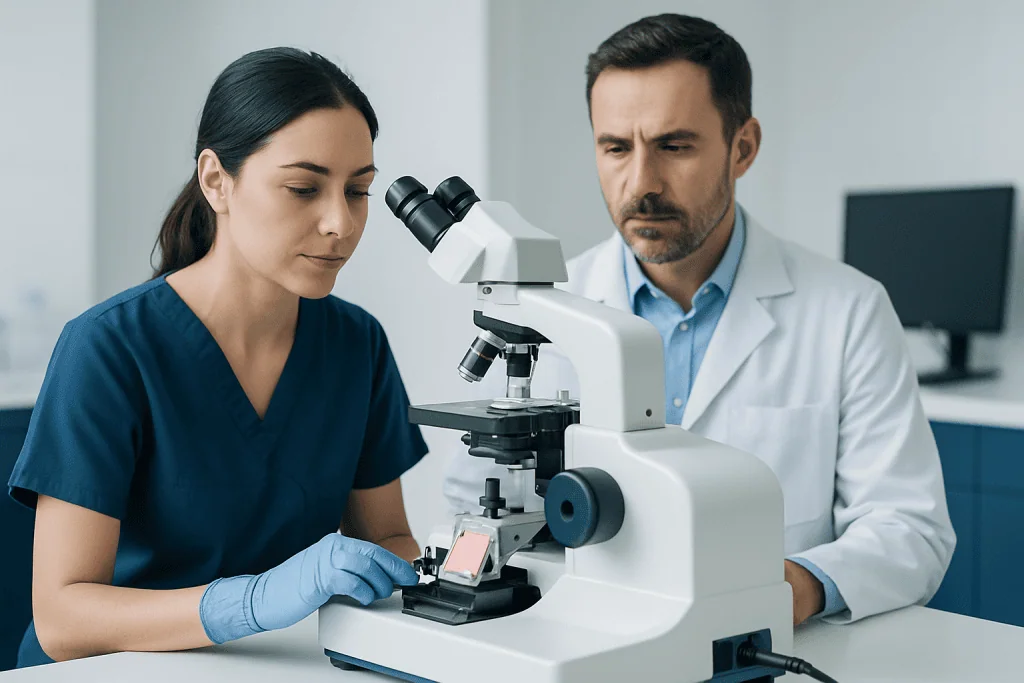
A histology test begins with collecting a tissue sample. This may happen during a needle biopsy, skin biopsy, endoscopy, colonoscopy, bronchoscopy, or surgery. The sample is placed in a preservative solution so that the tissue keeps its structure while it is sent to the laboratory.
In the lab, the tissue goes through several careful steps. It is processed, embedded in paraffin wax, sliced into extremely thin sections, and placed on glass slides. These slides are then stained with dyes, most commonly hematoxylin and eosin, so that different parts of the cells and tissue can be seen more clearly under the microscope.
A pathologist examines the slides to assess the size, shape, and arrangement of cells and the overall tissue pattern. If needed, additional techniques such as special stains, immunohistochemistry, or molecular testing may be used to clarify the diagnosis. This can be especially useful when doctors are evaluating suspected cancer or trying to distinguish between similar-looking conditions.
In some operations, doctors may request a rapid preliminary review called a frozen section. This gives the surgical team quick information while the procedure is still underway, although the final histology report is usually more complete and more accurate because it is based on fully processed tissue.
What histology can show
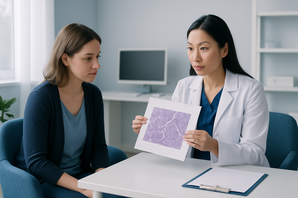
Histology can reveal many different types of tissue changes. It may show normal tissue, irritation, inflammation, scarring, infection, or cell injury. It can also identify whether a growth is benign, precancerous, or malignant. Because tissues from different organs have distinct microscopic patterns, histology helps pinpoint not only that something is abnormal, but often what kind of abnormality it is.
For example, histology may help explain digestive symptoms by identifying inflammation in the stomach or intestines after endoscopy. It can help clarify skin rashes or moles after a skin biopsy, and it can evaluate breast, thyroid, liver, lung, or prostate tissue when imaging has found a suspicious area. In some cases, histology also helps show how active or advanced a disease process is.
Common questions that histology may help answer include:
- Is the tissue normal or abnormal?
- Is there inflammation, infection, or immune-related damage?
- Are the changes benign or cancerous?
- If cancer is present, what type does it appear to be?
- Do the findings match the patient’s symptoms and imaging results?
Histology is usually interpreted together with other information. A tissue pattern alone may not tell the full story, so doctors compare the microscopic findings with symptoms, imaging, endoscopic appearance, blood tests, and medical history before making decisions about treatment.
When doctors order histology
Doctors may order histology whenever they need a closer look at tissue to understand the cause of symptoms or an abnormal test result. This is common when imaging detects a lump, thickening, ulcer, polyp, or enlarged lymph node, or when symptoms have continued despite initial treatment.
Histology is especially useful when a diagnosis could change treatment. For example, it may distinguish an inflammatory condition from infection, or a harmless growth from a tumor that needs further care. It is also used after surgery to examine removed tissue and confirm what was found.
Situations where histology may be considered include:
- A breast, thyroid, skin, or soft tissue lump
- Persistent stomach pain, bleeding, or altered bowel habits requiring colonoscopy or upper endoscopy
- Abnormal cells found on screening tests
- Suspicious findings on ultrasound, CT, MRI, or mammography
- Follow-up of known inflammatory or autoimmune diseases
- Checking whether a tumor has been fully removed during surgery
Not every symptom requires a biopsy, and not every biopsy leads to major treatment. In many cases, histology helps rule out serious disease or confirms a condition that can be managed with medication, monitoring, or minor procedures.
Understanding the pathology report
After histology is reviewed, the findings are summarized in a pathology report. This document may include the type of specimen, where it came from, what the tissue looked like under the microscope, and a final diagnosis or interpretation. Some reports are short and straightforward, while others are more detailed, especially when cancer or complex inflammatory disease is being assessed.
Medical terms in a pathology report can sound technical. Words such as benign, atypia, dysplasia, chronic inflammation, necrosis, margins, grade, or malignant each have specific meanings. Some terms describe change without confirming cancer, while others help estimate how aggressive a known cancer may be. This is one reason patients should review the report with the doctor who ordered the test.
If the report concerns cancer, histology may help identify the tumor type and may guide next steps such as biopsy review, surgery, additional imaging, medication, or oncology care. If the report shows inflammation or infection, treatment may instead focus on medicines, lifestyle changes, or monitoring. Sometimes the result is inconclusive, and another sample or a second opinion may be recommended.
Patients often find it helpful to ask simple questions: What was found? Is it serious? Does it explain the symptoms? Are more tests needed? What treatment, if any, is recommended now? A clear conversation can make the report easier to understand and reduce unnecessary worry.
Limits of histology and why results can take time
Histology is a powerful diagnostic tool, but it has limits. The result depends on the quality and size of the tissue sample, the exact area sampled, and whether the abnormality was captured. A very small biopsy may not fully represent a larger lesion, and some diseases have patchy changes that can be missed in one area.
Results can also take time because tissue processing is detailed and precise. If special stains, immunohistochemistry, or molecular tests are needed, this may add several more days. While waiting can be stressful, these extra steps are often important for making the diagnosis more accurate.
In some cases, histology does not give a complete answer on its own. The report may say that findings are nonspecific, indeterminate, or suggestive rather than definitive. This does not necessarily mean something is being missed; it may simply reflect the real complexity of the tissue changes. Doctors may then combine the histology with repeat imaging, clinical follow-up, or another procedure.
Patients who have questions about uncertainty can ask whether the sample was adequate, whether specialist review is needed, and whether the findings fit with the rest of the medical picture. These are reasonable questions and often helpful in planning the next step.
What patients can do before and after tissue sampling
There is no special preparation for histology itself, but preparation may be needed for the procedure used to collect the tissue. For example, fasting may be required before endoscopy, or blood-thinning medicines may need review before some biopsies. The healthcare team will explain what is needed based on the organ involved and the type of procedure planned.
After a biopsy or surgery, patients should follow aftercare instructions carefully. This may include wound care, temporary activity restrictions, and watching for signs of bleeding or infection. Most tissue sampling procedures are routine, but even minor procedures deserve proper follow-up.
Helpful self-care steps include:
- Keeping follow-up appointments to review results
- Asking for a copy of the pathology report if desired
- Bringing a list of questions to the visit
- Telling the doctor about new symptoms after the procedure
- Avoiding online self-diagnosis based on isolated pathology terms
If further evaluation is needed, care may involve several specialists. Multidisciplinary review is particularly helpful for complex digestive, breast, skin, or oncologic findings. Near the end of the care pathway, patients may also seek coordinated evaluation at centers such as Acibadem International, where multidisciplinary specialists in JCI-accredited hospitals diagnose and treat conditions that require biopsy, pathology review, and advanced planning for international patients.
When to seek medical care
Medical care should be sought if a doctor has recommended a biopsy or tissue examination and symptoms are ongoing, worsening, or unexplained. Histology is often the next appropriate step when there is a persistent lump, unexplained bleeding, a nonhealing sore, abnormal imaging, or long-lasting digestive or respiratory symptoms.
Urgent medical attention is important after a biopsy or procedure if there is heavy bleeding, fever, worsening redness, severe pain, shortness of breath, chest pain, or signs of infection. Patients should also contact their doctor if they have not received results within the expected timeframe, especially when a treatment decision depends on them.
It is also reasonable to ask for further review if a pathology result is unclear or if the diagnosis does not seem to match symptoms. In selected cases, a second opinion from an experienced pathologist or specialist can be helpful, especially for rare conditions or complex tumor findings.
Frequently asked questions
What is histology in simple terms?
Histology is the study of body tissues under a microscope. In patient care, it usually means examining a biopsy or surgical sample to look for disease and help confirm a diagnosis.
Is histology the same as a biopsy?
Not exactly. A biopsy is the procedure used to remove a small piece of tissue, while histology is the laboratory examination of that tissue after it has been collected. The two are closely connected, but they are different parts of the diagnostic process.
Does an abnormal histology result always mean cancer?
No. Histology can show many noncancerous changes, including inflammation, infection, scarring, and benign growths. A doctor must interpret the result in context before deciding what it means.
How long do histology results usually take?
Timing varies by hospital, the type of tissue, and whether extra testing is needed. Some routine results are available within a few days, while more complex cases can take longer if special stains or additional review are required.
Why would a doctor order histology?
Doctors order histology when they need more definite information about abnormal tissue. It can help explain symptoms, clarify imaging findings, confirm a diagnosis, and guide treatment decisions.
Can histology be wrong?
Histology is highly valuable, but no test is perfect. Results can be limited by sample size, sampling location, or tissue quality, and some cases remain uncertain. If needed, doctors may recommend repeat sampling, additional tests, or a second opinion.
References
- National Cancer Institute
- MedlinePlus
- College of American Pathologists
- Mayo Clinic
- World Health Organization
This article is for general information only and is not a substitute for professional medical advice. Please consult a qualified doctor about your individual situation.
Oncology care in Turkey — second opinion and treatment plan
JCI-accredited · board-certified surgeons · reply within 24h
Add us as a Preferred Source to see more of our trusted health content across Google Search, AI Overviews and Discover.

More from the Health Library

Eyebrow Twitching — Explained by Medical Evidence, Not Myths

Watermelon Juice Benefits — Explained by Medical Evidence, Not Myths

Bruises on Black Skin: A Complete Medical Overview

Charcot — Explained by Medical Evidence, Not Myths
Scalene Muscles — Explained by Medical Evidence, Not Myths