How Neuro-Ophthalmologists Diagnose Vision Problems Related to the Brain

Neuro-ophthalmologists specialize in visual symptoms caused by problems affecting the optic nerve, eye muscles, nerves, or brain. Diagnosis often starts with a careful review of symptoms, medical history, and a focused eye and neurologic exam.
Key Takeaways
- Neuro-ophthalmologists specialize in visual symptoms caused by problems affecting the optic nerve, eye muscles, nerves, or brain.
- Diagnosis often starts with a careful review of symptoms, medical history, and a focused eye and neurologic exam.
- Tests may include visual field testing, imaging such as MRI or CT, blood tests, and other targeted studies.
- Common symptoms include double vision, vision loss, eye movement problems, eyelid drooping, and unexplained headaches with visual changes.
- Treatment depends on the underlying cause and may involve medication, rehabilitation, surgery, or urgent neurologic care.
Neuro-ophthalmologists evaluate visual problems that may be linked to the optic nerve, eye movement pathways, or the brain. Their assessment combines detailed history-taking, eye and neurologic examination, and selected tests to find the cause and guide treatment.
Overview: What Neuro-Ophthalmologists Do
Neuro-ophthalmologists are doctors who diagnose and manage vision problems related to the optic nerve, the nerves that move the eyes, and the parts of the brain that process vision. They work at the point where ophthalmology and neurology meet, helping patients whose symptoms cannot be explained by a routine eye problem alone.
Many people are referred to a neuro-ophthalmologist after noticing sudden or gradual vision loss, double vision, abnormal eye movements, visual field defects, drooping eyelids, or episodes of temporary blindness. Some patients also have headaches, dizziness, facial symptoms, weakness, or numbness, which can point to a neurologic cause.
The goal of the visit is not only to check how well the eyes see, but also to understand how the visual system is functioning from the eye to the brain. This is why the evaluation often includes both a detailed eye examination and a focused neurologic assessment.
Symptoms That May Suggest a Brain-Related Vision Problem
Brain-related vision problems can appear in different ways. Some symptoms are obvious, such as sudden loss of vision in one or both eyes. Others are more subtle, including difficulty reading, missing part of the visual field, problems judging depth, or feeling that the eyes are not working together properly.
Double vision is a common reason for referral. It may happen when the muscles that move the eyes are not coordinated because of a problem in the nerves, brainstem, or pathways controlling eye movement. Patients may also describe eye strain, head tilting, or needing to close one eye to see clearly.
Other symptoms that may raise concern include:
- Blurred or dim vision not explained by glasses alone
- Loss of side vision
- Pain with eye movement
- Color vision changes
- Episodes of temporary visual blackout
- Drooping eyelid or unequal pupils
- Visual disturbances with headache
These symptoms do not always mean there is a serious neurologic disease. However, they do deserve proper assessment, especially if they begin suddenly, worsen, or occur together with other neurologic symptoms.
How the Diagnostic Process Begins

The first and often most important step is taking a careful history. The neuro-ophthalmologist usually asks when the symptoms started, whether they came on suddenly or gradually, whether one or both eyes are affected, and whether the changes are constant or intermittent. The pattern of symptoms can provide important clues about the location and cause of the problem.
Questions also cover general health, past eye conditions, migraine history, autoimmune disease, diabetes, infections, recent trauma, medications, and family history. For example, visual symptoms linked to multiple sclerosis, stroke, thyroid disease, myasthenia gravis, or raised pressure around the brain can present in very different ways.
Because some visual complaints can reflect broader neurologic conditions, the doctor may also ask about weakness, imbalance, numbness, speech changes, swallowing difficulty, memory concerns, or headaches. This helps identify whether the visual problem is isolated or part of a larger condition.
Eye and Neurologic Examinations
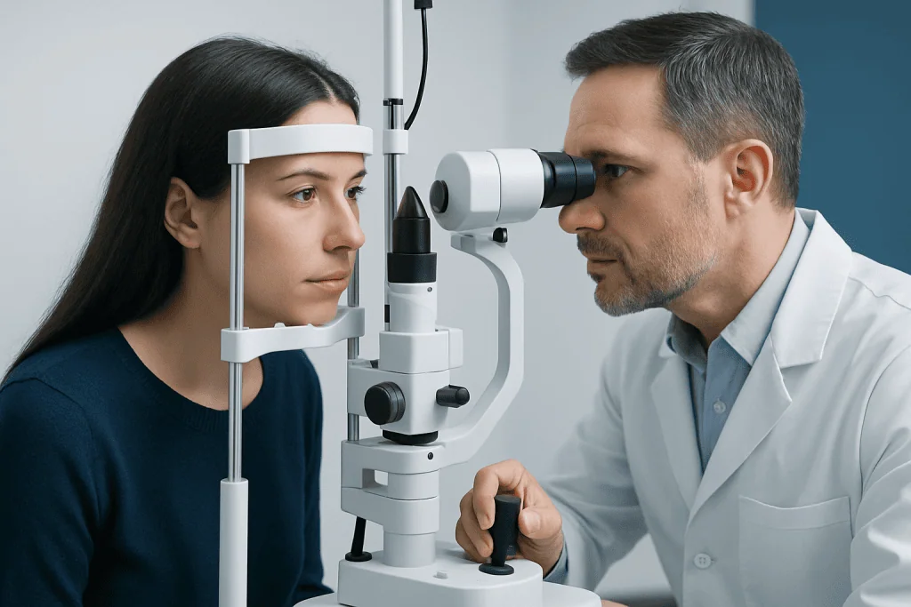
After the history, the doctor performs a detailed examination. This usually includes checking visual acuity, color vision, pupil reactions, eyelid position, eye alignment, and how the eyes move in different directions. The appearance of the optic nerve and retina is also examined by looking into the back of the eye.
Visual field testing is an important part of neuro-ophthalmic assessment. It helps show whether any areas of vision are missing and whether the pattern matches a problem in the optic nerve, optic chiasm, optic tract, or visual cortex. Certain field defects can strongly suggest a lesion in a specific part of the visual pathway.
The neurologic examination may assess facial movement, sensation, balance, coordination, reflexes, and strength. Although this may feel broader than a standard eye visit, it is helpful because visual symptoms may be the first sign of a condition affecting the nervous system more generally.
In some cases, the examination points to disorders such as optic neuritis or raised intracranial pressure with swelling of the optic nerves. When that happens, the next step is selecting the most appropriate tests to confirm the diagnosis and look for the cause.
Tests Used to Confirm the Cause
Neuro-ophthalmologists use tests selectively, based on what the history and examination suggest. Many patients have imaging of the brain or orbits, especially when symptoms indicate possible inflammation, stroke, tumor, nerve compression, or demyelinating disease. MRI scanning is often especially useful because it shows the optic nerves, brain, and surrounding structures in detail. In urgent settings, CT scan may also be used, particularly when bleeding, fracture, or acute stroke is being considered.
Visual field testing may be done with automated machines that map areas of reduced vision. Optical coherence tomography can measure the thickness of the retinal nerve fiber layer and optic nerve structures, which helps monitor optic nerve damage over time. Blood tests may be ordered to look for infection, inflammation, autoimmune disease, vitamin deficiency, thyroid problems, or other systemic causes.
Additional studies are sometimes needed. These may include lumbar puncture to measure cerebrospinal fluid pressure, vascular imaging if blood vessel problems are suspected, or electrophysiologic testing to assess how visual signals travel from the eyes to the brain. If double vision is the main concern, blood tests or bedside tests may help evaluate conditions such as myasthenia gravis.
Not every patient needs every test. A key part of neuro-ophthalmology is knowing which investigations are most likely to provide useful answers while avoiding unnecessary procedures.
Common Conditions Neuro-Ophthalmologists Diagnose
Neuro-ophthalmologists assess a wide range of disorders. Some involve the optic nerve, such as optic neuritis, ischemic optic neuropathy, compressive optic neuropathy, and papilledema. Others affect eye movements, including cranial nerve palsies, internuclear ophthalmoplegia, thyroid eye disease, and myasthenia gravis.
They also diagnose visual problems caused by brain conditions such as stroke, tumors, multiple sclerosis, migraines, traumatic brain injury, and increased intracranial pressure. For example, a person who loses one side of the visual field in both eyes may have a lesion in the visual pathway behind the optic chiasm, while someone with painful vision loss and reduced color vision may have optic nerve inflammation.
Some symptoms are linked to urgent neurologic problems and require rapid care. Sudden new visual field loss, painful pupil changes, double vision with severe headache, or vision changes with signs of stroke should be assessed promptly. When necessary, a neuro-ophthalmologist works with emergency physicians, neurologists, neuroradiologists, and neurosurgeons.
Treatment, Follow-Up, and Multidisciplinary Care
Treatment depends entirely on the cause of the visual problem. In some cases, treatment is directed at inflammation or autoimmune disease. In others, care may focus on controlling vascular risk factors, managing migraine, treating infection, reducing intracranial pressure, or addressing a structural problem such as a tumor or aneurysm.
Patients with eye movement disorders may benefit from temporary prism glasses, patching, rehabilitation exercises, or treatment of the underlying neurologic disorder. Some structural causes require coordinated care with surgery or other procedures. If a mass, nerve compression, or another anatomical problem is found, the team may consider brain tumor surgery or another targeted intervention when appropriate.
Follow-up is often important because some conditions change over time. Repeat visual fields, optic nerve imaging, or neurologic examination can show whether treatment is working or whether the diagnosis needs to be reconsidered. Even when symptoms are distressing, a step-by-step approach often leads to clarity and a practical treatment plan.
For international patients who need coordinated evaluation, Acibadem International’s multidisciplinary specialists and JCI-accredited hospitals diagnose and treat neuro-ophthalmic conditions with support from ophthalmology, neurology, radiology, and neurosurgery teams.
When to Seek Medical Attention
People should seek prompt medical attention for sudden vision loss, new double vision, severe headache with visual symptoms, or visual changes accompanied by weakness, speech difficulty, facial drooping, or confusion. These symptoms may signal a condition that needs urgent evaluation.
It is also wise to arrange medical review for gradual but unexplained changes in vision, drooping eyelids, persistent eye movement problems, episodes of temporary vision loss, or recurring visual field defects. Even when symptoms are not painful, they can still reflect an issue affecting the optic nerve or brain.
Keeping a brief record of symptoms can help. Patients may note when symptoms started, whether one or both eyes are affected, whether the problem comes and goes, and whether headaches or neurologic symptoms occur at the same time. This information can make the evaluation more precise and efficient.
Frequently asked questions
What is the difference between an ophthalmologist and a neuro-ophthalmologist?
An ophthalmologist diagnoses and treats eye diseases in general, including cataracts, glaucoma, and retinal problems. A neuro-ophthalmologist focuses on vision problems caused by the optic nerve, eye movement nerves, and the brain. Patients are often referred when symptoms seem to involve both the eyes and the nervous system.
Why would someone be referred to a neuro-ophthalmologist?
A referral may happen when vision loss, double vision, abnormal pupils, eyelid drooping, or visual field loss cannot be fully explained by a routine eye exam. It is also common when headaches, weakness, numbness, or other neurologic symptoms occur with visual changes. The specialist helps determine where in the visual pathway the problem is happening.
Do brain-related vision problems always mean a serious disease?
No. Some causes are temporary, treatable, or not dangerous, such as certain migraine-related visual symptoms. However, because some visual changes can be linked to stroke, inflammation, nerve disorders, or increased pressure around the brain, proper evaluation is important.
What tests are commonly used in neuro-ophthalmology?
Common tests include visual acuity testing, pupil assessment, eye movement examination, visual field testing, and imaging such as MRI or CT when needed. Some patients also need blood tests, optic nerve imaging, or lumbar puncture. The exact tests depend on the pattern of symptoms and exam findings.
Can double vision come from the brain and not the eye itself?
Yes. Double vision often happens when the eyes do not move together properly, which can be caused by problems in the nerves, brainstem, or muscles controlling eye movement. A neuro-ophthalmologist helps determine whether the source is neurologic, muscular, or related to the eye socket.
When should sudden vision changes be treated as an emergency?
Sudden vision loss, sudden double vision, or visual changes with severe headache, weakness, facial drooping, or speech difficulty should be treated urgently. These symptoms can be related to stroke, acute optic nerve problems, bleeding, or other conditions that need fast care. Seeking prompt medical attention is the safest approach.
References
- American Academy of Ophthalmology
- North American Neuro-Ophthalmology Society
- National Eye Institute
- National Institute of Neurological Disorders and Stroke
- Mayo Clinic
This article is for general information only and is not a substitute for professional medical advice. Please consult a qualified doctor about your individual situation.
Explore treatments in Turkey — costs, top hospitals & a free quote
JCI-accredited · board-certified surgeons · reply within 24h
Add us as a Preferred Source to see more of our trusted health content across Google Search, AI Overviews and Discover.