

How Stroke Is Diagnosed: Brain Imaging, Vessel Tests, and Urgent Evaluation
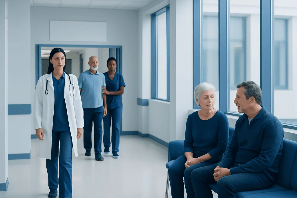
Stroke diagnosis is time-sensitive because early treatment can protect brain tissue. A brain CT scan is often the first imaging test to quickly check for bleeding.
Key Takeaways
- Stroke diagnosis is time-sensitive because early treatment can protect brain tissue.
- A brain CT scan is often the first imaging test to quickly check for bleeding.
- MRI and blood vessel imaging can provide more detail about the type, location, and cause of stroke.
- Doctors also assess heart rhythm, blood pressure, blood sugar, and other medical factors that may mimic or contribute to stroke.
- Anyone with sudden facial drooping, arm weakness, speech trouble, vision loss, or severe imbalance should seek emergency care immediately.
Stroke diagnosis begins with urgent medical evaluation because treatment depends on how quickly the cause is identified. Doctors use a focused neurological exam, brain imaging, blood tests, and vessel studies to confirm whether symptoms are caused by a stroke and whether it is due to a blocked or bleeding blood vessel.
Overview: Why Stroke Diagnosis Must Happen Quickly
Stroke diagnosis is an emergency process designed to answer several urgent questions at once: Is the person having a stroke, what type of stroke is it, when did symptoms begin, and is there a treatable blockage or bleeding in the brain? These answers matter because the right treatment depends on the cause. A clot blocking blood flow is treated differently from bleeding inside or around the brain.
A stroke happens when part of the brain is injured because its blood supply is interrupted or because a blood vessel leaks or bursts. The main categories are ischemic stroke, caused by a blocked artery, and hemorrhagic stroke, caused by bleeding. A transient ischemic attack, sometimes called a mini-stroke, causes temporary symptoms but still needs urgent evaluation because it can warn of a future stroke.
Diagnosis usually begins as soon as emergency personnel or hospital staff recognize stroke warning signs. The medical team quickly checks breathing, circulation, blood pressure, blood sugar, and neurological function while arranging urgent imaging. This rapid, organized approach helps avoid delays and allows specialists to decide whether time-sensitive treatments may help.
Common Stroke Symptoms That Need Emergency Assessment
Stroke symptoms often begin suddenly. They may include facial drooping, weakness or numbness on one side of the body, slurred speech, trouble understanding language, sudden vision loss or double vision, severe dizziness, poor coordination, or difficulty walking. Some people also develop a sudden, unusually severe headache, especially if bleeding is involved.
These symptoms do not confirm a stroke on their own, because other conditions can look similar. Low blood sugar, seizures, migraine with aura, infections, and other neurological disorders can sometimes mimic stroke. Even so, symptoms should never be ignored, because only urgent medical testing can reliably tell the difference.
A helpful public message is FAST: Face drooping, Arm weakness, Speech difficulty, Time to call emergency services. New confusion, sudden imbalance, or abrupt vision changes also deserve immediate attention. During evaluation, the time symptoms started or the last time the person was known to be well is especially important, because it helps guide treatment decisions.
The First Steps in Urgent Stroke Evaluation

When a patient arrives with possible stroke symptoms, the healthcare team performs a focused but rapid assessment. Doctors ask when symptoms began, what symptoms occurred, whether they have improved, and whether the person has a history of high blood pressure, diabetes, atrial fibrillation, smoking, prior stroke, or recent surgery or bleeding. They also review medications, especially blood thinners.
A neurological examination follows. This checks alertness, speech, facial movement, limb strength, sensation, vision, balance, and coordination. Many hospitals use a standardized scale, such as the NIH Stroke Scale, to measure stroke severity and track changes over time. This exam does not replace imaging, but it helps identify which parts of the brain may be affected.
At the same time, basic bedside tests are done quickly. Blood sugar is checked immediately because low or very high glucose can cause stroke-like symptoms. Vital signs, oxygen levels, and heart rhythm are monitored, and blood samples may be taken to look at clotting, blood counts, kidney function, and other factors relevant to diagnosis and treatment.
Brain Imaging: CT and MRI in Stroke Diagnosis
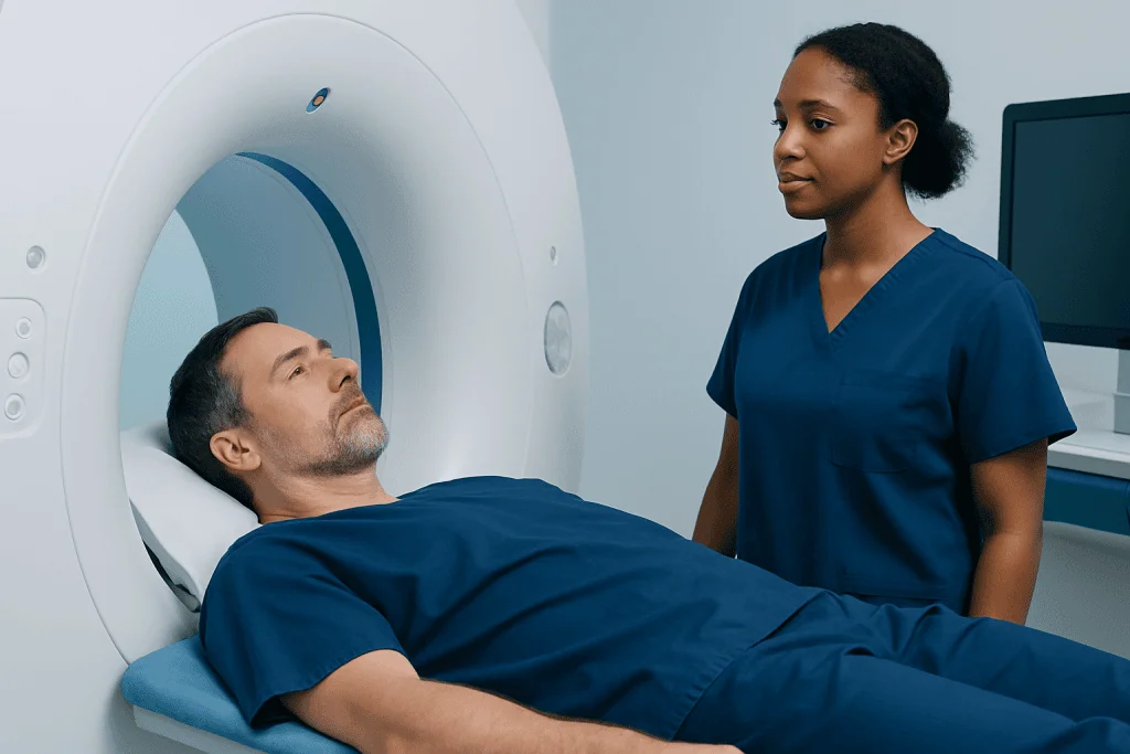
Brain imaging is central to stroke diagnosis. In many emergency departments, a non-contrast CT scan is the first test because it is fast, widely available, and especially useful for detecting bleeding in the brain. It can quickly distinguish between hemorrhagic stroke and most ischemic strokes, which is critical because treatments for a clot should not be given if bleeding is present.
Although an early ischemic stroke may not always appear clearly on the first CT scan, CT still plays a major role in the first minutes of evaluation. In some cases, doctors may also use CT perfusion imaging to assess how much brain tissue is already injured and how much may still be saved if blood flow is restored.
MRI provides more detailed images of brain tissue and can be very sensitive for early ischemic injury. Diffusion-weighted MRI is particularly useful for detecting recent stroke. MRI may also help clarify smaller strokes, strokes in the back of the brain, or uncertain cases where symptoms are subtle. Depending on the situation, specialists may recommend advanced imaging through MRI scanning or urgent CT imaging to support fast decision-making.
The choice between CT and MRI depends on the patient’s condition, timing, availability, and the clinical question. In emergencies, CT is often first because it can be completed very quickly. MRI may follow when more detail is needed or when the diagnosis remains uncertain.
Vessel Tests and Heart Checks to Find the Cause
Once doctors know whether a stroke is due to a blockage or bleeding, they often need to identify the source. Blood vessel imaging helps show whether an artery in the brain or neck is narrowed, blocked, torn, or malformed. Common tests include CT angiography, MR angiography, and carotid ultrasound. These studies can show large vessel occlusions, carotid artery disease, or other structural problems that affect treatment planning.
Some patients may need additional specialized vascular studies, particularly when a procedure is being considered or when the anatomy is complex. In selected cases, doctors may use angiography to obtain a detailed view of the blood vessels. This can help evaluate narrowed or blocked arteries, aneurysms, arteriovenous malformations, or other vascular conditions linked to stroke.
Because some strokes begin in the heart, cardiac testing is also important. An electrocardiogram can detect atrial fibrillation or other rhythm problems. Heart monitoring during hospitalization may identify intermittent irregular rhythms that a single test misses. Echocardiography may be used to look for clots, valve disease, or structural heart problems that could send emboli to the brain.
These tests are part of a broader search for stroke cause, sometimes called an etiologic workup. Knowing the cause helps reduce the chance of another stroke by guiding long-term prevention. For example, treatment may differ for a stroke caused by carotid narrowing, atrial fibrillation, or a small-vessel disease process such as stroke related to longstanding vascular risk factors.
Blood Tests and Other Conditions That Can Mimic Stroke
Blood tests do not diagnose stroke by themselves, but they provide important supporting information. Doctors may check blood sugar, complete blood count, electrolytes, kidney function, and clotting measures. These tests help rule out common mimics, identify medical complications, and ensure that possible treatments can be given as safely as possible.
Stroke mimics are conditions that can produce sudden neurological symptoms similar to stroke. Examples include seizures, migraine, low blood sugar, inner ear problems, brain tumors, infections, medication effects, and functional neurological disorders. The medical team uses the history, exam, and imaging together to sort through these possibilities.
In some cases, further testing is needed after the emergency phase. This may include repeat imaging, longer heart rhythm monitoring, or blood tests for less common clotting or inflammatory disorders in selected patients. The goal is to reach the most accurate diagnosis and tailor prevention to the individual.
How Diagnosis Guides Treatment and Next Steps
The purpose of rapid stroke diagnosis is not only to name the problem, but also to guide immediate treatment. If imaging shows an ischemic stroke and the patient meets criteria, doctors may consider clot-dissolving medication. If a large artery is blocked, some patients may benefit from catheter-based removal of the clot through interventional neuroradiology techniques. If bleeding is found, treatment focuses on controlling blood pressure, managing complications, and identifying the bleeding source.
After the emergency stage, the team develops a prevention plan based on the stroke type and cause. This may include medicines to reduce clotting risk, treatment for atrial fibrillation, blood pressure control, diabetes management, cholesterol-lowering therapy, or procedures for significant carotid disease. Patients may also need rehabilitation for speech, mobility, balance, or daily activities.
Recovery and secondary prevention are closely linked. Someone who has had a transient ischemic attack or confirmed stroke may need follow-up with neurology, cardiology, rehabilitation, or vascular specialists. In selected cases, the diagnostic pathway may overlap with the evaluation of brain aneurysm or other cerebrovascular disorders if bleeding or unusual vessel findings are suspected.
Near the end of the diagnostic journey, patients and families often have many practical questions about prognosis and recurrence risk. Clear communication is important. Acibadem International’s multidisciplinary specialists in JCI-accredited hospitals diagnose and treat stroke and related cerebrovascular conditions for international patients, with evaluation tailored to each patient’s needs.
Reducing Risk and Knowing When to Seek Help
Although not every stroke can be prevented, many risk factors can be managed. Important steps include controlling high blood pressure, treating diabetes, lowering high cholesterol, stopping smoking, staying physically active, maintaining a healthy weight, and following medical advice for atrial fibrillation or other heart disease. Limiting excess alcohol and taking prescribed medicines regularly also support stroke prevention.
People who have had a transient ischemic attack should treat it as an emergency warning sign, even if symptoms resolve. A TIA can signal a high short-term risk of a larger stroke. Rapid assessment often includes the same core diagnostic tools used for stroke, because identifying the cause quickly can prevent permanent brain injury later.
Emergency care is needed right away for sudden weakness, numbness, facial drooping, speech difficulty, sudden trouble seeing, abrupt severe dizziness, or a sudden severe headache unlike usual headaches. It is safer to seek urgent help and be told it is not a stroke than to wait and lose valuable treatment time.
Frequently asked questions
What is the first test usually done for stroke diagnosis?
A non-contrast CT scan of the brain is often the first test in emergency stroke evaluation. It is fast and especially useful for detecting bleeding, which helps doctors choose the safest treatment.
Can an MRI diagnose a stroke better than a CT scan?
MRI can be more sensitive than CT for detecting early ischemic stroke and smaller areas of brain injury. However, CT is often used first because it is quicker and highly effective for identifying brain bleeding in emergencies.
Why do doctors need to know when stroke symptoms started?
The timing of symptom onset helps determine whether time-sensitive treatments may be appropriate. If the exact start time is unknown, doctors often use the last time the person was known to be well.
What tests check the blood vessels during stroke evaluation?
Doctors may use CT angiography, MR angiography, carotid ultrasound, or, in selected cases, catheter angiography. These tests show whether arteries are narrowed, blocked, torn, or otherwise abnormal.
Can something other than a stroke cause the same symptoms?
Yes. Low blood sugar, seizures, migraine, infections, and several other conditions can mimic stroke. That is why urgent medical evaluation and brain imaging are important whenever symptoms appear suddenly.
Is a transient ischemic attack diagnosed the same way as a stroke?
A transient ischemic attack is evaluated urgently with many of the same tools used for stroke, including neurological assessment and brain and vessel imaging. Even though symptoms may go away, a TIA can be an important warning sign of future stroke risk.
References
- World Stroke Organization
- American Stroke Association
- National Institute of Neurological Disorders and Stroke
- Centers for Disease Control and Prevention
- European Stroke Organisation
This article is for general information only and is not a substitute for professional medical advice. Please consult a qualified doctor about your individual situation.
Explore treatments in Turkey — costs, top hospitals & a free quote
JCI-accredited · board-certified surgeons · reply within 24h
Add us as a Preferred Source to see more of our trusted health content across Google Search, AI Overviews and Discover.
Check your numbers in seconds
BMI, calories, due date, blood pressure and 30+ more clinical calculators — free, instant, doctor-reviewed ranges.

More from the Health Library



Related Specialists
Dr. Tülay Tekin
Acibadem Life Clinical Service
Prof. Dr. Mehmet Serdar Binnet
Orthopedic Surgery & Traumatology
Assoc. Prof. Dr. Fatma Tokat
Breast Clinic Clinical Service
Prof. Dr. Ferda Kahveci
Anesthesiology