Interventional Radiology Ablation: Procedure, Recovery and Results
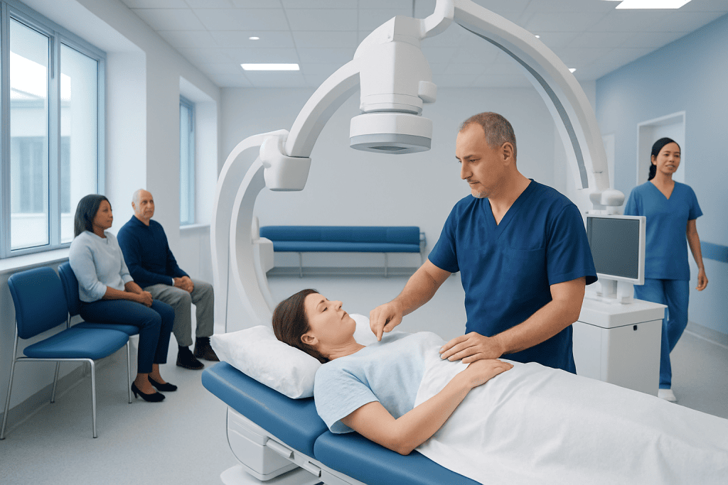
Interventional radiology ablation uses imaging to guide a needle-like probe directly into a treatment target. Heat-based methods such as microwave and radiofrequency ablation, as well as cryoablation using cold, may be used.
Key Takeaways
- Interventional radiology ablation uses imaging to guide a needle-like probe directly into a treatment target.
- Heat-based methods such as microwave and radiofrequency ablation, as well as cryoablation using cold, may be used.
- It is usually performed through a small skin puncture rather than a large surgical incision.
- Most people need a short period of observation and limited activity after treatment, but recovery varies by procedure.
- Follow-up imaging is essential to assess whether the treated tissue has been fully controlled and to monitor for recurrence.
Interventional radiology ablation is a minimally invasive, image-guided treatment that destroys carefully selected areas of abnormal tissue, most often tumors, without open surgery. Recovery is commonly faster than after conventional surgery, although the experience and expected results depend on the treated organ, lesion size, technique, and overall health.
Overview: What Is Interventional Radiology Ablation?
Interventional radiology ablation is a minimally invasive procedure that uses imaging guidance to destroy abnormal tissue in a precise location. It is most often used to treat selected tumors in organs such as the liver, kidneys, lungs, bones, or adrenal glands. Depending on the clinical situation, it may be used with curative intent, to control a tumor, to relieve symptoms, or alongside surgery, systemic therapy, or radiation treatment.
During the procedure, an interventional radiologist guides a thin probe through the skin and into the target using ultrasound, computed tomography (CT), magnetic resonance imaging (MRI), or fluoroscopy. The probe delivers energy that heats tissue or, in cryoablation, creates extreme cold. The goal is to treat the target plus a small surrounding margin while protecting nearby healthy structures.
Because ablation is performed through a small puncture, it is generally considered a minimally invasive procedure rather than open surgery. It is not the right approach for every tumor or every patient. A multidisciplinary team considers imaging findings, pathology, tumor location, size, number of lesions, previous treatment, and a person’s general health before recommending it.
How Interventional Radiology Ablation Works
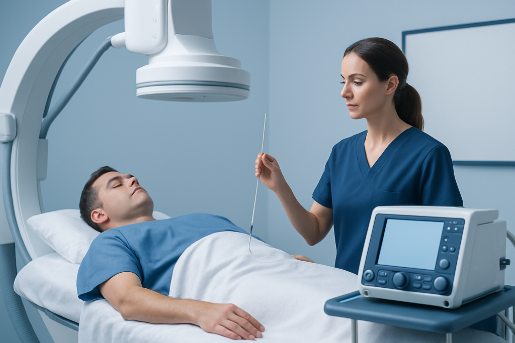
The term ablation describes the controlled destruction of tissue. Heat-based techniques include radiofrequency ablation, which uses electrical current to create heat, and microwave ablation, which uses microwave energy to heat tissue. Cryoablation works differently by freezing tissue. The most suitable technique depends on the organ involved, the target’s size and shape, its proximity to sensitive structures, and the team’s clinical assessment.
Imaging guidance is central to the treatment. It helps the doctor plan the safest path to the lesion, place the probe accurately, and evaluate the treatment zone. In some cases, contrast imaging is performed during or after ablation to help confirm that the intended area has been covered.
Ablation can be particularly useful when surgery would carry substantial risk, when preserving as much organ function as possible is important, or when a person has a limited number of lesions that can be targeted directly. For example, carefully selected kidney tumors may be managed with image-guided treatment as part of an individualized plan for kidney cancer.
The procedure treats a defined target rather than the whole body. It therefore does not replace systemic treatments, such as chemotherapy, immunotherapy, or targeted medicines, when these are needed to address cancer cells beyond the visible lesion.
Who May Be a Candidate for Ablation?
Candidacy for interventional radiology ablation is individualized. The procedure may be considered for people with small or limited tumors, tumors that are difficult to remove surgically, recurrent lesions after prior treatment, or symptoms caused by a focal lesion. It may also be an option for people whose medical conditions make a larger operation less suitable.
Doctors review detailed scans to assess the number, size, and position of lesions. A tumor close to a major blood vessel, bowel, bile duct, airway, nerve, or other important structure can be more challenging to treat safely. In certain cases, protective techniques may be used to move or shield nearby tissue, but some locations may still be better treated by another approach.
Before treatment, the team usually checks blood tests, kidney function, medications, allergies, and bleeding risk. Blood-thinning medicines, diabetes medications, and some supplements may need special instructions before the procedure. Patients should not stop prescribed medicines on their own; the treating team should provide individualized guidance.
Decisions are often made in a tumor board or multidisciplinary meeting involving interventional radiology, oncology, surgery, diagnostic imaging, pathology, and other relevant specialties. This approach helps compare ablation with alternatives such as radiofrequency ablation, surgery, radiation therapy, or medical treatment.
What Happens During the Procedure?
Before ablation, patients receive instructions about fasting, regular medicines, transportation home, and whether an overnight stay may be needed. On arrival, a clinical team confirms the treatment plan, reviews consent, places an intravenous line, and monitors blood pressure, oxygen level, and heart rhythm. Sedation, anesthesia, or both may be used depending on the treated area and the complexity of the procedure.
After the skin is cleaned and numbed, the interventional radiologist makes a very small puncture and advances the ablation probe while viewing real-time or repeated images. Once the probe is correctly positioned, energy is delivered for a planned period. Some procedures require more than one probe placement or treatment cycle to cover the full target.
The team then removes the probe and applies a small dressing. Patients are monitored in a recovery area for pain, nausea, bleeding, changes in vital signs, and other early concerns. The total visit can vary from several hours to longer observation, depending on the organ treated, the anesthesia used, and individual recovery.
Although the skin opening is small, ablation is a significant medical intervention. The care team will explain the specific goals of treatment, expected immediate effects, and the plan for follow-up scans. These scans are important because treatment success is evaluated over time, not by symptoms alone.
Recovery Timeline and What to Expect
Recovery after interventional radiology ablation varies, but many patients go home the same day or after one night of observation. Mild pain or tenderness near the puncture site and in the treated area is common. Tiredness, a low-grade fever, temporary nausea, or flu-like symptoms can also occur, particularly after treatment of larger areas; clinicians may call this a post-ablation syndrome.
For the first few days, patients are commonly advised to rest, drink fluids if permitted, and avoid strenuous exercise, heavy lifting, and driving until cleared by their team. Most return gradually to usual daily activities within several days to one or two weeks, although more extensive treatment or complications can lengthen recovery. The discharge instructions provided by the treating center take priority because restrictions differ by procedure.
How long is bed rest after an ablation? Bed rest is often limited to a few hours after an image-guided ablation while staff monitor the puncture site and recovery from sedation or anesthesia. Some patients need longer observation, especially after treatment involving the kidney, lung, liver, or a difficult-to-reach target. The care team will advise when it is safe to stand, walk, eat, and travel home.
How painful is ablation recovery? Recovery discomfort is often mild to moderate and is commonly managed with the pain-relief plan provided by the care team. Pain depends on the treatment site, amount of tissue treated, and individual pain sensitivity. New, severe, worsening, or uncontrolled pain should be reported promptly, as it may need assessment.
What is the recovery like after interventional radiology? After many interventional radiology procedures, people experience a brief observation period followed by temporary activity limits and a gradual return to routine. Ablation may cause more fatigue or localized discomfort than a simple diagnostic procedure because tissue has been intentionally treated. Follow-up contact and imaging are part of recovery, even when a person feels well.
Benefits, Risks, and Expected Results
Ablation may offer important benefits for appropriately selected patients. It avoids a large incision, may preserve more healthy tissue than surgical removal, and can often involve a shorter hospital stay and quicker physical recovery than open surgery. It can also be repeated in some circumstances if new suitable lesions develop or if further local treatment is required.
As with any invasive procedure, there are risks. These can include bleeding, infection, bruising, damage to nearby organs or structures, blood clots, reactions to sedation or contrast material, and incomplete treatment of the target. Organ-specific risks may include a collapsed lung after treatment near the lung, urine leakage after some kidney procedures, or injury to bile ducts after certain liver procedures. The interventional radiologist will explain the risks most relevant to the planned treatment.
Is ablation considered a major or minor surgery? Ablation is generally classified as a minimally invasive procedure, not major open surgery, because it is performed through a small skin entry point. However, it should not be viewed as minor in the sense of being risk-free. It requires careful planning, anesthesia or sedation in many cases, imaging expertise, and structured follow-up.
Results depend on the type of disease, treatment goal, lesion characteristics, and whether all visible disease can be treated. Follow-up CT, MRI, ultrasound, or other imaging is usually scheduled at defined intervals to assess the ablation zone. If scans suggest remaining viable tumor or recurrence, the multidisciplinary team may discuss repeat ablation or another treatment option.
Self-Care and When to Seek Medical Care
After discharge, patients should follow their personalized instructions on wound care, showering, eating, activity, pain medicines, and follow-up appointments. Keeping the dressing clean and dry as instructed, avoiding activities that strain the treated area, and arranging support at home for the first day can make recovery more comfortable. It is also important to attend all planned imaging appointments, even if symptoms have improved.
Patients should contact their care team for increasing redness, warmth, swelling, drainage, or persistent bleeding at the puncture site; fever that is persistent or worsening; new shortness of breath; chest pain; severe abdominal or flank pain; fainting; confusion; or an inability to keep fluids down. These symptoms do not always indicate a serious complication, but they should be assessed without delay.
When to seek medical care Urgent medical care is appropriate for severe difficulty breathing, heavy bleeding, severe chest pain, loss of consciousness, or symptoms of a severe allergic reaction, such as swelling of the face or throat. For less urgent concerns, patients should contact the interventional radiology or oncology team using the number included in their discharge information.
Acibadem International’s multidisciplinary specialists and JCI-accredited hospitals provide assessment and treatment planning for international patients who may be considered for image-guided ablation and related cancer care.
Frequently asked questions
What is interventional radiology ablation used for?
Interventional radiology ablation is most commonly used to treat selected tumors by destroying them through a needle-like probe guided with medical imaging. It may also be used for some non-cancerous conditions, depending on the tissue involved and local expertise. The reason for treatment and expected benefit should be discussed with the multidisciplinary care team.
Is interventional radiology ablation the same as surgery?
It is not open surgery because it is usually performed through one or more very small skin punctures. It is still an invasive medical procedure that requires careful planning, imaging guidance, and monitoring. In some situations it may be an alternative to surgery, while in others it is used alongside surgery or other treatments.
How long does an ablation procedure take?
The procedure time varies widely based on the organ treated, the number and size of targets, imaging technique, and anesthesia needs. Some straightforward treatments may take about an hour, while more complex cases take longer. Additional recovery-room observation is needed after the procedure.
Can a tumor come back after ablation?
A treated tumor may be fully controlled, but local residual tumor or recurrence can occur. New tumors may also develop elsewhere, depending on the underlying condition. Scheduled follow-up imaging is therefore essential after ablation.
Will I need anesthesia for image-guided ablation?
Many patients receive sedation, local anesthetic, general anesthesia, or a combination, depending on the procedure and treatment location. The anesthesia plan is tailored to patient safety and comfort. An anesthesia or sedation team will review medical history and explain preparation requirements.
When can I return to work after ablation?
Some people return to light work within a few days, while others need one or two weeks or longer depending on the treated organ, physical demands of work, and how they feel. Heavy lifting and strenuous activity may need to be avoided temporarily. The treating clinician can provide the safest timeframe for an individual situation.
References
- Society of Interventional Radiology
- American College of Radiology
- National Cancer Institute
- European Society for Medical Oncology
- Radiological Society of North America
This article is for general information only and is not a substitute for professional medical advice. Please consult a qualified doctor about your individual situation.
Oncology care in Turkey — second opinion and treatment plan
JCI-accredited · board-certified surgeons · reply within 24h
Add us as a Preferred Source to see more of our trusted health content across Google Search, AI Overviews and Discover.
Check your numbers in seconds
BMI, calories, due date, blood pressure and 30+ more clinical calculators — free, instant, doctor-reviewed ranges.