Kennedy Ulcer Pictures — Explained by Medical Evidence, Not Myths
A Kennedy terminal ulcer is a term used for a sudden skin breakdown that may occur in some people approaching the end of life. Pictures can show color, shape, and location, but they cannot establish the cause of a wound or predict prognosis.
Key Takeaways
- A Kennedy terminal ulcer is a term used for a sudden skin breakdown that may occur in some people approaching the end of life.
- Pictures can show color, shape, and location, but they cannot establish the cause of a wound or predict prognosis.
- These skin changes are often described as red, purple, maroon, or black and may develop quickly over the sacrum, buttocks, hips, or other pressure-prone areas.
- Pressure reduction, gentle skin care, pain relief, and comfort-focused wound care remain important even when healing is unlikely.
- New skin discoloration or an open wound should be reviewed by a nurse, doctor, or wound-care professional.
Kennedy ulcer pictures can help explain why these wounds may look different from typical pressure injuries, but a photograph cannot diagnose a Kennedy terminal ulcer or determine how long a person may live. A qualified clinician should assess any new, rapidly changing, painful, or discolored skin area, especially in a person receiving palliative or end-of-life care.
What Kennedy Ulcer Pictures Can Show
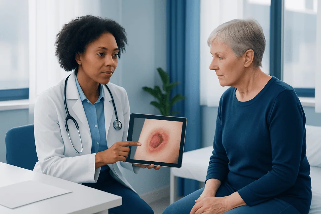
Kennedy ulcer pictures may show a rapidly developing area of skin discoloration or breakdown in a person who is very unwell and nearing the end of life. The area may appear red, purple, maroon, blue-black, or black. It can be shaped like a butterfly, pear, horseshoe, or irregular patch, and it often occurs over the sacrum, coccyx, buttocks, hips, or another bony area.
However, a picture cannot confirm that a wound is a Kennedy terminal ulcer. Lighting, skin tone, camera quality, and the stage of the wound can all change its appearance. More importantly, clinicians need to consider the person’s overall health, mobility, circulation, nutrition, continence, medications, medical history, and the speed at which the area changed.
The term Kennedy terminal ulcer is used in some clinical and palliative-care settings, but its definition and classification are not fully standardized. It should not be used as a diagnosis based only on appearance, and it should never be interpreted as a precise prediction of death. The most useful role of photographs is to support careful monitoring alongside in-person clinical assessment.
How These Skin Changes May Look
Images commonly associated with Kennedy ulcers show a skin area that changes quickly, sometimes over hours or days. In lighter skin tones, this may begin as deep red or purple discoloration. In darker skin tones, early changes may be less visibly red and may instead look darker than nearby skin, ashen, violaceous, gray, or unusually shiny. The area can feel warmer, cooler, firmer, softer, or more painful than surrounding skin.
As the skin injury progresses, it may form a blister, become open, or develop dark tissue. Some wounds have an uneven border rather than the more rounded, localized pattern often associated with sustained pressure. Still, no individual visual feature is unique to a Kennedy ulcer. Similar appearances may occur with deep tissue pressure injury, bruising, reduced blood flow, infection, moisture-related skin damage, or inflammatory skin conditions.
Changes in darker skin can be particularly difficult to recognize in photographs. Looking for differences in temperature, texture, swelling, tenderness, and firmness is as important as looking for color. Families and caregivers should avoid repeatedly pressing or rubbing fragile skin to check it, as this can cause discomfort or further damage.
- Document the date, location, and visible changes if a clinician advises this.
- Use consistent lighting and include only the wound area when taking care-related photographs.
- Share images securely with the clinical team only when requested and in line with privacy guidance.
Kennedy Ulcer or Pressure Injury: Why Pictures Are Not Enough
Pressure injuries develop when prolonged pressure, often combined with friction or shear, damages skin and underlying tissue. They commonly occur when a person cannot change position independently. A Kennedy ulcer may resemble a pressure injury because it often occurs in similar places, but it is described as appearing in the setting of severe systemic decline near the end of life.
Clinicians do not rely on shape alone to distinguish these conditions. They assess whether pressure and shear were present, whether repositioning was possible and tolerated, whether the person has poor circulation or multiple organ dysfunction, and how quickly the wound appeared. They also consider whether a preventive care plan was in place. Even with attentive care, skin can become vulnerable when the body is no longer able to maintain circulation and tissue repair effectively.
It is important not to assume that every wound in a dying person is unavoidable, or that every pressure injury is a Kennedy ulcer. Both assumptions can delay appropriate assessment and care. Pressure relief, moisture management, nutritional support where appropriate, and comfort measures should continue to be considered for every individual.
Other conditions can also cause dark or damaged skin. These include bruising from trauma or blood-thinning medicines, arterial or venous circulation problems, diabetic foot complications, vasculitis, and skin infections. A clinician can decide whether additional investigation or treatment is needed based on the person’s goals of care and clinical circumstances.
Why Kennedy Ulcers May Develop Near the End of Life
Near the end of life, the body may gradually direct blood flow and energy toward essential organs. Circulation to the skin can decrease, and the skin may become less able to tolerate pressure, moisture, friction, and minor injury. Reduced food and fluid intake, immobility, fever, swelling, anemia, low blood pressure, and serious infection can further reduce tissue resilience.
For this reason, a wound may develop despite regular turning, supportive surfaces, and good nursing care. This does not mean preventive measures are unimportant. Rather, it recognizes that skin failure can occur when the body’s ability to repair and protect tissue is profoundly reduced. The clinical team should explain the likely contributing factors in a clear, compassionate way.
Not everyone receiving hospice, palliative care, or treatment for advanced illness develops these wounds. When they do occur, the care plan usually focuses on comfort, dignity, reducing pain and odor, protecting surrounding skin, and avoiding burdensome procedures that do not match the person’s wishes or overall goals of care.
How Clinicians Assess a New Wound
Assessment starts with the whole person, not just the wound. A doctor, nurse, or wound-care specialist may ask when the skin change was first noticed, how quickly it developed, whether it is painful, and whether there has been pressure, friction, incontinence, injury, or a recent change in mobility. They will also review serious illnesses, circulation, medications, nutritional intake, and the person’s care priorities.
The wound itself is assessed for location, size, depth, color, drainage, odor, temperature, edges, and surrounding skin changes. In some situations, clinicians may check for signs of infection or impaired blood flow. Tests are not always necessary, particularly if a person is receiving comfort-focused end-of-life care and results would not change management.
Clinical documentation can help identify whether the area is stable, worsening, or responding to care. It also supports communication between family members, home-care teams, hospitals, and hospice services. If there is uncertainty, a wound-care consultation may help clarify whether the lesion is more consistent with pressure damage, another skin condition, or end-of-life skin failure.
Comfort-Focused Wound Care and Prevention
Care is individualized. When possible and comfortable, relieving pressure remains important. This may involve gentle repositioning, pillows or positioning devices, a pressure-redistributing mattress, and avoiding dragging the skin across sheets. In advanced illness, frequent turning can sometimes cause pain, breathlessness, or distress, so the plan should balance skin protection with comfort.
Dressings may be used to protect the wound, absorb drainage, reduce friction, and limit discomfort during care. The best dressing depends on the wound’s condition and the person’s skin sensitivity. Strong antiseptics, adhesive products, and frequent dressing changes can irritate fragile skin, so wound products should be chosen and changed by trained professionals whenever possible.
Pain should be actively assessed, including pain during movement and dressing changes. The care team may recommend medication, gentle handling, timing care around pain relief, and minimizing unnecessary disturbance. If odor or drainage is distressing, clinicians can suggest practical measures that support comfort and dignity.
Family caregivers should not feel they have failed if skin damage occurs during a loved one’s final illness. Asking what can be done to improve comfort, what changes to expect, and when to call for help can make care more manageable. Acibadem International’s multidisciplinary specialists and JCI-accredited hospitals can assess complex wounds and support international patients according to their individual clinical needs.
When to Seek Medical Care
A new dark, purple, red, or black area of skin should be reported promptly to the person’s doctor, nurse, hospice team, or wound-care professional. This is especially important if the area develops quickly, becomes open, causes pain, produces drainage, or is located over a bony prominence. Early assessment can identify reversible contributors and guide appropriate comfort measures.
Urgent medical advice is needed if there are signs that may suggest infection or another acute problem, such as spreading redness, warmth, swelling, pus-like drainage, fever, sudden severe pain, rapidly spreading discoloration, or a marked decline in alertness. These symptoms do not always mean infection, but they need timely clinical review.
For a person receiving end-of-life care at home, the hospice or palliative-care contact should generally be the first call. They can advise on immediate comfort steps and whether an in-person visit is needed. If a person is not known to be near the end of life, a rapidly appearing dark skin lesion should not be assumed to be a Kennedy ulcer and should be evaluated by a healthcare professional.
Frequently asked questions
Can a photo diagnose a Kennedy ulcer?
No. A photograph can document the color, location, and visible progression of a wound, but it cannot determine its cause. A clinician needs to assess the person’s health, circulation, mobility, pressure exposure, and goals of care.
What does a Kennedy terminal ulcer usually look like?
It may appear as a sudden red, purple, maroon, dark brown, or black area of skin injury, often over the sacrum, buttocks, or hips. It can have an irregular, pear-shaped, butterfly-like, or horseshoe-like appearance. These features are not exclusive to Kennedy ulcers.
Does a Kennedy ulcer mean death is imminent?
A Kennedy ulcer is described in association with advanced illness and the dying process, but it cannot reliably predict exactly when a person will die. The timing and course vary greatly between individuals. Families should discuss prognosis with the clinician who knows the person’s overall condition.
Can a Kennedy ulcer be prevented?
Pressure reduction, skin care, moisture management, nutrition support when appropriate, and regular assessment may lower the risk of skin damage. However, in severe end-of-life illness, the skin may fail despite attentive preventive care. The care team can help set realistic, comfort-centered goals.
Are Kennedy ulcers painful?
They can be painful, although some people may have reduced sensation or be unable to communicate discomfort. Pain may increase during movement, cleaning, or dressing changes. Clinicians can adjust wound care and pain management to reduce distress.
What should a caregiver do after noticing a new skin wound?
The caregiver should notify the person’s nurse, doctor, hospice team, or wound-care provider promptly. Until advice is received, they should avoid massaging the area, minimize friction, keep the person comfortable, and follow any existing repositioning and skin-care plan. They should seek urgent advice if there is fever, spreading redness, pus-like drainage, severe pain, or rapid deterioration.
This article is for general information only and is not a substitute for professional medical advice. Please consult a qualified doctor about your individual situation.
Persistent digestive symptoms? Get evaluated in Turkey
JCI-accredited · board-certified surgeons · reply within 24h
Add us as a Preferred Source to see more of our trusted health content across Google Search, AI Overviews and Discover.

More from the Health Library

Neurophysiologist — Explained by Medical Evidence, Not Myths
Feces Softener: An Evidence-Based Guide for Patients

Hypersensitivity: What Patients Need to Know

Forager: A Complete Medical Overview
Supraclavicular: An Evidence-Based Guide for Patients
Status Asthmaticus: A Complete Medical Overview
Gastroenterology Specialists at Acibadem

Prof. Dr. Ethem Tankurt
Gastroenterology
Prof. Dr. Fatih Oğuz Önder
Gastroenterology
Dr. Fatma Seçil Kırdök
Gastroenterology
Prof. Dr. Ferdane Pirinççi Sapmaz
Gastroenterology