Kidney MRI: Preparation, Procedure and Results

A kidney MRI uses magnets and radio waves, not X-rays or radiation, to create detailed images. Preparation depends on whether contrast is planned, so patients should follow the imaging center’s individual instructions.
Key Takeaways
- A kidney MRI uses magnets and radio waves, not X-rays or radiation, to create detailed images.
- Preparation depends on whether contrast is planned, so patients should follow the imaging center’s individual instructions.
- Most people can return to usual activities immediately after a kidney MRI unless they received sedation.
- MRI contrast is generally well tolerated, but kidney function and allergy history should be reviewed before it is used.
- A radiologist interprets the images and sends a report to the clinician who ordered the scan.
A kidney MRI is a noninvasive imaging test that uses a magnetic field and radio waves to produce detailed pictures of the kidneys, urinary tract, and nearby tissues. It can help clinicians investigate symptoms, assess known kidney conditions, and plan care without using ionizing radiation.
Kidney MRI overview: what the scan shows
A kidney MRI, also called renal magnetic resonance imaging, is a scan that creates detailed images of the kidneys using a powerful magnet, radio waves, and a computer. It does not use ionizing radiation. The examination may include images of the kidneys themselves, the blood vessels supplying them, the urine-collecting system, the adrenal glands, and nearby abdominal structures.
Clinicians may request this test when they need more detail than other imaging examinations can provide, or when a person should avoid radiation exposure. A kidney MRI can help assess a kidney mass or cyst, infection, blockage, congenital difference, blood-vessel problem, injury, or a change seen on ultrasound or CT. It may also support evaluation of related conditions such as kidney cancer when clinically appropriate.
The images are reviewed by a radiologist, a doctor trained in medical imaging. The radiologist’s report is then shared with the referring clinician, who explains what the findings mean in the context of symptoms, examination findings, blood tests, and other investigations.
How kidney MRI works and who may benefit
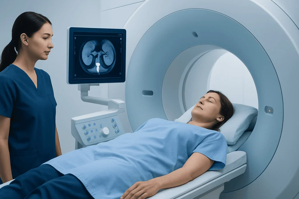
During MRI, the body is placed within a strong magnetic field. Radiofrequency pulses briefly change the behavior of hydrogen atoms in body tissues; when the atoms return to their usual state, they release signals that a computer converts into images. Different image sequences highlight fluid, blood vessels, solid tissues, inflammation, and other features in different ways.
Some kidney MRI examinations use a gadolinium-based contrast agent injected into a vein. Contrast can make blood vessels and certain tissues easier to distinguish, helping to characterize a lesion or assess blood flow. Not every kidney MRI requires contrast, and the imaging team decides this based on the clinical question and the person’s medical history.
Kidney MRI may be useful for adults and children, including people who need detailed soft-tissue imaging or repeated follow-up scans. However, MRI is not suitable for everyone. Certain implanted devices, metal fragments, some clips or pumps, and severe difficulty lying still in an enclosed space may require special planning or a different test. Modern MRI-compatible devices are common, but compatibility must always be verified.
What should I do to prepare for a kidney MRI?

Patients should carefully follow the instructions provided by their imaging center, because preparation can differ according to the reason for the scan and whether contrast or sedation is planned. They should tell the team about kidney disease, dialysis, pregnancy or possible pregnancy, prior contrast reactions, asthma or severe allergies, surgery, and all implanted or external medical devices.
Before entering the MRI room, patients must remove metal items such as jewelry, watches, hearing aids, removable dental work, hairpins, credit cards, and clothing with metal fasteners. They may be asked to change into a gown. It is important to mention tattoos, cosmetic permanent makeup, medication patches, and any past workplace or injury-related exposure to metal fragments.
If contrast may be used, the clinician may request a recent blood test to assess kidney function, especially for people with known kidney disease, diabetes, older age, or other relevant health concerns. Patients should bring information about prior imaging, medications, and implants. They should not stop prescribed medicines unless their clinician specifically tells them to do so.
What to do the night before an MRI? Most people can eat, drink, and take regular medication as usual the night before. If fasting, bowel preparation, or sedation is planned, the imaging center will provide specific instructions; these should take priority over general advice. Preparing identification, referral documents, prior scan reports, and a list of medications the evening before can make the appointment smoother.
What happens if I drink water before an MRI?
For many kidney MRI examinations, drinking water before the scan is allowed and may be encouraged, particularly when no sedation is planned. Good hydration can make an intravenous line easier to place and is usually sensible unless a clinician has advised fluid restriction for heart failure, advanced kidney disease, or another medical reason.
However, some abdominal MRI protocols ask patients not to eat or drink for several hours beforehand. Fasting may reduce motion from bowel activity and improve image quality. Other protocols may ask a patient to arrive with a comfortably full bladder. Because these instructions can differ, the safest approach is to follow the written directions from the imaging department rather than changing fluid intake independently.
If a person accidentally drinks water despite being instructed to fast, they should contact the imaging center before the appointment. The staff can advise whether the examination can proceed, needs a short delay, or should be rescheduled. Patients should not feel embarrassed about asking; accurate preparation helps produce the clearest possible images.
Kidney MRI procedure: step by step
On arrival, a radiology professional reviews the safety questionnaire and confirms the reason for the scan. An intravenous cannula may be placed if contrast is expected. The patient lies on a movable table, usually on their back, and a receiving device called a coil is positioned around the abdomen to capture detailed signals.
The table moves slowly into the scanner. The MRI machine makes repeated loud tapping, knocking, or humming sounds while images are acquired, so earplugs or headphones are provided. The patient can communicate with the radiographer throughout the scan through an intercom and is given a call button. Remaining still is important, and brief breath-holds are commonly requested to reduce motion in abdominal images.
A kidney MRI often takes about 30 to 60 minutes, though the duration varies with the number of image sequences, use of contrast, and the clinical question. If contrast is used, it is administered through the IV partway through the examination. A cool sensation at the injection site or a temporary unusual taste can occur, but serious reactions are uncommon.
Some people experience anxiety in the scanner because the space can feel enclosed. Patients should mention this when booking or arriving. The team may offer practical support, such as communication throughout the scan, music where available, or discussion of whether a prescribed calming medicine or sedation is appropriate. Sedation requires advance planning and someone to accompany the patient home.
Is an MRI hard on the kidneys? Benefits, risks and recovery
An MRI scan itself is not hard on the kidneys. The magnetic field and radio waves do not damage kidney function. The main kidney-related consideration is gadolinium-based contrast, which is filtered from the body largely through the kidneys. For most people with normal or mildly reduced kidney function, modern approved gadolinium contrast agents are used safely when clinically indicated.
For people with severe kidney impairment or acute kidney injury, the care team considers contrast particularly carefully. In rare situations, certain gadolinium agents have been associated with nephrogenic systemic fibrosis, a serious condition affecting the skin and other tissues. Screening for kidney function and selecting an appropriate agent, dose, or non-contrast alternative helps minimize this already uncommon risk.
The benefits of kidney MRI include excellent soft-tissue detail, assessment of blood vessels without radiation, and the ability to obtain several types of images in one examination. Limitations include the need to remain still, scan noise, longer examination times than some other imaging tests, and restrictions related to some metal implants. The ordering clinician weighs these factors against the expected value of the information.
Recovery is usually immediate. Patients who do not receive sedation can generally eat, drink, drive, and return to normal activities after the scan. Drinking fluids after contrast is reasonable for most people, unless they have been given fluid limits. People who receive sedation need an escort and should avoid driving, alcohol, important decisions, and operating machinery for the period advised by their care team.
Kidney MRI results and what happens next
A radiologist reviews the MRI images and prepares a report describing relevant findings. The report may comment on kidney size and structure, cysts or masses, swelling of the urine drainage system, blood vessels, signs of inflammation or infection, and surrounding organs. A result that identifies an abnormality does not automatically mean cancer or permanent kidney damage; many imaging findings are benign or need only monitoring.
Results are commonly sent to the referring clinician within a few days, although timing varies by facility and urgency. The clinician may discuss the results by phone, through a patient portal, or at a follow-up appointment. They may recommend no further action, repeat imaging, laboratory tests, referral to a urologist or nephrologist, or additional evaluation depending on the findings.
Further care may include kidney-function assessment or investigation of urinary symptoms. Where a structural concern requires specialist treatment, a urology team may discuss options such as kidney stone treatment or another tailored approach. At Acibadem International, multidisciplinary specialists in JCI-accredited hospitals assess kidney and urinary conditions for international patients when further evaluation or treatment is needed.
When to seek medical care
A kidney MRI is usually arranged as a planned test, but certain symptoms warrant timely medical advice rather than waiting for a routine scan. These include visible blood in the urine, persistent flank or back pain, fever with urinary symptoms, new swelling, markedly reduced urine output, or unexplained weight loss. A clinician can determine whether urgent assessment, blood and urine testing, or imaging is appropriate.
Emergency care is important for severe pain with fever or vomiting, inability to pass urine, confusion, fainting, or symptoms that rapidly worsen. These can indicate conditions such as severe infection or urinary obstruction that may require prompt treatment. People with a single kidney, known significant kidney disease, or a kidney transplant should seek advice promptly if they develop concerning urinary or systemic symptoms.
After an MRI with contrast, patients should seek urgent medical attention for breathing difficulty, facial or throat swelling, widespread hives, or severe dizziness, as these may indicate a serious allergic reaction. Delayed concerns, such as persistent rash or problems around an IV site, should be discussed with a healthcare professional. Most people have no lasting effects from the examination.
Frequently asked questions
How long does a kidney MRI take?
A kidney MRI commonly takes around 30 to 60 minutes. The exact time depends on whether contrast is needed, how many image sequences are required, and whether breath-hold images are included. Patients should allow extra time for registration and safety screening.
Do I need contrast for a kidney MRI?
Not always. Contrast may be used when it can improve evaluation of a kidney mass, blood vessels, inflammation, or other specific concerns. The radiologist and referring clinician decide based on the reason for the scan and the patient’s kidney function and medical history.
Can I have a kidney MRI if I have kidney disease?
Often, yes. A non-contrast MRI may be suitable, and contrast can sometimes still be used after careful assessment. Patients should tell the imaging team about any kidney disease, dialysis, transplant, or recent kidney-function test results.
Can I eat before a kidney MRI?
Many people can eat normally before a kidney MRI, but some abdominal MRI protocols require fasting for a few hours. The imaging center’s instructions are the most reliable guide because they are based on the planned examination. Patients should ask in advance if they have not received clear instructions.
Is a kidney MRI painful?
The scan itself is painless. Lying still may become uncomfortable for some people, and an IV contrast injection can cause a brief pinch or cool sensation. The scanner noise can be loud, but ear protection is provided.
When will I receive kidney MRI results?
A radiologist first reviews the images and creates a formal report. Results are often available to the referring clinician within a few days, although urgent studies may be reported sooner. The clinician who ordered the test is best placed to explain how the findings relate to the patient’s health.
References
- Radiological Society of North America
- American College of Radiology
- National Institute of Diabetes and Digestive and Kidney Diseases
- National Kidney Foundation
- U.S. Food and Drug Administration
This article is for general information only and is not a substitute for professional medical advice. Please consult a qualified doctor about your individual situation.
Oncology care in Turkey — second opinion and treatment plan
JCI-accredited · board-certified surgeons · reply within 24h