Kidney Stones: Warning Signs, Causes, and Treatment

Kidney stones may cause sharp flank pain, blood in the urine, nausea, and urinary urgency, but small stones can sometimes pass with few symptoms. Common causes include low fluid intake, high urine mineral levels, certain diets, family history, urinary tract problems, and some medical conditions.
Key Takeaways
- Kidney stones may cause sharp flank pain, blood in the urine, nausea, and urinary urgency, but small stones can sometimes pass with few symptoms.
- Common causes include low fluid intake, high urine mineral levels, certain diets, family history, urinary tract problems, and some medical conditions.
- Diagnosis usually involves a medical history, urine and blood tests, and imaging such as ultrasound or CT scan.
- Treatment depends on stone size, location, symptoms, infection risk, and kidney function; options range from observation to minimally invasive procedures.
- Prevention often includes drinking enough fluids, adjusting salt and animal protein intake, and tailoring diet or medication to the stone type.
Kidney stones are hard mineral deposits that form in the urinary tract and can cause severe pain when they move or block urine flow. With timely diagnosis, appropriate treatment, and preventive lifestyle steps, most people recover well and can lower the risk of future stones.
Overview
Kidney stones, also called renal calculi or urinary stones, are solid crystals that form when minerals and salts in the urine become too concentrated. They may develop in one or both kidneys and can travel into the ureter, the narrow tube that carries urine from the kidney to the bladder. Some stones are as small as grains of sand, while others can grow large enough to block urine flow.
The most common stones contain calcium, usually calcium oxalate or calcium phosphate. Other types include uric acid stones, struvite stones related to certain infections, and cystine stones linked to a rare inherited condition. Knowing the stone type is important because prevention advice and medical treatment can differ.
Kidney stones are common and often treatable. A small stone may pass naturally with fluids and symptom control, while larger stones or stones causing complications may need a procedure. The main goals are to relieve pain, protect kidney function, treat any infection, remove or help pass the stone, and reduce the chance of recurrence.
Kidney Stone Symptoms and Warning Signs
Kidney stone symptoms often begin when a stone moves from the kidney into the ureter or causes blockage. The classic symptom is sudden, severe pain in the side or back below the ribs, known as renal colic. This pain may come in waves and may spread to the lower abdomen, groin, or genital area as the stone moves.
Other symptoms can include nausea, vomiting, restlessness, sweating, frequent urination, burning with urination, cloudy or foul-smelling urine, and visible blood in the urine. Some people notice only pink, red, or brown urine, while others have blood detected only on a urine test. If the stone is near the bladder, urinary urgency and a feeling of incomplete emptying may occur.
Symptoms that deserve prompt medical attention include fever, chills, persistent vomiting, inability to pass urine, severe pain that does not improve, or symptoms in someone with one kidney, known kidney disease, pregnancy, or a weakened immune system. A stone combined with infection or obstruction needs urgent evaluation because treatment may be needed to drain the urinary system and protect the kidneys.
Causes and Risk Factors

Kidney stones form when the balance of water, minerals, acids, and protective substances in the urine changes. If urine is too concentrated, crystals can develop and gradually grow into stones. Low fluid intake, heavy sweating, hot climates, and dehydration from vomiting or diarrhea can increase urine concentration and raise stone risk.
Diet can contribute, although it is rarely the only cause. High salt intake can increase calcium in the urine. A diet very high in animal protein may raise uric acid and lower urinary citrate, a natural inhibitor of stone formation. Foods high in oxalate, such as spinach, rhubarb, beets, nuts, and some teas, may matter for people who form calcium oxalate stones, especially if calcium intake is too low or fluids are insufficient.
Medical and personal factors also play a role. Risk may be higher in people with a family history of stones, previous kidney stones, recurrent urinary tract infections, gout, obesity, inflammatory bowel disease, certain bowel surgeries, hyperparathyroidism, or structural urinary tract problems. Some medicines and supplements can contribute in selected patients, so a clinician may review prescriptions, over-the-counter products, and vitamin use as part of prevention planning.
- Calcium stones are the most frequent type and may be linked to urine calcium, oxalate, citrate, and fluid levels.
- Uric acid stones can be associated with acidic urine, gout, metabolic conditions, or high purine intake.
- Struvite stones may form after infections caused by specific bacteria.
- Cystine stones occur in people with cystinuria, an inherited condition affecting amino acid transport.
Diagnosis
Diagnosis begins with a medical history and physical examination. The doctor will ask about pain location and timing, urinary symptoms, fever, previous stones, family history, diet, fluid intake, medications, and medical conditions. The examination may include checking temperature, blood pressure, abdominal tenderness, and flank tenderness.
Urine testing can show blood, crystals, infection signs, pH level, and other clues about stone type. Blood tests may assess kidney function, calcium, uric acid, infection markers, and electrolyte balance. If a stone is passed or removed, laboratory stone analysis is very useful because it identifies the stone composition and guides prevention.
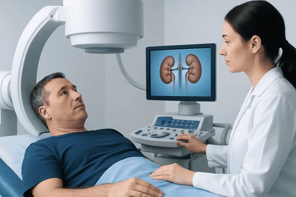
Imaging helps confirm the stone, locate it, estimate its size, and check whether it is causing obstruction. Ultrasound is often used because it avoids radiation and can show swelling of the kidney. A non-contrast CT scan is highly accurate for many urinary stones and may be recommended when the diagnosis is uncertain, symptoms are severe, or treatment planning requires detailed information. X-rays may be helpful for follow-up of certain calcium-containing stones.
For people with repeated stones, stones at a young age, one kidney, complex medical conditions, or high recurrence risk, a more detailed metabolic evaluation may be recommended. This can include one or more 24-hour urine collections to measure urine volume, calcium, oxalate, citrate, uric acid, sodium, and other factors.
Treatment Options
Treatment depends on the stone’s size, location, type, symptoms, whether infection is present, and how well the kidneys are functioning. Small stones that are likely to pass may be managed with observation, adequate fluids, and pain control under medical guidance. Some patients may be prescribed medication to relax the ureter and help a stone pass, depending on the stone location and individual suitability.
Medical care is especially important if pain is intense, symptoms continue, or urine flow is blocked. Doctors may use anti-nausea medicines, pain-relief medicines, and hydration support when needed. Antibiotics are used only when there is evidence or strong suspicion of infection; they do not dissolve ordinary kidney stones.
If a stone is too large to pass, causes ongoing obstruction, is associated with infection, or threatens kidney function, a procedure may be recommended. Options include shock wave lithotripsy, which uses sound waves to break selected stones into smaller pieces; ureteroscopy, in which a small scope is passed through the urinary tract to remove or fragment the stone; and percutaneous nephrolithotomy, a minimally invasive surgical approach often used for larger or complex kidney stones. A temporary ureteral stent may be placed to help urine drain and allow healing.
After treatment, follow-up is important. The care team may confirm that the stone has passed or been fully treated, check kidney drainage, review stone analysis, and discuss prevention. People treated at Acibadem International can access multidisciplinary evaluation through urology, nephrology, radiology, and laboratory services in JCI-accredited hospitals for diagnosis and management of kidney stones.
Prevention and Self-Care
Prevention usually starts with improving urine dilution. Many adults are advised to drink enough fluids to produce pale urine through the day, unless a doctor has restricted fluids for heart, kidney, or other medical reasons. Water is generally the best choice. People who sweat heavily, exercise intensely, or live in hot climates may need extra fluids.
Dietary advice should be individualized, but several general steps help many stone formers. Reducing excess salt can lower urinary calcium. Eating normal dietary calcium with meals, rather than avoiding calcium completely, may help bind oxalate in the gut for people prone to calcium oxalate stones. Very high intake of animal protein may need moderation, especially in uric acid or calcium stone formers.
Some people benefit from limiting high-oxalate foods, increasing fruits and vegetables, or adjusting citrus intake to raise urinary citrate. However, restrictive diets are not appropriate for everyone and can be difficult to maintain without guidance. A registered dietitian or physician can tailor recommendations to the stone type, urine test results, nutrition needs, and other conditions such as diabetes, gout, or chronic kidney disease.
- Do not stop prescribed medicines or start supplements for stone prevention without medical advice.
- Keep follow-up appointments after a stone episode, even if symptoms improve.
- Try to collect a passed stone for analysis if the doctor recommends it.
- Seek individualized advice if stones recur, occur in childhood, or occur with kidney disease.
When to See a Doctor
A person should contact a healthcare professional for suspected kidney stone symptoms, especially new severe side or back pain, blood in the urine, painful urination, or pain that travels toward the groin. Even when a stone seems likely, similar symptoms can occur with urinary tract infection, appendicitis, gallbladder disease, gynecologic conditions, or other problems that require different care.
Urgent medical care is needed if kidney stone symptoms occur with fever, chills, persistent vomiting, faintness, confusion, inability to urinate, or severe pain that is not controlled. Prompt evaluation is also important for pregnant patients, people with a single kidney, transplant recipients, those with known kidney disease, and people with immune system suppression.
Patients who have had one stone should ask about prevention, because recurrence risk can often be reduced. Follow-up may include imaging, blood tests, urine tests, and stone analysis. With a clear plan, most people can manage an acute stone safely and take practical steps to protect urinary health over time.
Frequently asked questions
Can kidney stones pass on their own?
Yes, some small kidney stones pass naturally through the urine, especially if they are located lower in the ureter and are not causing complications. However, stone size, position, pain level, infection risk, and kidney function all affect whether observation is safe. A doctor can advise whether home management, medication, or a procedure is appropriate.
What does kidney stone pain feel like?
Kidney stone pain is often sharp, cramping, and severe, usually starting in the side or back below the ribs. It may come in waves and move toward the lower abdomen or groin. Some people also have nausea, vomiting, urinary urgency, burning, or blood in the urine.
Does drinking more water dissolve kidney stones?
Drinking enough water can help small stones pass and is one of the most important ways to prevent new stones. However, water usually does not dissolve most established calcium stones. Some uric acid stones may be treated by changing urine chemistry under medical supervision.
Are kidney stones always caused by too much calcium?
No. Although many stones contain calcium, the cause is usually a combination of urine concentration, salt intake, oxalate levels, citrate levels, metabolism, genetics, and other health factors. Avoiding all calcium is not recommended for most people and may even increase calcium oxalate stone risk if dietary calcium becomes too low.
When is kidney stone surgery needed?
A procedure may be needed if a stone is large, unlikely to pass, causes persistent pain, blocks urine flow, is linked to infection, or affects kidney function. Common treatments include shock wave lithotripsy, ureteroscopy, and percutaneous nephrolithotomy. The best option depends on stone size, location, type, anatomy, and the patient's overall health.
How can someone reduce the chance of another kidney stone?
Prevention often includes drinking enough fluids, reducing excess salt, moderating animal protein, and following stone-specific diet advice. Stone analysis and 24-hour urine testing can identify personal risk factors. Some patients also need prescription medication, but this should be guided by a qualified clinician.
This article is for general information only and is not a substitute for professional medical advice. Please consult a qualified doctor about your individual situation.
Explore treatments in Turkey — costs, top hospitals & a free quote
JCI-accredited · board-certified surgeons · reply within 24h
Add us as a Preferred Source to see more of our trusted health content across Google Search, AI Overviews and Discover.
Check your numbers in seconds
BMI, calories, due date, blood pressure and 30+ more clinical calculators — free, instant, doctor-reviewed ranges.

Urology Specialists at Acibadem





More from the Health Library
Kidney Stones: Flank Pain, CT Diagnosis, and Prevention

Kidney Stones: Flank Pain, Urine Tests, and Prevention Strategies

Peripheral Neuropathy: Numbness, Tingling, and Nerve Testing

Tonsillitis: Sore Throat, Fever, and When Surgery Is Considered

Condition vs Disorder vs Syndrome: What Is the Difference?