Knee Joint Anatomy Made Simple: Bones, Cartilage, Ligaments, and Common Injuries
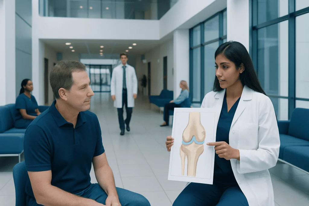
The knee is a large hinge-like joint that allows bending, straightening, and slight rotation. Its main bones are the femur, tibia, and patella, which work together to support movement.
Key Takeaways
- The knee is a large hinge-like joint that allows bending, straightening, and slight rotation.
- Its main bones are the femur, tibia, and patella, which work together to support movement.
- Cartilage and menisci cushion the joint and help distribute weight smoothly.
- Ligaments provide stability, while tendons and muscles help the knee move.
- Common knee injuries include ligament sprains, meniscus tears, tendon problems, and cartilage wear.
- Persistent pain, swelling, locking, or instability should be assessed by a qualified doctor.
Knee joint anatomy can seem complex, but it becomes easier to understand when broken into its main parts: bones, cartilage, ligaments, tendons, and muscles. Each structure has a specific role in helping the knee bend, bear weight, and stay stable during daily activities and sports.
Overview of the Knee Joint
The knee is one of the body’s largest and most important joints. It connects the thigh bone to the shin bone and helps a person walk, climb stairs, sit, stand, run, and jump. Because it carries body weight and is used frequently, the knee is exposed to considerable daily stress.
Knee joint anatomy is often described as a hinge joint, but it is slightly more complex than a simple door hinge. In addition to bending and straightening, the knee also allows a small amount of rotation. This combination of movement makes normal activities possible while also requiring strong support from surrounding tissues.
The knee works well because several structures function together. Bones form the framework, cartilage reduces friction, menisci act as shock absorbers, ligaments hold the joint steady, and tendons connect muscles to bone. When any of these parts are injured or worn down, symptoms such as pain, swelling, stiffness, or instability can develop.
Bones of the Knee
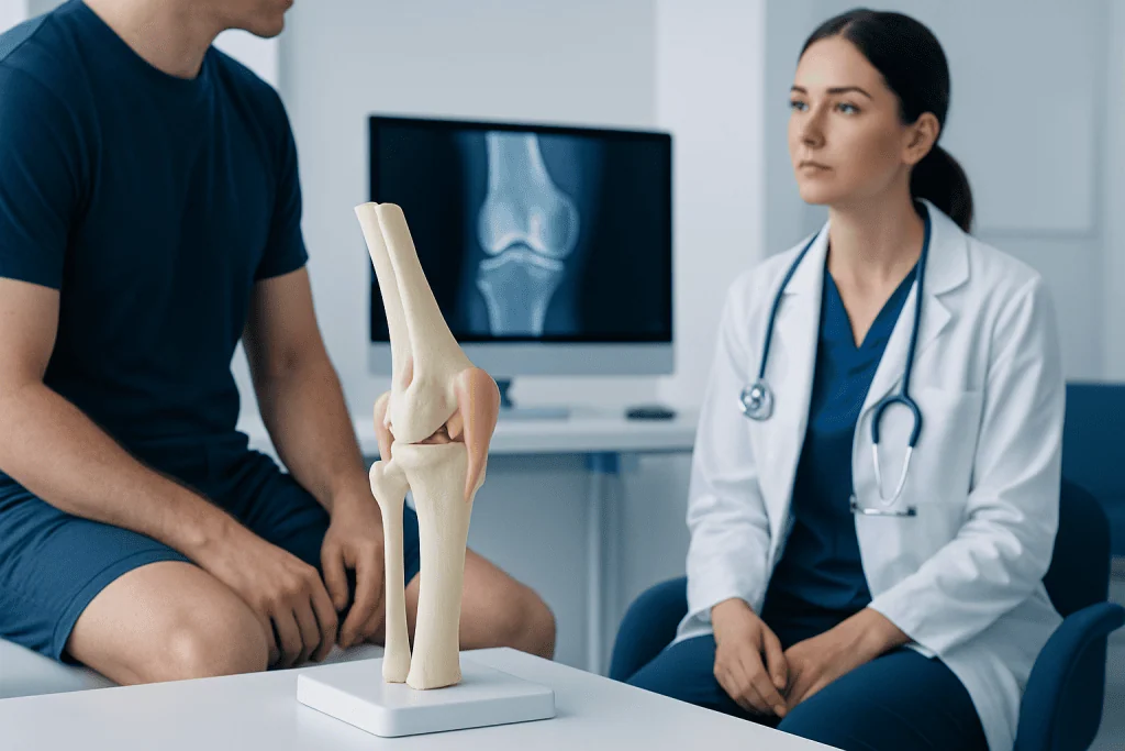
Three bones make up the main structure of the knee: the femur, tibia, and patella. The femur is the thigh bone, and its lower end forms the upper part of the knee joint. The tibia, or shin bone, forms the lower part of the joint and carries most of the body’s weight through the leg.
The patella, commonly called the kneecap, sits in front of the joint. It protects the knee and improves the leverage of the thigh muscles during straightening of the leg. This helps make movements such as standing from a chair or climbing stairs more efficient.
The fibula, a smaller bone on the outer side of the lower leg, is not directly part of the main knee joint surface, but it still contributes to nearby support. Together, these bones create a joint that is strong enough for weight-bearing while remaining flexible enough for smooth movement.
The ends of the femur and tibia are shaped to fit together, but they are not perfectly matched on their own. That is one reason the knee depends so much on cartilage, menisci, ligaments, and muscles for comfort and stability.
Cartilage, Menisci, and Other Cushions
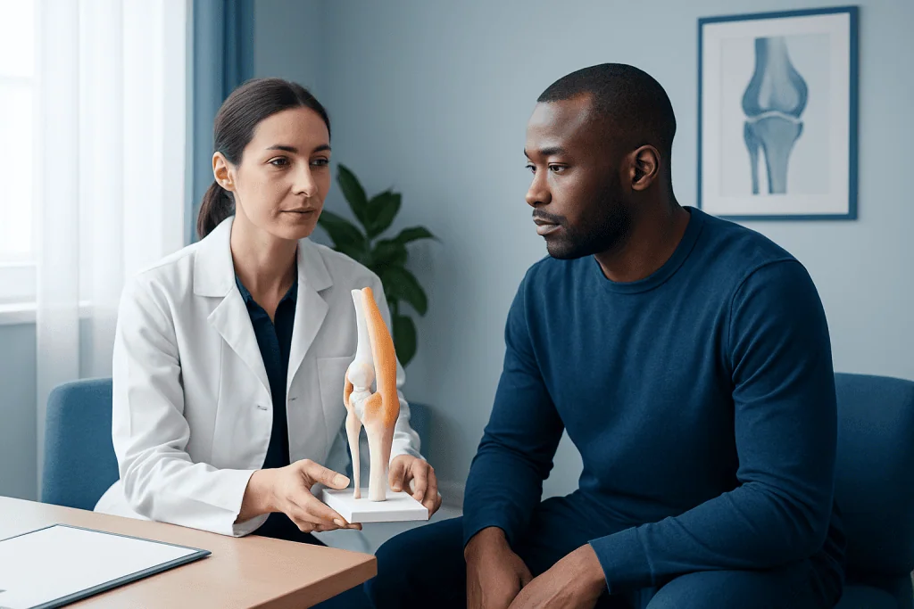
The surfaces of the femur, tibia, and the back of the patella are covered with articular cartilage. This smooth, slippery tissue allows the bones to glide against each other with very little friction. Healthy cartilage helps movements feel smooth and comfortable.
Inside the knee are two crescent-shaped pads called the menisci. There is a medial meniscus on the inner side of the knee and a lateral meniscus on the outer side. These structures help absorb shock, spread body weight across the joint, and improve stability.
Without the menisci, pressure on the knee would be concentrated over a smaller area. Over time, this can increase wear on the joint surfaces. Meniscus injuries are common in sports, but they can also happen during twisting movements in everyday life, especially as the tissue becomes less flexible with age.
The knee also contains small fluid-filled sacs called bursae, which reduce friction between tendons, skin, and bone. When bursae become irritated, they can cause localized swelling and tenderness. Cartilage wear and meniscus damage may also contribute to knee pain and reduced mobility.
Ligaments, Tendons, and Muscles
Ligaments are tough bands of tissue that connect bone to bone and help keep the knee stable. Four main ligaments are especially important:
- Anterior cruciate ligament (ACL): helps prevent the tibia from sliding too far forward.
- Posterior cruciate ligament (PCL): helps prevent the tibia from sliding too far backward.
- Medial collateral ligament (MCL): supports the inner side of the knee.
- Lateral collateral ligament (LCL): supports the outer side of the knee.
Tendons connect muscles to bones. The quadriceps tendon connects the large thigh muscles to the patella, and the patellar tendon connects the patella to the tibia. These structures are essential for straightening the knee, walking, rising from a seated position, and controlling movement during activity.
Muscles around the knee also play a major role. The quadriceps at the front of the thigh help straighten the leg, while the hamstrings at the back help bend the knee. Muscles in the hips and calves also influence knee alignment and function. Strong, balanced muscles can reduce stress on the joint and improve stability.
When ligaments are stretched or torn, the knee may feel loose or unstable. When tendons become inflamed or injured, pain often appears during movement. Problems in these support structures can range from mild strain to significant injury that may require rehabilitation or specialist care.
How the Knee Moves and Stays Stable
The knee mainly performs flexion and extension, which means bending and straightening. It also allows a small amount of rotation, especially when the knee is bent. This subtle rotation is important for natural walking, changing direction, and adjusting to uneven ground.
Stability in the knee comes from both passive and active support. Passive support includes the bones, ligaments, joint capsule, and menisci. Active support comes from the muscles and tendons that control movement and help keep the joint aligned during activity.
Good knee function depends on timing and coordination. For example, when a person walks down stairs, the muscles help control descent while the cartilage and menisci absorb forces. During sports, the ligaments protect the knee from excessive twisting or shifting while the muscles react to rapid changes in direction.
If one part of this system is weakened or injured, other parts may have to work harder. This can lead to pain, overuse, or further injury. That is why understanding knee anatomy is useful not only for injury recovery but also for prevention and long-term joint health.
Common Knee Injuries and Conditions
Common knee injuries affect different parts of the joint. Ligament injuries, especially ACL and MCL sprains or tears, often occur during sudden twisting, pivoting, or direct impact. Meniscus tears may happen from a forceful twist or from gradual age-related degeneration. Tendon problems, such as patellar tendinopathy, are more common with repetitive jumping or overuse.
Another frequent problem is damage to the cartilage behind the kneecap or on the joint surfaces. Over time, wear and tear can lead to osteoarthritis, a condition in which joint cartilage breaks down and the knee becomes painful, stiff, or swollen. Some people also develop bursitis, which causes irritation in the small fluid sacs around the knee.
Typical symptoms vary depending on the injury, but often include pain, swelling, stiffness, clicking, locking, or a feeling that the knee may give way. Some injuries happen suddenly, while others develop gradually over weeks or months. Recurrent symptoms should not be ignored, especially if they interfere with walking, exercise, or sleep.
Evaluation may include a physical examination and imaging tests such as X-rays or MRI. In some cases, treatment may involve rehabilitation, medication, bracing, or surgery. For selected structural problems, a specialist may discuss knee arthroscopy or, for severe joint damage, knee replacement as part of an individualized care plan.
Diagnosis and Treatment Options
Doctors diagnose knee problems by combining symptoms, medical history, and physical examination. They may ask how the pain started, where it is located, whether there was a twisting injury or direct blow, and if the knee feels unstable, swollen, or locked. A careful examination can show which structures may be involved.
Imaging tests are often helpful. X-rays can show fractures, alignment issues, and arthritis-related changes, while MRI is especially useful for soft tissues such as ligaments, menisci, cartilage, and tendons. Ultrasound may sometimes help assess fluid, bursae, or superficial tendon problems.
Treatment depends on the cause and severity of the problem. Many mild to moderate knee injuries improve with rest, activity modification, ice, compression, elevation, and guided physical therapy. Pain relief medicines may be recommended by a doctor when appropriate. Strengthening, stretching, and balance work are important for restoring function and reducing future strain.
Some cases need more advanced care. A complete ligament tear, a displaced meniscus tear, or advanced joint degeneration may require orthopedic assessment. Depending on the diagnosis, treatment could include injections, bracing, rehabilitation, or surgery such as ACL surgery. Near the end of the care pathway, multidisciplinary specialists at Acibadem International’s JCI-accredited hospitals also evaluate and treat knee conditions for international patients.
Prevention, Self-care, and When to See a Doctor
Not every knee problem can be prevented, but healthy habits can lower risk. Regular exercise that strengthens the quadriceps, hamstrings, hips, and core helps support the knee. Warm-ups before sports, gradual increases in training, supportive footwear, and attention to technique may also reduce strain.
Weight management can be helpful because excess body weight increases pressure on the knee joint. It is also wise to avoid sudden overloading, repeated deep twisting, or pushing through pain. After minor overuse, early self-care with rest and temporary activity adjustment may help prevent symptoms from worsening.
A doctor should assess knee pain that is severe, persistent, or associated with significant swelling, inability to bear weight, locking, or instability. Medical advice is also important after a major injury, a fall, or a sports accident, especially if a pop was felt or heard at the time of injury.
Prompt evaluation can make treatment more effective and may help prevent long-term joint damage. People with repeated knee symptoms, reduced range of motion, or concerns about arthritis should seek professional guidance rather than relying only on home remedies.
Frequently asked questions
What are the main parts of knee joint anatomy?
The main parts of the knee are the femur, tibia, and patella, along with cartilage, menisci, ligaments, tendons, and surrounding muscles. These structures work together to allow movement, absorb shock, and keep the joint stable.
What is the function of the meniscus in the knee?
The menisci act like cushions between the femur and tibia. They help distribute weight, absorb shock, and improve the fit and stability of the joint during movement.
Which ligaments are most important in the knee?
The four major ligaments are the ACL, PCL, MCL, and LCL. Together, they help control forward, backward, and side-to-side motion, preventing the knee from moving in unsafe ways.
Why is the knee prone to injury?
The knee bears body weight while also allowing movement and direction changes, which places it under frequent stress. Sports, sudden twisting, falls, overuse, and age-related wear can all make injury more likely.
What are common symptoms of a knee injury?
Common symptoms include pain, swelling, stiffness, clicking, locking, and a feeling that the knee may give way. The exact symptoms depend on whether the problem affects cartilage, ligaments, tendons, or bone.
Can knee injuries heal without surgery?
Many knee injuries improve with non-surgical treatment such as rest, physical therapy, and activity modification. Surgery is usually considered when there is significant structural damage, ongoing instability, or symptoms that do not improve with conservative care.
References
- American Academy of Orthopaedic Surgeons
- National Institute of Arthritis and Musculoskeletal and Skin Diseases
- MedlinePlus
- Mayo Clinic
- NHS
This article is for general information only and is not a substitute for professional medical advice. Please consult a qualified doctor about your individual situation.
Joint and spine care in Turkey — expert assessment & treatment
JCI-accredited · board-certified surgeons · reply within 24h
Add us as a Preferred Source to see more of our trusted health content across Google Search, AI Overviews and Discover.

More from the Health Library



Related Specialists

Dr. Elvan Cevizci Akkılıç
Neurology
Assoc. Prof. Dr. Bahadir Osman Bozkirli
General Surgery
Assoc. Prof. Dr. Hande Şenem Deveci
Otorhinolaryngology
Assoc. Prof. Dr. Oğuz Durakbaşa
Orthopedic Surgery & Traumatology