Left Femoral Neck: A Complete Medical Overview
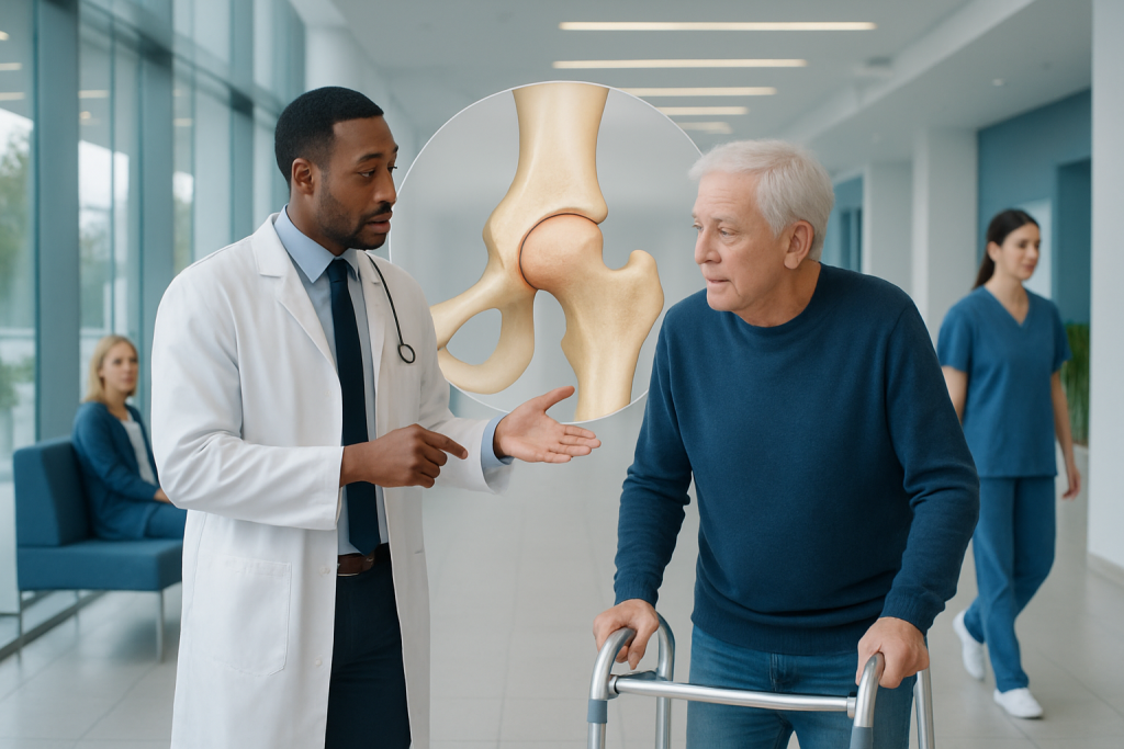
The left femoral neck is part of the upper femur and lies just below the femoral head, or ball of the hip joint. A left femoral neck fracture commonly causes groin or hip pain and difficulty standing or walking after a fall or injury.
Key Takeaways
- The left femoral neck is part of the upper femur and lies just below the femoral head, or ball of the hip joint.
- A left femoral neck fracture commonly causes groin or hip pain and difficulty standing or walking after a fall or injury.
- Some fractures are not clearly visible on an initial X-ray, so CT or MRI may be needed when symptoms strongly suggest a fracture.
- Treatment depends on fracture displacement, age, bone health, mobility, and the person’s overall health.
- Urgent assessment is important after a fall when there is severe hip pain, inability to bear weight, or an outwardly turned or shortened leg.
- Fall prevention, muscle-strengthening activity, adequate nutrition, and bone-health assessment can help reduce fracture risk.
The left femoral neck is the narrow bridge of bone connecting the ball of the left hip joint to the main shaft of the femur. It is important because fractures in this area can affect hip stability and blood supply to the femoral head, particularly after a fall or in people with weakened bones.
Overview: What Is the Left Femoral Neck?
The left femoral neck is a short, angled section of bone at the top of the left femur, or thigh bone. It connects the femoral head—the rounded “ball” that fits into the hip socket—to the longer shaft of the femur. This structure helps transfer body weight from the pelvis to the leg during standing, walking, and climbing stairs.
In medical records, the phrase “left femoral neck” may appear in an imaging report, examination note, or diagnosis. It identifies the location on the left side, rather than a condition by itself. The term may be used when describing normal anatomy, pain around the hip, a bone lesion, or a suspected or confirmed fracture.
The femoral neck has particular clinical importance because blood vessels that support the femoral head run close to it. A significant fracture can disrupt this blood supply, which may affect healing and, in some cases, damage bone tissue in the femoral head. For this reason, suspected femoral neck injuries are assessed carefully and treated promptly.
Why Injuries to This Area Matter
A femoral neck fracture is a break through this narrow part of the upper femur. It is a type of hip fracture, although not every hip problem involves the femoral neck. Fractures may be nondisplaced, meaning the bone pieces remain in their usual alignment, or displaced, meaning the pieces have shifted apart.
Displacement matters because it can make the hip less stable and can increase the likelihood of interrupted blood flow to the femoral head. The risk of healing problems is also influenced by the pattern of the fracture, the time since injury, smoking status, nutrition, existing medical conditions, and the person’s ability to follow rehabilitation guidance.
Femoral neck fractures occur most often in older adults after a low-energy fall, especially when osteoporosis has weakened the bones. In younger people, they are more likely to result from substantial trauma, such as a road traffic collision, sports injury, or fall from height. Less commonly, a weakened bone from another medical condition can fracture with minimal force.
Not all left hip pain is caused by a fracture. Muscle and tendon injuries, arthritis, bursitis, lower back conditions, and problems within the hip joint can produce similar discomfort. A clinician considers the symptoms, physical examination, and appropriate imaging together to identify the cause.
Symptoms and Signs of a Left Femoral Neck Fracture
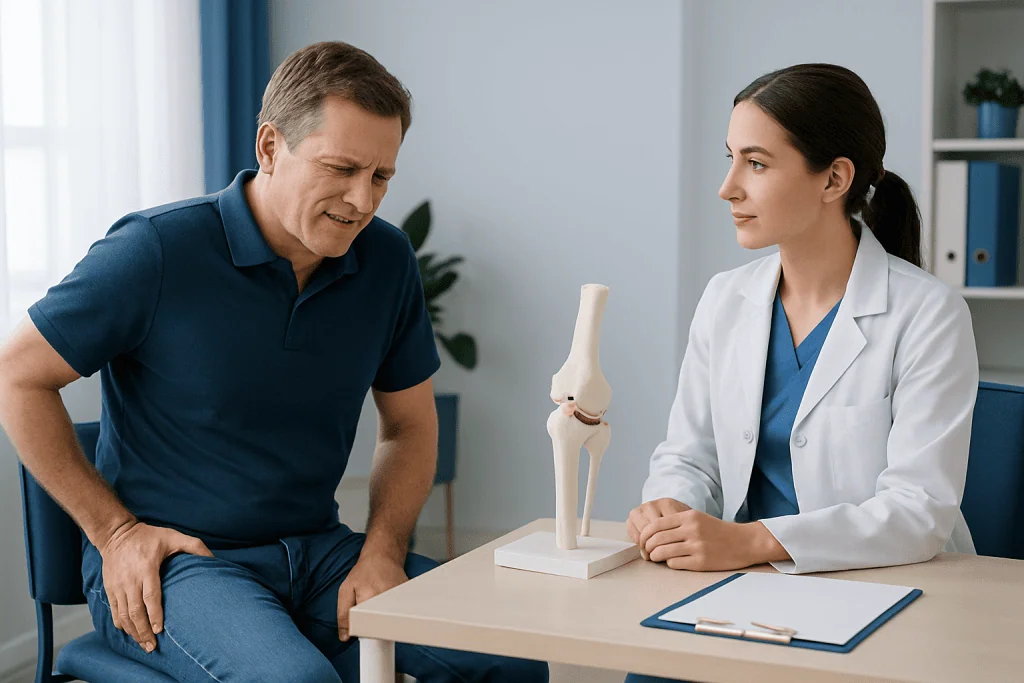
The most typical symptom is sudden pain in the groin, upper thigh, buttock, or outer left hip after a fall or other injury. Pain often becomes worse when trying to stand, move the leg, or place weight through the left side. Many people cannot walk normally after the injury.
With a displaced fracture, the left leg may look shorter than the right or may rest turned outward. Bruising and swelling may occur but are not always prominent. In some cases, especially with a small or nondisplaced fracture, a person may still be able to take a few steps, although this does not rule out a significant injury.
Older adults may not describe pain in a straightforward way. A sudden decline in mobility, refusal to stand, new confusion after a fall, or pain when the hip is gently moved can be important clues. A caregiver should seek medical assessment rather than assuming the symptoms are due to a minor bruise.
Long-standing hip or groin pain without a clear injury can have many causes. Persistent symptoms should be assessed, particularly if they are worsening, interrupt sleep, limit daily activity, or occur alongside fever, unexplained weight loss, or a known history of cancer.
Causes and Factors That Increase Risk
Falls are the leading cause of femoral neck fractures in later life. Balance problems, reduced vision, muscle weakness, sedating medicines, alcohol use, unsafe footwear, and hazards such as loose rugs or poor lighting can all increase the chance of falling. Conditions that affect coordination or sensation may also contribute.
Osteoporosis is a major risk factor because it reduces bone strength. Risk can rise with older age, menopause, a previous fragility fracture, low body weight, smoking, heavy alcohol intake, prolonged immobility, low calcium or vitamin D intake, and long-term use of certain medicines. Some endocrine, digestive, kidney, and inflammatory conditions may also affect bone health.
In physically active younger adults, repetitive impact loading may rarely lead to a femoral neck stress fracture. This may cause gradually increasing groin or hip pain rather than a sudden injury. Continuing high-impact exercise despite pain can allow a stress injury to worsen, so timely assessment is important.
Clinicians also consider whether a fracture could be pathological, meaning it occurred through bone already weakened by a condition such as a tumor, infection, or metabolic bone disease. This is less common, but it changes the investigation and treatment plan.
How the Left Femoral Neck Is Assessed
Assessment begins with a discussion of how symptoms started, whether there was a fall or trauma, current mobility, medicines, and medical history. A clinician examines the hip and leg carefully, checking pain with movement, leg position, circulation, sensation, and the ability to bear weight. They may also assess for injuries elsewhere after a significant accident.
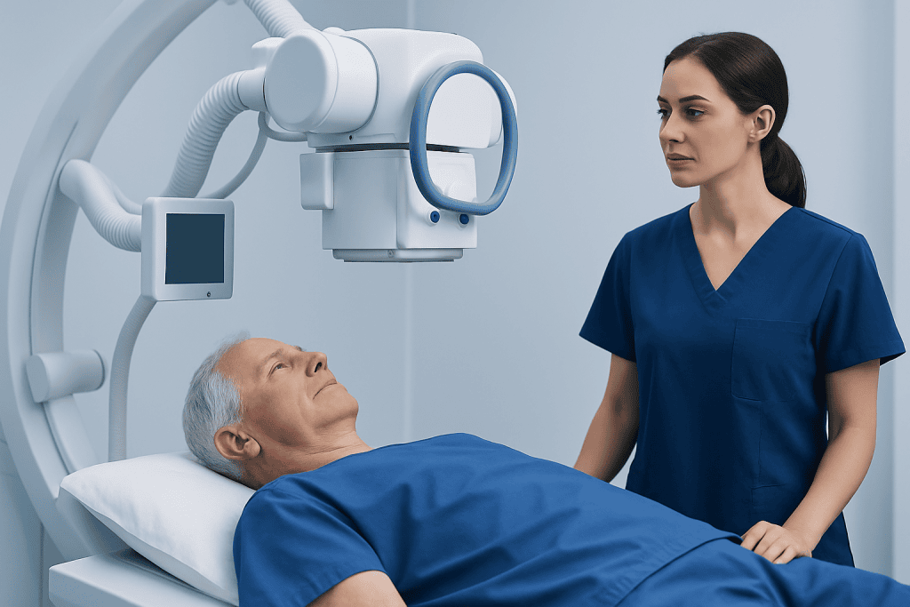
Plain X-rays of the pelvis and hip are usually the first imaging test for a suspected femoral neck fracture. They can often show the fracture location and whether the bone is displaced. However, an early or nondisplaced fracture may not be obvious on an X-ray.
If symptoms and examination findings strongly suggest a fracture but X-rays are normal or uncertain, MRI is often the most sensitive test for an occult, or hidden, fracture. CT may also be used in some circumstances, particularly when MRI is not suitable or when more detail is needed for surgical planning.
Blood tests may be performed before an operation or to assess overall health. After a low-impact fracture, a clinician may also recommend evaluation for osteoporosis and potentially reversible contributors to weak bones, helping guide future fracture prevention.
Treatment and Recovery Options
Treatment is individualized. The main goals are to relieve pain, restore safe mobility, support bone healing when possible, and reduce complications linked to immobility. The care team considers the fracture pattern, whether it is displaced, the person’s age, activity level, bone quality, cognitive status, and other health conditions.
Some nondisplaced femoral neck fractures may be treated with surgical fixation, using screws or other devices to hold the bone in position while it heals. Displaced fractures often need surgery. Depending on the circumstances, this may involve fixation or replacement of part or all of the hip joint, known as hemiarthroplasty or total hip replacement.
Non-surgical treatment is uncommon for most femoral neck fractures because prolonged bed rest can lead to complications and may not provide reliable healing. It may be considered only in carefully selected situations, such as when surgery is not medically appropriate. Decisions should be made with an orthopedic team after discussing likely benefits, limitations, and recovery needs.
Rehabilitation starts as early as safely possible. Pain management, physiotherapy, walking aids, exercises for strength and balance, wound care after surgery, and prevention of blood clots are important parts of recovery. Healing and return to activity vary considerably; following the rehabilitation plan and attending follow-up appointments can support the best possible functional outcome.
Protecting Bone Health and Reducing Future Risk
After a low-impact femoral neck fracture, preventing another fracture is an important part of care. A clinician may assess bone density and review nutrition, medicines, fall history, and medical conditions that may contribute to bone loss. When osteoporosis is diagnosed, treatment may include lifestyle measures and prescription medicines selected for the individual.
Regular weight-bearing and muscle-strengthening activity can help maintain bone and muscle health when it is safe to do so. Balance-focused exercise may reduce fall risk for some people. After a fracture or surgery, activity should progress under guidance from the treating clinician or physiotherapist rather than returning immediately to previous exercise levels.
A balanced diet with adequate protein, calcium, and vitamin D supports general bone health. People should ask their healthcare professional whether supplements are appropriate, particularly if they have kidney disease, take several medicines, or have dietary restrictions. Avoiding tobacco and limiting alcohol can also support bone health and healing.
Practical fall prevention includes improving lighting, removing tripping hazards, using handrails, wearing supportive shoes, having vision checked, and reviewing medicines that may cause dizziness or drowsiness. For people at higher risk, a personalized falls assessment can identify the most useful changes.
When to Seek Medical Care
Urgent medical evaluation is needed after a fall, collision, or other injury if there is severe left hip or groin pain, inability to stand or bear weight, a visibly shortened or outwardly rotated leg, numbness, weakness, or rapidly increasing swelling. The person should avoid walking on the injured leg unless a clinician has said it is safe to do so.
Emergency care is also appropriate if hip pain follows significant trauma, or if pain is accompanied by chest pain, breathing difficulty, fainting, confusion, or signs of shock. It is generally safer not to move a person with a possible hip fracture unless necessary for immediate safety; emergency services can advise on the next steps.
Medical advice should be arranged soon for persistent groin or hip pain after a fall, even if the person can still walk, because some fractures are subtle at first. Gradually worsening pain during running, military training, or another repetitive-impact activity also deserves assessment for a possible stress injury.
Acibadem International’s multidisciplinary specialists and JCI-accredited hospitals assess hip injuries and bone-health concerns for international patients. Evaluation should always be guided by a qualified clinician who can interpret symptoms, imaging findings, and individual health needs.
Frequently asked questions
Is the left femoral neck part of the hip?
Yes. The left femoral neck is the upper portion of the left thigh bone that connects the femoral head to the femoral shaft. It sits within the hip region and helps support movement and weight transfer through the hip joint.
Can a person walk with a left femoral neck fracture?
Some people, particularly those with a nondisplaced or stress fracture, may still be able to walk for a short time. However, walking does not rule out a fracture and may worsen an unstable injury. Hip or groin pain after a fall should be medically assessed promptly.
Will a femoral neck fracture show on an X-ray?
Many femoral neck fractures are visible on standard hip and pelvic X-rays. Small, early, or nondisplaced fractures can sometimes be missed initially. If clinical suspicion remains high, MRI or CT may be used to look for a hidden fracture.
Does a left femoral neck fracture always require surgery?
Most femoral neck fractures are treated surgically because surgery can improve stability and allow earlier mobilization. The exact approach depends on fracture displacement, bone quality, overall health, and personal functional needs. In limited situations, non-surgical management may be considered.
How long does recovery from femoral neck fracture treatment take?
Recovery varies widely based on the type of fracture, treatment received, age, health, and rehabilitation progress. People often begin supported movement soon after surgery, but strength, confidence, and independence can take weeks to months to improve. Regular follow-up helps the care team monitor healing and recovery.
Can osteoporosis cause a femoral neck fracture without a major accident?
Osteoporosis weakens bones, so a fracture may occur after a simple fall from standing height or another minor impact. A fracture after low-energy trauma is a reason to discuss bone-health testing and prevention strategies with a healthcare professional.
References
- American Academy of Orthopaedic Surgeons
- National Institute of Arthritis and Musculoskeletal and Skin Diseases
- National Institute for Health and Care Excellence
- International Osteoporosis Foundation
- American College of Radiology
This article is for general information only and is not a substitute for professional medical advice. Please consult a qualified doctor about your individual situation.
Joint and spine care in Turkey — expert assessment & treatment
JCI-accredited · board-certified surgeons · reply within 24h
Add us as a Preferred Source to see more of our trusted health content across Google Search, AI Overviews and Discover.