

Lung Nodules: When to Worry, What Tests Are Done, and Next Steps

Most lung nodules are benign and may be caused by old infections, inflammation, or scarring. CT scan is the main test used to evaluate and monitor a lung nodule.
Key Takeaways
- Most lung nodules are benign and may be caused by old infections, inflammation, or scarring.
- CT scan is the main test used to evaluate and monitor a lung nodule.
- Doctors assess size, shape, growth, smoking history, age, and other factors to estimate risk.
- Some nodules only need repeat imaging, while others need PET-CT, biopsy, or surgery.
- Any new breathing symptoms, coughing up blood, or unexplained weight loss should be discussed with a doctor promptly.
Lung nodules are small, round or oval spots seen on a chest X-ray or CT scan. Most are not cancer, but the next steps depend on the nodule’s size, appearance, growth, and the person’s risk factors.
Overview
Lung nodules, also called pulmonary nodules, are small spots in the lung seen on imaging tests. They are usually round or oval and are often found by chance when a chest X-ray or CT scan is done for another reason. In many people, a lung nodule causes no symptoms and does not lead to serious illness.
A lung nodule is generally considered small, often less than 3 centimeters across. Larger masses are evaluated differently because they may carry a higher chance of cancer. Even so, size alone does not give the full answer. Doctors also look at the nodule’s edges, density, location, and whether it changes over time.
Most lung nodules are benign. Common non-cancerous causes include healed infections, inflammation, small areas of scarring, and certain autoimmune conditions. A smaller number are related to early lung cancer or cancer that has spread from another part of the body, which is why careful evaluation matters.
Finding a lung nodule can be unsettling, but it does not automatically mean cancer. The goal is to identify which nodules are low risk and can simply be watched, and which need more testing. A structured follow-up plan helps avoid both unnecessary procedures and delayed diagnosis.
Symptoms and Signs
Most lung nodules do not cause symptoms, especially when they are small. They are commonly discovered incidentally during imaging for cough, chest pain, injury, preoperative checks, or screening. Because of this, many people feel completely well when the nodule is found.
When symptoms are present, they are often related to the underlying cause rather than the nodule itself. For example, an infection may cause fever, cough, mucus, or tiredness. Inflammatory conditions may be linked with joint pain, skin changes, or other symptoms elsewhere in the body.
Certain warning signs deserve prompt medical review because they can suggest a more serious condition. These include coughing up blood, ongoing shortness of breath, unexplained weight loss, chest pain, or a cough that does not improve. These symptoms do not always mean cancer, but they should not be ignored.
- Many lung nodules cause no symptoms at all.
- Symptoms may reflect infection or inflammation rather than the nodule itself.
- Red-flag symptoms include hemoptysis, persistent cough, and unexplained weight loss.
Causes and Risk Factors

Lung nodules can form for several reasons. Benign causes are common and include prior fungal or bacterial infections, tuberculosis exposure in some regions, inflamed tissue, and scar tissue left after illness or injury. Non-infectious causes may include rheumatoid arthritis, sarcoidosis, or other inflammatory disorders.
Some nodules are related to tumors. These may be benign growths such as hamartomas, or malignant growths such as early lung cancer. In some cases, a nodule may represent a spread of cancer from another organ. The chance of malignancy depends on a combination of imaging features and personal risk factors, not just the presence of a spot on the scan.
Risk factors that can increase concern include older age, current or past smoking, exposure to radon, asbestos, silica, or air pollution, and a personal or family history of cancer. A weakened immune system can also affect the range of possible causes, including certain infections.
Doctors pay close attention to the nodule’s pattern on CT. Smooth, stable nodules are often lower risk, while spiculated or irregular borders, larger size, upper-lung location, or interval growth may raise suspicion. Still, these features only guide risk assessment; they do not confirm a diagnosis on their own.
What Tests Are Done?
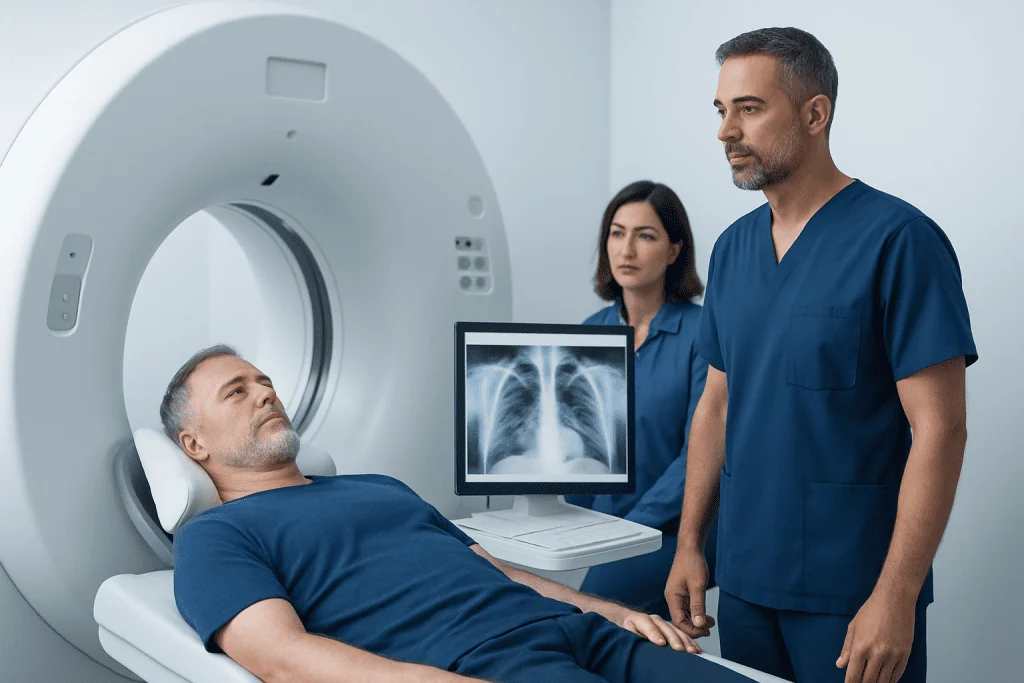
The first step is usually a careful review of the imaging study that found the nodule. A chest X-ray may show a spot, but a CT scan gives far more detail. CT helps measure the nodule precisely and assess its shape, density, and whether there are single or multiple nodules.
Doctors may compare the current scan with older images if available. Stability over two years can be reassuring for many solid nodules, while growth over time may require further evaluation. The doctor will also review symptoms, smoking history, travel history, prior infections, occupational exposures, and any history of cancer.
If the nodule appears intermediate or higher risk, further tests may be recommended. These can include a PET-CT scan to look for metabolic activity, blood tests in selected situations, or pulmonary function tests if a procedure or surgery is being considered. PET-CT is more useful for some nodules than others and may be less informative when nodules are very small.
In certain cases, tissue sampling is needed to make a firm diagnosis. This may be done with bronchoscopy, a needle biopsy through the chest wall, or surgery, depending on the nodule’s location and the person’s overall health. The best approach is individualized to balance accuracy, safety, and the need for timely answers.
How Doctors Decide Whether to Watch or Investigate
After the initial scan, clinicians estimate the likelihood that a lung nodule is benign or malignant. They use clinical judgment, guideline-based risk models, and the imaging pattern. Factors such as nodule size, solidity, calcification, edges, growth rate, and patient risk factors all contribute to the decision.
Low-risk nodules are often managed with surveillance imaging. This means repeat CT scans at set intervals to confirm that the spot stays stable or resolves. Many small nodules, especially in people without major risk factors, never require invasive testing.
Intermediate-risk nodules may need additional testing before a decision is made. PET-CT can help in some cases, and biopsy may be considered if the result will change management. High-risk nodules are more likely to move toward tissue diagnosis or removal, particularly if the imaging strongly suggests an early cancer.
These decisions are often discussed in a multidisciplinary setting involving chest physicians, radiologists, thoracic surgeons, and oncologists. This team approach can be especially helpful when the nodule has mixed features or when the person has other lung conditions such as COPD or emphysema that affect treatment planning.
Treatment Options and Next Steps
Treatment depends on the cause of the nodule. If the nodule is thought to be benign and low risk, no immediate treatment may be needed beyond follow-up imaging. If an infection is suspected, treatment may target that infection, while inflammatory causes may lead to assessment for an underlying immune or systemic condition.
When the nodule is suspicious for cancer, the next steps may include biopsy or surgical removal. A lung biopsy can provide tissue for diagnosis, helping doctors determine whether the nodule is malignant and what type of cells are present. This information is important for choosing the most appropriate care plan.
If cancer is confirmed, treatment may include surgery, radiation therapy, systemic therapy, or a combination of these. Early-stage disease found in a single nodule can sometimes be treated very effectively. In selected patients, thoracic surgeons may discuss lung cancer surgery when removal offers the best chance of cure or control.
Near the end of the diagnostic journey, some patients benefit from care in a specialist center. Acibadem International’s multidisciplinary specialists and JCI-accredited hospitals evaluate and treat lung nodules and related conditions for international patients, coordinating imaging, biopsy, and specialist consultation when needed.
Prevention and Self-care
Not all lung nodules can be prevented, but some steps can reduce the risk of serious lung disease. Avoiding tobacco is one of the most important measures. People who smoke can benefit from discussing smoking cessation support with a healthcare professional, as stopping smoking lowers future cancer and lung disease risk.
Reducing exposure to workplace or environmental inhaled hazards also matters. Depending on the setting, this may mean using proper protective equipment and following safety guidance around dust, chemicals, or asbestos. In some homes and regions, testing for radon may also be appropriate.
General lung health supports overall well-being. Staying up to date with recommended vaccinations, seeking care for persistent respiratory infections, and managing chronic conditions can all help. People who are eligible for lung cancer screening should ask whether regular low-dose CT screening is appropriate, since screening is different from nodule follow-up but may overlap in some individuals.
Self-care after a nodule is found mainly involves keeping follow-up appointments and bringing previous scan reports if possible. It can also help to write down questions before consultations, such as whether the nodule is solid or subsolid, what the estimated risk is, and when the next scan should be done.
When to See a Doctor
A person should see a doctor if a lung nodule has been reported on imaging, even if they feel well. Many nodules are harmless, but the report should be reviewed in context so the right follow-up plan can be made. Delaying review can lead to unnecessary uncertainty or missed surveillance.
Prompt medical attention is especially important if there are symptoms such as coughing up blood, ongoing chest pain, worsening shortness of breath, recurrent fever, or unexplained weight loss. These symptoms can have many causes, but they deserve timely assessment. People with a history of smoking or previous cancer should be especially careful not to skip follow-up imaging.
It is also sensible to seek specialist advice if scan findings are unclear, if the nodule changes over time, or if more than one test has been recommended and the options feel confusing. A clear explanation of risks, benefits, and alternatives can help patients make informed decisions. In most cases, a thoughtful step-by-step approach leads to the safest plan.
Anyone with significant anxiety about a newly discovered nodule should feel comfortable raising these concerns. Reassurance, explanation of the scan findings, and a written follow-up schedule often make the situation much easier to manage.
Frequently asked questions
Are lung nodules usually cancer?
No. Most lung nodules are benign, especially when they are small and found in people with lower risk profiles. Doctors look at the size, appearance, and growth pattern of the nodule, along with personal risk factors, to decide how concerned to be.
What size lung nodule is considered concerning?
In general, larger nodules deserve closer evaluation than smaller ones, but size is only one part of the picture. Border shape, density, growth over time, and a person’s smoking or cancer history are also very important.
How often are follow-up scans needed?
The timing depends on the nodule type, size, and estimated risk. Some nodules need a repeat CT in a few months, while others may only need longer-term observation or no further imaging at all.
Can a lung nodule go away on its own?
Yes, some nodules caused by infection or inflammation may shrink or disappear over time. Others remain stable for years without causing harm, which is why comparison with older scans and scheduled follow-up are so helpful.
Is biopsy always needed for a lung nodule?
No. Many lung nodules can be safely monitored with CT scans and do not require biopsy. Biopsy is usually considered when the nodule has suspicious features or when a tissue diagnosis is needed to guide treatment.
What is the difference between a chest X-ray and CT for lung nodules?
A chest X-ray may detect a spot, but CT provides much more detail. CT can measure the nodule more accurately and show features that help doctors estimate whether it is likely benign or malignant.
References
- American College of Chest Physicians
- Fleischner Society
- National Cancer Institute
- American Thoracic Society
- Radiological Society of North America
This article is for general information only and is not a substitute for professional medical advice. Please consult a qualified doctor about your individual situation.
Oncology care in Turkey — second opinion and treatment plan
JCI-accredited · board-certified surgeons · reply within 24h
Add us as a Preferred Source to see more of our trusted health content across Google Search, AI Overviews and Discover.

More from the Health Library



Related Specialists

Assoc. Prof. Dr. Baran Bozkurt
Neurosurgery
Assoc. Prof. Dr. Muhammet Ahmet Karakaya
Anesthesiology
Dr. Nevzat Demirhan
Radiology
Dr. Mehmet Yakupçebioğlu
Physical Medicine & Rehabilitation