Lymphangioma: Diagnosis, Outlook, and Modern Treatment Approaches

Lymphangioma is benign, but it can still cause symptoms depending on where it develops. Many lymphangiomas are diagnosed with physical examination and imaging such as ultrasound or MRI.
Key Takeaways
- Lymphangioma is benign, but it can still cause symptoms depending on where it develops.
- Many lymphangiomas are diagnosed with physical examination and imaging such as ultrasound or MRI.
- Treatment may include monitoring, sclerotherapy, surgery, or a combination of approaches.
- The best treatment plan depends on whether the lesion affects breathing, swallowing, appearance, comfort, or normal function.
- Prompt medical review is important if a lump grows quickly, becomes painful, or causes airway or feeding problems.
Lymphangioma is a non-cancerous malformation of the lymphatic system, most often present at birth or found in early childhood. Outlook is usually good, and treatment depends on the lesion’s size, location, symptoms, and effect on nearby structures.
Overview: What lymphangioma is
Lymphangioma is a benign growth-like lesion caused by abnormal development of lymphatic vessels. In current medical practice, many specialists describe it as a type of lymphatic malformation rather than a true tumor. It is made up of fluid-filled spaces or channels that form when part of the lymphatic system does not develop in the usual way.
Lymphangiomas are most often present at birth, although some are noticed later in infancy, childhood, or less commonly in adulthood. They can occur almost anywhere in the body, but the head and neck are among the most common locations. Some remain small and cause few problems, while others enlarge over time or press on nearby tissues.
A well-known subtype is cystic hygroma, which usually refers to a larger, cystic lymphatic malformation often seen in the neck. The words are sometimes used interchangeably in everyday discussions, but doctors may classify these lesions by their appearance as macrocystic, microcystic, or mixed. This helps guide treatment planning and expectations.
How lymphangioma can appear and feel
The way a lymphangioma looks depends on its size, depth, and location. A superficial lesion may appear as a soft swelling under the skin, while a deeper one may only be noticed after imaging. Some lesions are compressible and may feel spongy, while others are made up of many tiny channels and feel more firm or uneven.
Color can vary. Some lesions are skin-colored, while others may look slightly bluish if they are close to the surface. In the mouth or tongue, small lymphatic vesicles can create a clustered, pebbled appearance. A lesion may stay stable for long periods, then enlarge during infection, inflammation, or bleeding into the malformation.
Common symptoms can include:
- A painless soft lump or swelling
- Visible asymmetry of the face, neck, limb, or other area
- Discomfort, tightness, or pressure
- Repeated inflammation or infection in the affected area
- Bleeding or oozing from superficial lesions
- Problems with swallowing, speech, or breathing if the lesion is in the head and neck
Not every lymphangioma causes symptoms right away. Sometimes it is discovered incidentally during an examination for another concern, especially when it is small or deep.
Causes, types, and risk factors

Lymphangioma develops because part of the lymphatic system forms abnormally before birth. The lymphatic system helps manage tissue fluid and supports immune function. When lymphatic channels do not connect or drain normally, fluid can collect and form cysts or malformed spaces.
In most cases, there is no action during pregnancy or childhood that causes the condition. It is generally considered congenital, meaning it begins during fetal development. Some lesions become more noticeable only later, especially if they slowly enlarge or are triggered by infection, trauma, or hormonal changes.
Doctors often group lymphatic malformations into three broad patterns:
- Macrocystic: larger fluid-filled spaces, often easier to drain or treat with sclerotherapy
- Microcystic: many very small channels or cysts, sometimes involving skin or mucous membranes
- Mixed: features of both types
Some lymphatic malformations can occur alongside other congenital conditions or vascular anomalies. Because these disorders can overlap, doctors may consider related entities such as hemangioma or other vascular lesions when forming a diagnosis, although they are not the same condition.
How lymphangioma is diagnosed
Diagnosis usually begins with a medical history and careful physical examination. The doctor will ask when the swelling was first noticed, whether it changes in size, and whether it causes pain, infection, feeding difficulty, breathing changes, or other functional problems. In children, the timing of growth and any birth history can also be important.
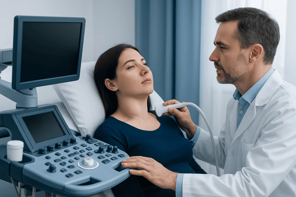
Imaging helps confirm the diagnosis and map the lesion’s full extent. Ultrasound is often a useful first test, especially for a superficial neck mass in a child. MRI is commonly used when a more detailed view is needed because it shows how the malformation relates to muscles, airways, nerves, and other nearby structures. In selected situations, CT may be used, but MRI is often preferred for soft tissue detail.
A biopsy is not always necessary. Many lymphangiomas have a typical clinical and imaging appearance. However, if the diagnosis is uncertain, or if another mass needs to be ruled out, the care team may recommend further evaluation. In complex cases, specialists in pediatrics, radiology, surgery, ENT, dermatology, and interventional treatments may work together to decide the best plan.
Because lesions in the mouth, throat, or neck can affect the airway or swallowing, evaluation may include additional examinations in these areas. This is one reason timely assessment matters, even though the condition itself is benign.
Modern treatment approaches
Treatment for lymphangioma is individualized. Small, stable lesions that are not causing symptoms may simply be monitored over time. When treatment is needed, the main goals are to reduce symptoms, protect function, lower the risk of complications, and improve appearance where possible. A single treatment may be enough for some people, while others benefit from staged or combined therapy.
Sclerotherapy is one of the most commonly used modern options, especially for macrocystic lesions. In this minimally invasive approach, a specialist places a needle into the cystic spaces, drains fluid when appropriate, and injects a medicine that irritates the lining so the spaces shrink over time. This may be especially helpful for lesions in the neck or other soft tissues and is part of broader interventional radiology care.
Surgery may be recommended when the lesion is well-defined, causes functional problems, recurs after other treatment, or can be removed safely with a reasonable expectation of benefit. Complete removal is not always possible, particularly when the malformation extends around nerves, blood vessels, or other important structures. Even after surgery, some lesions can recur if microscopic abnormal channels remain.
Other supportive or targeted treatments may be considered in selected cases, especially for microcystic or mixed lesions. These can include laser treatment for surface involvement, medicines to reduce inflammation, treatment of infection, and planning with pediatric surgery or ENT care when the airway, tongue, or swallowing are affected. If a lesion is part of a broader vascular anomaly picture, specialists may also evaluate related conditions such as vascular malformation during treatment planning.
Outlook, follow-up, and daily self-care
The outlook for lymphangioma is often very good, especially when the lesion is diagnosed clearly and managed by an experienced team. Because it is benign, the main concerns are usually local effects rather than spread to other parts of the body. Still, the course can vary. Some lesions remain stable, some shrink after treatment, and some need more than one intervention over time.
Follow-up is important because lymphatic malformations can change with growth, infection, trauma, or hormonal shifts. Recurrence or regrowth can happen, particularly after incomplete removal or in complex microcystic disease. Regular review helps the care team decide whether observation remains appropriate or whether another treatment step would be helpful.
At home, practical self-care measures may include protecting the area from repeated trauma, watching for sudden swelling, and seeking treatment early for skin breakdown or signs of infection. Families of children with lymphangioma may also benefit from guidance on feeding, oral care, and how to monitor breathing if the lesion involves the head and neck.
Near the end of the treatment journey, many people want reassurance about long-term care. Multidisciplinary follow-up can be especially useful for lesions affecting appearance, speech, swallowing, or mobility. Acibadem International’s multidisciplinary specialists and JCI-accredited hospitals diagnose and treat lymphatic malformations for international patients when coordinated expert care is needed.
When to seek medical care
Medical review is advisable for any new or persistent soft-tissue lump, especially in a baby or child. Although lymphangioma is benign, a proper diagnosis is important because other neck, skin, or soft-tissue masses can look similar. Early evaluation is also helpful when a lesion is near the airway, mouth, eye, or major nerves.
More urgent care is needed if swelling grows quickly, becomes red or painful, or is associated with fever, bleeding, trouble swallowing, noisy breathing, or shortness of breath. These changes can suggest infection, bleeding into the lesion, or pressure on nearby structures. In infants and young children, feeding difficulty or sleep-related breathing changes should also be assessed promptly.
People should also contact a doctor if a previously diagnosed lymphangioma starts changing in size or function, or if treatment side effects are concerning. Clear follow-up with a qualified specialist can help prevent avoidable complications and support the best possible outcome.
Frequently asked questions
Is lymphangioma cancer?
No. Lymphangioma is considered a benign lymphatic malformation, not a cancer. Even so, it can still need treatment if it causes swelling, discomfort, cosmetic concerns, or pressure on nearby structures.
Can adults get lymphangioma?
Yes, although most lymphangiomas are present at birth or found in childhood. Some are diagnosed later in life because they were small, deep, or only became noticeable after enlargement or inflammation.
Does every lymphangioma need surgery?
No. Some lesions can be monitored if they are small and not causing symptoms. Others respond well to sclerotherapy or a combination of treatments, so surgery is only one of several possible options.
Can lymphangioma come back after treatment?
It can. Recurrence is more likely when the malformation is extensive or cannot be fully removed or fully treated in one session. Regular follow-up helps detect changes early and guide additional care if needed.
What is the difference between lymphangioma and cystic hygroma?
Cystic hygroma is generally used to describe a cystic type of lymphangioma, often in the neck. In modern practice, both are commonly understood within the broader category of lymphatic malformations.
How is lymphangioma confirmed?
Doctors usually confirm it with a physical examination and imaging, most often ultrasound and MRI. These tests help show the size, depth, and relationship to nearby tissues, which also supports treatment planning.
References
- National Organization for Rare Disorders
- American Academy of Pediatrics
- Society for Vascular Surgery
- Radiological Society of North America
- MedlinePlus
This article is for general information only and is not a substitute for professional medical advice. Please consult a qualified doctor about your individual situation.
Oncology care in Turkey — second opinion and treatment plan
JCI-accredited · board-certified surgeons · reply within 24h
Add us as a Preferred Source to see more of our trusted health content across Google Search, AI Overviews and Discover.