Lymphoscintigraphy Procedure: An Evidence-Based Patient Guide
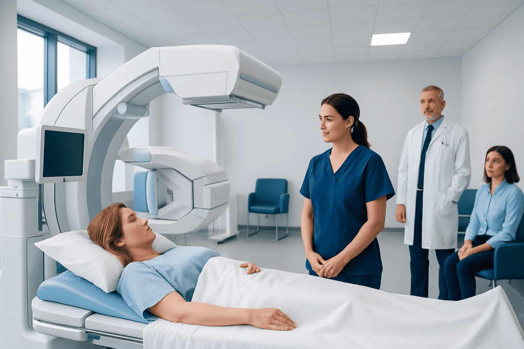
Lymphoscintigraphy uses a very small amount of radioactive tracer and a gamma camera to map lymphatic pathways. It is commonly performed before sentinel lymph node biopsy in melanoma and some breast cancers.
Key Takeaways
- Lymphoscintigraphy uses a very small amount of radioactive tracer and a gamma camera to map lymphatic pathways.
- It is commonly performed before sentinel lymph node biopsy in melanoma and some breast cancers.
- The injections can cause brief discomfort, but the scan itself is generally painless and does not require surgery.
- Timing varies by reason for testing, but imaging and preparation commonly take several hours.
- Patients should tell the care team about pregnancy, breastfeeding, allergies, medicines, and prior surgery near the test area.
A lymphoscintigraphy procedure is a specialized nuclear medicine scan that shows how lymphatic fluid travels from a specific area of the body to nearby lymph nodes. It is most often used to identify sentinel lymph nodes before surgery for certain cancers, and may also help assess lymphatic drainage problems.
What Is a Lymphoscintigraphy Procedure?
A lymphoscintigraphy procedure is an imaging examination performed in a nuclear medicine department. A clinician injects a tiny amount of radiotracer into or near the area being evaluated. The tracer enters lymphatic vessels, and a special gamma camera detects its movement to create images of lymphatic drainage and lymph nodes.
The examination is especially important for sentinel lymph node mapping. A sentinel node is the first lymph node, or group of nodes, most likely to receive lymph drainage from a tumor area. Identifying these nodes can help surgeons plan a targeted lymph node biopsy rather than removing many nodes routinely.
Lymphoscintigraphy may also be used when clinicians need to investigate suspected lymphatic obstruction, leakage, or abnormal drainage. The exact imaging method, injection location, and timing depend on why the test has been requested.
How It Works and Who May Need It
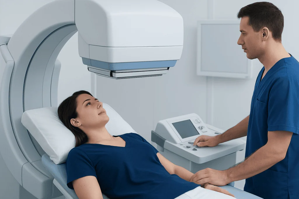
The lymphatic system is a network of vessels and lymph nodes that helps return fluid to the bloodstream and supports immune function. After injection, the radiotracer travels along lymphatic channels in the same general direction as lymph fluid. Images reveal where the tracer travels, whether it reaches expected nodes, and which nodes may be relevant for surgery.
Sentinel lymph node mapping is commonly considered for people having surgery for melanoma or selected breast cancers. In these settings, the result helps guide sentinel lymph node biopsy and may support more individualized surgical planning. Whether it is appropriate depends on the cancer type, location, stage, previous treatment, and the planned operation.
For lymphatic drainage concerns, the scan may be considered in people with unexplained limb swelling, suspected lymphedema, recurrent fluid collections, or possible lymphatic leakage. Other tests may be needed as well, because symptoms such as swelling can have many causes, including vein, heart, kidney, or medication-related conditions.
How Do I Prepare for Lymphoscintigraphy?

Preparation is usually straightforward, but patients should follow the instructions provided by their nuclear medicine team because requirements differ by examination. Many people can eat, drink, and take usual medicines normally. However, when the scan is coordinated with surgery, the surgical team may provide separate instructions about fasting, medicines, and arrival time.
Before the appointment, patients should tell the team if they are pregnant, might be pregnant, are breastfeeding, have had prior surgery or radiation in the area, or have experienced reactions during previous imaging procedures. They should also provide a current medication list and report any infection, wound, or significant skin problem near the planned injection site.
Comfortable clothing is useful, and jewelry or metal items near the area being imaged may need to be removed. Patients should bring relevant medical records if requested. For scans of a limb, the team may ask the person to avoid restrictive clothing or compression garments until staff advise otherwise.
What Happens During the Procedure?
The process usually begins with a short review of the medical history and an explanation of the scan. A nuclear medicine physician or trained clinician administers the radiotracer by small injections. For cancer-related sentinel node mapping, injections are commonly placed in the skin around the tumor site, biopsy scar, breast, or another location chosen by the clinical team.
After the injection, imaging may start immediately or after a short waiting period. The patient lies still while a gamma camera is positioned close to the body. The camera does not press tightly in the way some other imaging tests do, and it does not produce radiation; it records signals emitted by the tracer already in the body.
Images may be taken at several time points to follow lymphatic flow. The specialist may mark the skin over an identified sentinel node to help the surgeon locate it later. In some surgical pathways, an additional tracer or dye may be used in the operating room; this is separate from the imaging examination and is explained by the surgical team.
For people being assessed for limb swelling, the team may take images before and after gentle movement or exercise, depending on local protocol. Patients should remain in the department until staff confirm that the required images have been completed.
How Painful Is Lymphoscintigraphy?
Lymphoscintigraphy is generally well tolerated. The part most likely to cause discomfort is the radiotracer injection, which may feel like a brief sting, pressure, or burning sensation. Injections into sensitive areas, including the breast or skin near a surgical site, can be more uncomfortable for some people, but the discomfort is usually temporary.
The imaging itself is painless. The main challenge may be staying in one position while images are obtained. Patients who have pain, limited mobility, anxiety in medical settings, or difficulty lying flat should mention this in advance so the team can discuss positioning and comfort measures.
Patients should not hesitate to tell staff if they feel faint, unusually uncomfortable, or unwell during the appointment. The nuclear medicine team can pause, adjust positioning, and provide appropriate support.
How Long Does a Lymphoscintigraphy Test Typically Take?
The total appointment commonly takes a few hours, although the exact duration varies considerably. The injection itself is brief, while the time needed for tracer movement and imaging depends on the body area and the clinical question. Some people have images soon after injection, followed by additional images later the same day.
When lymphoscintigraphy is performed before cancer surgery, it may be scheduled on the day of surgery or the day before. The nuclear medicine and surgical teams coordinate the timing so the sentinel node information can be used during the planned operation.
Tests for suspected lymphatic drainage disorders can take longer because delayed images may be needed to observe how the tracer moves over time. The department can provide a more individualized schedule before the visit, including whether the patient should plan for breaks between image sets.
Results, Benefits, Recovery and Possible Risks
A nuclear medicine physician interprets the images and sends a report to the referring clinician or surgeon. The report may describe lymphatic channels, the location of sentinel nodes, delayed drainage, or other findings. Results are interpreted alongside the physical examination, pathology, surgical plan, and other imaging; a scan alone does not diagnose cancer spread or explain every cause of swelling.
The principal benefit is precise mapping of lymphatic drainage. For appropriate cancer surgery, this can support a focused approach to node sampling and help avoid unnecessarily extensive lymph node surgery. In lymphatic disorders, it can provide information that helps clinicians decide whether further assessment, conservative care, or a specialist referral is appropriate.
Most people return to routine activities immediately after the scan unless they are also having surgery or have been given other instructions. The radiotracer amount is small and typically leaves the body naturally, mainly through urine. Drinking fluids and urinating regularly after the test may help clear it, if medically appropriate for the individual.
Risks are low but include temporary soreness, bruising, or mild irritation at injection sites. Allergic reactions are uncommon. The radiation exposure is generally low and is justified only when the expected diagnostic or surgical-planning benefit is meaningful. Pregnant or breastfeeding patients need individualized advice before proceeding.
When to Seek Medical Care
Patients should contact the nuclear medicine department or their clinician if they develop persistent redness, increasing swelling, severe pain, fever, rash, trouble breathing, or feel unwell after the procedure. These symptoms are uncommon and may not necessarily be caused by the scan, but they should be assessed promptly.
Independent of lymphoscintigraphy, sudden swelling of one limb, chest pain, shortness of breath, or coughing blood requires urgent medical assessment because these symptoms can have serious causes. Persistent or progressive swelling, recurrent skin infections, unexplained lumps, or changes near a known cancer site should also be discussed with a qualified doctor.
For international patients, Acibadem International’s multidisciplinary specialists and JCI-accredited hospitals can coordinate nuclear medicine imaging, surgery, oncology, and follow-up when lymphoscintigraphy is part of an individualized care plan.
Frequently asked questions
Is lymphoscintigraphy the same as a CT scan or MRI?
No. Lymphoscintigraphy is a nuclear medicine scan that tracks a radiotracer through lymphatic vessels and nodes. CT and MRI use different technologies and provide different types of anatomical information; they may sometimes be used alongside lymphoscintigraphy.
What kind of doctor does a lymphoscintigraphy?
A nuclear medicine physician typically oversees and interprets lymphoscintigraphy. Nuclear medicine technologists perform much of the imaging, while radiologists, surgeons, oncologists, or other referring specialists use the results in care planning.
Can I drive after a lymphoscintigraphy procedure?
Most people can drive and return to normal daily activities after the scan because it does not usually involve sedation. If the appointment is followed by surgery, sedation, or another procedure, the surgical team may give different instructions.
How much radiation is used in lymphoscintigraphy?
The test uses a small amount of radiotracer designed for diagnostic imaging. The nuclear medicine team selects the smallest appropriate amount and recommends the examination only when the expected clinical value outweighs the limited radiation exposure.
Will lymphoscintigraphy show whether cancer has spread?
The scan identifies lymphatic drainage pathways and sentinel lymph nodes, but it does not by itself prove whether cancer is present in a node. If needed, a surgeon removes the mapped sentinel node for laboratory examination by a pathologist.
Can lymphoscintigraphy diagnose lymphedema?
It can provide useful evidence about impaired lymphatic drainage and may support evaluation for lymphedema. Diagnosis also relies on symptoms, examination findings, medical history, and sometimes additional tests to rule out other causes of swelling.
References
- Society of Nuclear Medicine and Molecular Imaging
- American College of Radiology
- National Cancer Institute
- American Cancer Society
- European Association of Nuclear Medicine
This article is for general information only and is not a substitute for professional medical advice. Please consult a qualified doctor about your individual situation.
Explore treatments in Turkey — costs, top hospitals & a free quote
JCI-accredited · board-certified surgeons · reply within 24h
Add us as a Preferred Source to see more of our trusted health content across Google Search, AI Overviews and Discover.