Mammo Screening W Tomo Bilateral: How It Works, Results and What to Expect
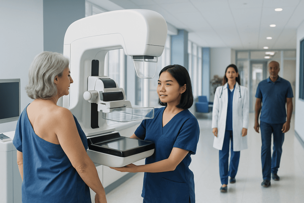
A bilateral mammogram examines both breasts; tomosynthesis adds detailed 3D-style image slices. The examination usually takes about 15 to 30 minutes, including positioning and image checks.
Key Takeaways
- A bilateral mammogram examines both breasts; tomosynthesis adds detailed 3D-style image slices.
- The examination usually takes about 15 to 30 minutes, including positioning and image checks.
- Breast compression is brief and necessary to produce clear images while keeping radiation exposure low.
- Tomosynthesis may be especially useful when breast tissue is dense, but the best screening test depends on individual risk and local recommendations.
- An abnormal screening result does not by itself mean cancer; additional imaging is commonly used to clarify a finding.
Mammo screening w tomo bilateral means a screening mammogram of both breasts that includes digital breast tomosynthesis, often called 3D mammography. It combines standard mammogram views with thin, layered images that can help radiologists assess breast tissue more clearly.
Mammo Screening W Tomo Bilateral: Overview
A mammo screening w tomo bilateral is a routine breast imaging examination that evaluates both breasts using mammography and tomosynthesis. Tomosynthesis, also called 3D mammography, takes a series of low-dose X-ray images from different angles. A computer then reconstructs them into thin image layers, allowing the radiologist to look through breast tissue in greater detail.
The purpose is to find breast changes before they cause symptoms. These changes may include small masses, distortions in breast architecture, or calcifications. Screening is performed in people who do not have a new breast concern; a diagnostic mammogram is used when there is a symptom, such as a lump, nipple discharge, or a change in breast skin.
“Bilateral” simply means that both the left and right breast are imaged. Most screening mammograms are bilateral because comparing both sides and comparing current images with earlier mammograms can help the radiologist interpret normal tissue patterns and identify new changes.
How It Works and What Images Show
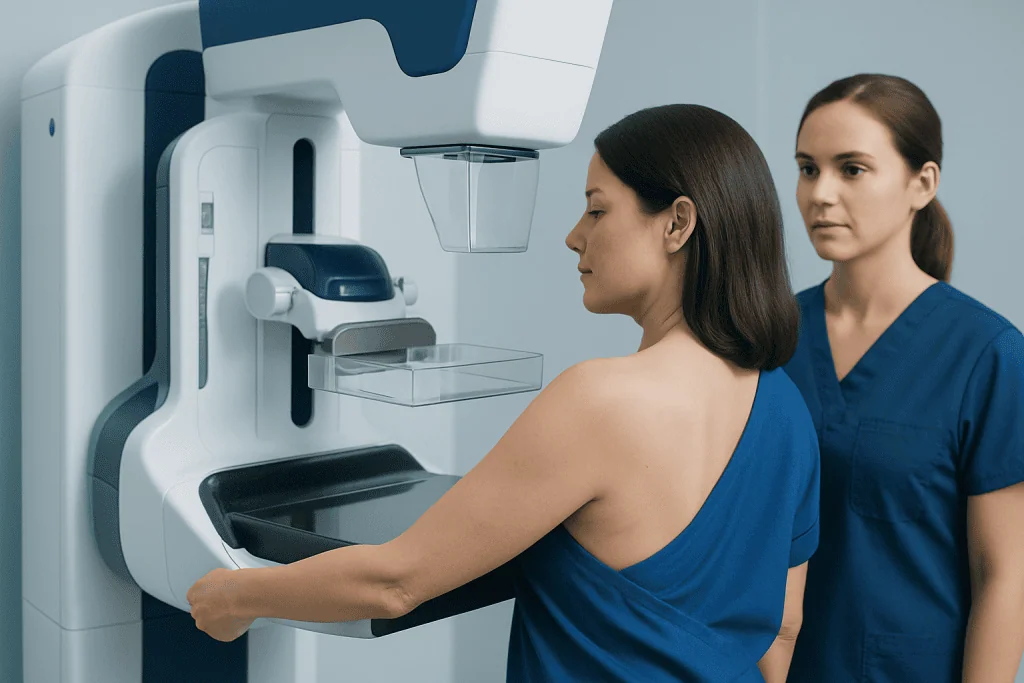
During standard digital mammography, each breast is usually photographed in two positions. One view is taken from above, while the other is taken from the side at an angle. With tomosynthesis, the X-ray tube moves in a small arc over the compressed breast and captures multiple images during the same positioning.
The radiologist reviews the reconstructed slices alongside the mammogram images. Looking through tissue layer by layer can reduce the effect of overlapping normal tissue, which may otherwise hide a finding or create an appearance that needs further assessment. Tomosynthesis does not replace clinical awareness: a new lump or other breast symptom should be assessed even if a recent screening examination was normal.
The radiation dose from modern mammography is low. Depending on the system and technique used, tomosynthesis may be performed with synthesized 2D images or with standard 2D images as well. Imaging teams use established quality and radiation-safety standards to obtain useful images with the lowest reasonable exposure.
Who May Benefit From Screening With Tomosynthesis
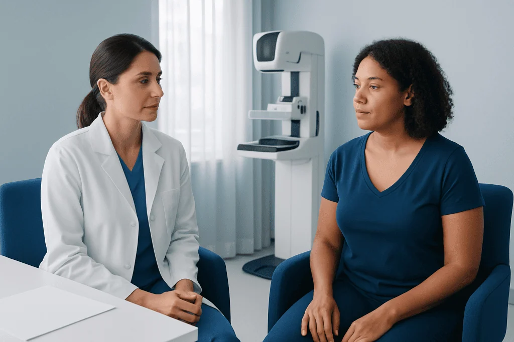
Screening recommendations depend on age, personal history, family history, breast density, genetic factors, prior chest radiation, and the guidance followed in a person’s country or healthcare system. Many people at average risk are offered regular mammography from midlife, while those at higher risk may need screening earlier or with additional imaging. A clinician can help create an individualized plan.
Tomosynthesis can be considered for people having routine mammography, including those with dense breasts. Dense breast tissue is common and is not a disease. It can make mammogram interpretation more difficult because both dense tissue and some abnormalities appear light on X-ray images. Breast density also has implications for risk assessment, which should be discussed with a doctor or breast imaging specialist.
People with a prior history of breast cancer, a known genetic mutation associated with higher breast cancer risk, or strong family history may need a tailored surveillance program. This may involve mammography, breast MRI, ultrasound, or other tests at specific intervals. The choice is based on risk and clinical circumstances rather than density alone.
What Happens During the Procedure
Before the appointment, the imaging center may ask about prior breast surgery, implants, pregnancies, breastfeeding, hormone use, family history, and previous breast imaging. Bringing prior mammogram images or ensuring they are available to the new center is valuable because comparison can prevent unnecessary callbacks and improve interpretation.
On the day, the person undresses from the waist up and stands at the mammography unit. A technologist positions one breast on a detector plate and gently compresses it with a second plate for a few seconds while images are taken. The process is then repeated in the required views for the other breast.
It is usually helpful to avoid deodorant, powder, lotion, or perfume on the underarms or breasts immediately before the examination, as some products can create image artifacts. The technologist should be told about pain, limited shoulder movement, breast implants, pregnancy, or possible pregnancy so positioning and imaging can be planned appropriately.
- Check-in and preparation are followed by a brief review of relevant history.
- Each breast is positioned and compressed for several short image acquisitions.
- The technologist checks image quality before the appointment ends.
- A radiologist interprets the images and sends the result to the referring clinician or directly to the patient according to local practice.
How Long Does a Mammogram With Tomo Take?
A mammogram with tomosynthesis commonly takes about 15 to 30 minutes from start to finish. The actual X-ray exposure lasts only seconds for each view; most of the appointment involves careful positioning, compression, and confirming that the images are clear enough to interpret.
The appointment can take longer if a person has implants, mobility limitations, breast tenderness, prior surgery, or needs extra views. A screening examination may also lead to a separate diagnostic appointment if the radiologist needs more images to clarify an area. This is a common part of breast screening and does not automatically indicate cancer.
After the exam, most people can return to normal activities immediately. There is no recovery period, anesthesia, injection, or driving restriction. Mild, short-lived breast tenderness from compression can occur, particularly if the breasts are already sensitive.
What Does Bilateral Mammogram With Tomo Mean?
A bilateral mammogram with tomo means that both breasts are examined with mammography that includes tomosynthesis. “Bilateral” refers to the two breasts, and “tomo” is shorthand for tomosynthesis, the technology that creates multiple thin image slices from a series of low-dose X-ray images.
This wording is generally used for screening when no specific new concern has been identified. If a patient has a symptom or an area that requires a closer look, the order may instead specify diagnostic mammography, targeted ultrasound, or additional specialized views. The terminology on an imaging request can vary by institution, but the radiology report should explain what was performed and the next recommended step, if any.
Results are often reported using the Breast Imaging Reporting and Data System, known as BI-RADS. A negative or benign result generally supports returning to routine screening. A result needing further evaluation means more imaging is recommended to reach a clearer conclusion; it is not a cancer diagnosis.
Should I Get a Tomosynthesis Instead of Just a Mammogram?
Tomosynthesis is usually performed as part of a mammogram rather than as a completely separate alternative. Compared with 2D mammography alone, it provides additional layered images that may improve the assessment of overlapping breast tissue and may reduce the chance that a person is called back for extra images in some screening settings.
However, the best screening approach is individual. Availability, image quality, breast density, previous imaging findings, personal risk, and national screening guidance all matter. For some people at substantially increased risk, mammography with tomosynthesis may be combined with breast MRI or another test rather than used alone.
A doctor or breast radiologist can explain whether tomosynthesis is appropriate within a personalized breast screening plan. It remains important to attend recommended screening even when a person feels well, while also reporting any new breast symptom promptly rather than waiting for the next routine mammogram.
Results, Benefits, Limitations and Possible Risks
The main benefit of mammography with tomosynthesis is earlier identification of some breast abnormalities, often before they can be felt. Layered imaging can make it easier to distinguish a true finding from normal tissue overlap. It may be particularly helpful for evaluating complex tissue patterns, although no imaging test detects every breast cancer.
Limitations are important to understand. Mammograms can miss cancers, especially in dense breasts, and they can identify findings that turn out to be benign. This can lead to additional mammogram views, ultrasound, MRI, or occasionally a biopsy. These follow-up steps are designed to clarify the result and are a standard part of careful breast assessment.
Risks include temporary discomfort from compression and low exposure to ionizing radiation. The benefit of appropriate screening generally outweighs this small exposure for people who meet screening recommendations. Anyone who is pregnant, could be pregnant, or is breastfeeding should inform the imaging team so they can advise on the most appropriate timing and technique.
If further assessment is needed, imaging may include a breast cancer evaluation and treatment pathway where appropriate. If cancer is diagnosed, care planning typically involves a multidisciplinary team and may include surgery, systemic therapy, and radiation treatment based on the specific diagnosis.
Can a Radiologist Tell if It Is Breast Cancer on Ultrasound?
Ultrasound can provide valuable information about a breast finding, but it usually cannot confirm breast cancer by appearance alone. It can help distinguish fluid-filled cysts from solid areas, assess the shape and margins of a lesion, evaluate a palpable area, and guide a needle biopsy when tissue sampling is needed.
A radiologist may identify ultrasound features that are more or less suspicious and recommend the appropriate next step. When imaging findings are concerning, a biopsy is generally needed to determine whether cancer is present and, if so, to identify the type and biological features. Imaging and pathology are interpreted together for an accurate diagnosis.
Ultrasound is not routinely a replacement for screening mammography for most average-risk individuals. It is commonly used as an additional test after a mammogram or to assess a symptom. For people with dense breasts or elevated risk, the role of ultrasound should be decided with a qualified clinician as part of an overall screening strategy.
When to Seek Medical Care
A person should arrange medical assessment promptly for a new breast lump, persistent focal breast pain, nipple discharge that is bloody or occurs without squeezing, nipple inversion that is new, breast skin dimpling, redness that does not settle, swelling, or a change in breast size or shape. Most breast changes are not cancer, but they deserve professional evaluation.
It is also important to contact a clinician after receiving a screening result that recommends additional imaging or consultation. Timely follow-up helps clarify the finding and supports appropriate next steps. People should not rely on a normal prior mammogram to dismiss a new or changing symptom.
Acibadem International’s multidisciplinary specialists and JCI-accredited hospitals support international patients who need breast imaging assessment, diagnosis, and treatment planning. A breast specialist can explain screening results in the context of personal risk, symptoms, and prior imaging.
Frequently asked questions
Is mammo screening w tomo bilateral painful?
Compression can feel uncomfortable or briefly painful for some people, but it lasts only a few seconds for each image. The pressure is needed to spread breast tissue, reduce movement, and produce clear images. Telling the technologist about discomfort can help them adjust positioning when possible.
Do I need to prepare for a bilateral tomosynthesis mammogram?
Most people do not need special preparation. It is generally advisable to avoid deodorant, powder, creams, or perfume on the breasts and underarms on the day because these can interfere with the images. Prior mammograms should be available for comparison whenever possible.
What happens if a 3D mammogram shows something abnormal?
The radiologist may recommend additional mammogram views, targeted breast ultrasound, or sometimes MRI. Many recalled findings are ultimately benign or due to normal overlapping tissue. If imaging remains suspicious, a biopsy may be recommended to establish a diagnosis.
Can I have a mammogram with tomosynthesis if I have breast implants?
Yes, people with implants can usually have mammography and tomosynthesis. The technologist uses specialized positioning and may take extra views to visualize as much breast tissue as possible. The imaging center should be informed about implants when the appointment is booked.
Is tomosynthesis appropriate for dense breasts?
Tomosynthesis may be useful for dense breasts because it provides layered images that can reduce the effect of tissue overlap. It does not eliminate the limitations of mammography in dense tissue or replace individualized risk assessment. A clinician can discuss whether supplemental imaging is appropriate.
How quickly are bilateral mammogram with tomo results available?
Timing varies by imaging center and whether prior images need to be reviewed. Some facilities provide results quickly, while others send a report within several days. If further imaging is recommended, the center or referring clinician should explain how and when to arrange it.
References
- World Health Organization
- American Cancer Society
- U.S. Food and Drug Administration
- American College of Radiology
- National Cancer Institute
This article is for general information only and is not a substitute for professional medical advice. Please consult a qualified doctor about your individual situation.
Explore treatments in Turkey — costs, top hospitals & a free quote
JCI-accredited · board-certified surgeons · reply within 24h
Add us as a Preferred Source to see more of our trusted health content across Google Search, AI Overviews and Discover.