

Mediastinal Lymph Nodes: What Patients Need to Know

Mediastinal lymph nodes are normal immune structures in the central chest. Enlargement does not automatically mean cancer; infections and inflammatory conditions are also common causes.
Key Takeaways
- Mediastinal lymph nodes are normal immune structures in the central chest.
- Enlargement does not automatically mean cancer; infections and inflammatory conditions are also common causes.
- Imaging tests such as chest CT often detect enlarged mediastinal lymph nodes, but imaging alone may not identify the exact cause.
- Doctors may recommend follow-up scans, blood tests, bronchoscopy, or biopsy depending on the size, location, and clinical context.
- Persistent symptoms such as cough, chest pain, fever, weight loss, or shortness of breath should be medically evaluated.
Mediastinal lymph nodes are lymph nodes located in the center of the chest, near the lungs, heart, and airways. They are part of the immune system, and while they may enlarge during infection or inflammation, they can also be affected by other conditions, so the cause usually needs careful medical assessment.
Overview: what mediastinal lymph nodes are
Mediastinal lymph nodes are small, bean-shaped immune organs located in the mediastinum, the area in the center of the chest between the lungs. This space contains important structures such as the heart, major blood vessels, windpipe, esophagus, and nearby tissues. Like lymph nodes elsewhere in the body, mediastinal lymph nodes help filter lymphatic fluid and support the body’s defense against infection and disease.
Patients often hear about mediastinal lymph nodes after a chest X-ray or CT scan mentions that they are “prominent,” “enlarged,” or show “mediastinal lymphadenopathy.” This finding is descriptive rather than diagnostic. In other words, it tells the doctor that the lymph nodes look larger than expected, but it does not by itself explain why.
In many cases, enlarged mediastinal lymph nodes are related to a temporary immune response, such as a recent respiratory infection. In other situations, they may be associated with inflammatory conditions, chronic lung disease, or cancers involving the lungs, blood, or lymphatic system. Because the possible causes vary widely, the next step is usually to interpret the imaging result alongside symptoms, medical history, and other test findings.
Why these lymph nodes may become enlarged
Mediastinal lymph nodes can enlarge when immune cells inside them become more active. This often happens when the body is responding to infection, irritation, or inflammation in nearby areas such as the lungs or airways. For example, pneumonia, viral chest infections, tuberculosis, and some fungal infections may lead to enlarged chest lymph nodes.
Noninfectious inflammatory conditions can also affect the mediastinum. Sarcoidosis is one well-known example, and autoimmune or chronic inflammatory diseases may also cause lymph node enlargement. In some people, the change is discovered incidentally during imaging done for another reason and may not be linked to a serious illness.
Doctors also consider cancers in the differential diagnosis. These include lung cancer, lymphoma, leukemia, and cancers that have spread from another part of the body. The overall concern depends on factors such as the patient’s age, smoking history, symptoms, immune status, the appearance of the nodes on imaging, and whether there are related findings in the lungs or elsewhere. If needed, evaluation for conditions such as lung cancer or lymphoma may become part of the workup.
Symptoms and what patients may notice

Mediastinal lymph nodes themselves do not always cause symptoms, especially when they are only mildly enlarged. In many people, they are found incidentally on imaging performed for another reason, such as evaluating a cough, chest discomfort, trauma, or preoperative assessment.
When symptoms are present, they are usually related either to the underlying condition or to pressure on nearby structures. Possible symptoms may include cough, shortness of breath, chest pain or tightness, wheezing, fever, fatigue, night sweats, or unintentional weight loss. Hoarseness or difficulty swallowing can occur in some cases if enlarged nodes affect nearby nerves or the esophagus.
It is important to remember that these symptoms are not specific to mediastinal lymph nodes alone. They can also occur with common infections, asthma, inflammatory lung disease, or malignancy. This is why doctors look at the whole clinical picture rather than relying on one symptom or one scan result.
- Often no symptoms are present
- Cough or persistent respiratory symptoms may prompt testing
- Fever or night sweats may suggest infection or inflammation
- Weight loss or ongoing fatigue may require more detailed evaluation
How doctors investigate mediastinal lymph nodes
Evaluation usually begins with a detailed medical history and physical examination. A doctor may ask about recent respiratory infections, smoking, exposure to tuberculosis, travel history, occupational exposures, autoimmune symptoms, and personal or family history of cancer. Blood tests may be ordered to look for signs of infection, inflammation, or blood disorders.
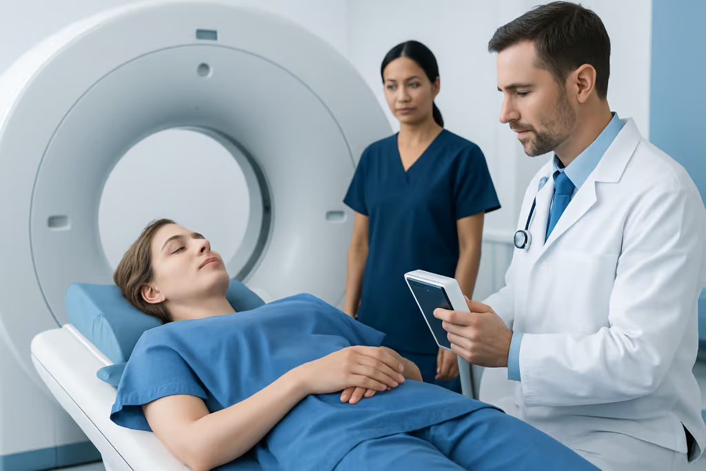
Imaging plays a central role. A chest CT scan gives a more detailed view than a plain chest X-ray and helps assess the size, number, and location of mediastinal lymph nodes. It can also show whether there are associated lung nodules, masses, scarring, or fluid around the lungs. In selected cases, PET-CT may be used to assess metabolic activity, although increased uptake can occur with both cancer and inflammation.
If imaging and clinical findings do not clearly explain the enlargement, tissue sampling may be recommended. This can often be done with bronchoscopy combined with endobronchial ultrasound-guided needle biopsy, which allows specialists to reach certain chest lymph nodes through the airways. In other cases, procedures such as mediastinoscopy or image-guided biopsy may be needed to obtain a sample for laboratory analysis.
The aim of biopsy is not simply to confirm that a node is enlarged, but to identify the exact cause. Pathology testing may show infection, granulomatous inflammation, lymphoma, metastatic cancer, or benign reactive changes. This information guides the next treatment decisions.
Treatment depends on the cause
There is no single treatment for enlarged mediastinal lymph nodes because the nodes are usually a sign of an underlying process rather than a disease on their own. If the cause is a bacterial infection, the doctor may recommend appropriate antimicrobial treatment. If a viral illness is responsible, supportive care and monitoring may be sufficient. Inflammatory conditions may require referral to a specialist and disease-specific medication.
When cancer is diagnosed, treatment planning depends on the type and stage of the disease. For example, enlarged mediastinal lymph nodes can influence staging and treatment options in lung cancer and lymphoma. Care may involve medical oncology, pulmonology, thoracic surgery, radiation oncology, pathology, and radiology working together.
Some patients only need observation. If the lymph nodes are slightly enlarged and the person has a recent infection or no concerning symptoms, a doctor may suggest repeat imaging after a period of time to see whether the nodes return to normal. This approach helps avoid unnecessary procedures while still monitoring for important change.
For patients who require advanced chest evaluation, multidisciplinary teams may also use procedures such as thoracic surgery when biopsy or treatment cannot be performed with less invasive methods. The right approach is individualized and should be based on a specialist’s assessment.
What the scan report may mean for patients
Reading a radiology report can be stressful, especially when terms such as “adenopathy,” “mass,” or “suspicious” appear without explanation. Patients should know that radiology language is designed to describe images carefully, not to provide a final diagnosis. A report noting mediastinal lymph nodes or mediastinal lymphadenopathy is one part of the overall evaluation.
Size matters, but context matters just as much. Some nodes are only mildly prominent and may reflect a recent illness. Others may have features that prompt closer follow-up, such as marked enlargement, clustering, asymmetry, or associated abnormalities in the lungs. A doctor interprets these details together with symptoms and test results before deciding whether the finding is likely benign, needs short-term repeat imaging, or requires biopsy.
Patients can help by asking practical questions: What did the scan show? How large are the nodes? Are they likely related to infection, inflammation, or something else? Is repeat imaging enough, or is tissue sampling advised? Clear communication often reduces anxiety and helps patients understand why a specific next step is recommended.
When to seek medical care
Medical care is advisable if a scan shows enlarged mediastinal lymph nodes and the finding has not yet been explained. Prompt assessment is especially important if there are symptoms such as persistent cough, chest pain, worsening shortness of breath, fever that does not improve, coughing up blood, significant fatigue, or unintentional weight loss.
People with a history of smoking, prior cancer, immune suppression, tuberculosis exposure, or ongoing inflammatory disease should also follow up carefully with a qualified doctor. Even when the cause turns out to be benign, evaluation helps make sure that a treatable problem is not missed.
Emergency care may be needed for severe breathing difficulty, new confusion, bluish lips, or other signs of urgent respiratory distress. For planned evaluation and treatment, centers with pulmonology, radiology, pathology, oncology, and thoracic surgery expertise can coordinate the workup efficiently. Acibadem International’s multidisciplinary specialists and JCI-accredited hospitals diagnose and treat chest conditions for international patients.
Frequently asked questions
Are mediastinal lymph nodes normal?
Yes. Mediastinal lymph nodes are a normal part of the immune system and are naturally present in the central chest. Concern usually arises only when they appear enlarged or abnormal on imaging.
Do enlarged mediastinal lymph nodes always mean cancer?
No. Many cases are caused by infection, inflammation, or a temporary immune response. Cancer is one possible cause, but imaging findings need to be interpreted together with symptoms, history, and sometimes biopsy results.
Can a chest infection cause enlarged mediastinal lymph nodes?
Yes. Respiratory infections such as pneumonia and some viral, bacterial, fungal, or tuberculosis-related illnesses can make mediastinal lymph nodes enlarge. In some cases, the nodes become smaller again after the infection improves.
How are mediastinal lymph nodes checked?
Doctors usually begin with imaging, especially a chest CT scan, and may also order blood tests. If the cause remains unclear or there are suspicious features, bronchoscopy with needle biopsy or another sampling procedure may be recommended.
Can enlarged mediastinal lymph nodes cause symptoms?
They can, but not always. Some people have no symptoms at all, while others may develop cough, shortness of breath, chest discomfort, fever, night sweats, or weight loss, usually because of the underlying condition.
Do mediastinal lymph nodes need treatment on their own?
Usually not. Treatment is aimed at the underlying cause rather than the lymph nodes themselves. Depending on the diagnosis, this may involve monitoring, infection treatment, anti-inflammatory therapy, or cancer care.
References
- American Cancer Society
- American Thoracic Society
- National Cancer Institute
- Centers for Disease Control and Prevention
- Radiological Society of North America
This article is for general information only and is not a substitute for professional medical advice. Please consult a qualified doctor about your individual situation.
Explore treatments in Turkey — costs, top hospitals & a free quote
JCI-accredited · board-certified surgeons · reply within 24h

More from the Health Library



Related Specialists

Prof. Dr. Samet Yardımcı
General Surgery
Dr. Demet Matben
Pediatrics
Asst. Prof. Dr. Tarkan İkizoğlu
Pediatrics
Dr. Iretiola Albert
Inpatient Clinic Physicians Clinical Service