Melanoma: Changing Skin Spots, Biopsy, and Treatment Choices

Melanoma may appear as a new spot or a changing mole, and the ABCDE rule can help identify warning signs. A skin biopsy is the only way to confirm melanoma and provides information needed for staging and treatment planning.
Key Takeaways
- Melanoma may appear as a new spot or a changing mole, and the ABCDE rule can help identify warning signs.
- A skin biopsy is the only way to confirm melanoma and provides information needed for staging and treatment planning.
- Surgery is the main treatment for early melanoma, while advanced melanoma may be treated with immunotherapy, targeted therapy, radiation, or a combination.
- Risk factors include ultraviolet exposure, sunburn history, many or unusual moles, fair skin, family history, and weakened immunity.
- Regular skin self-checks and dermatologist visits are important after diagnosis because melanoma can recur or a new melanoma can develop.
Melanoma is a type of skin cancer that can often be treated successfully when found early. Recognizing changing skin spots, having a timely biopsy, and understanding treatment choices help patients take informed next steps with their medical team.
Overview
Melanoma is a cancer that begins in melanocytes, the pigment-producing cells that give skin its color. It most often develops on the skin, but it can also occur in less common sites such as the eye, under a nail, or on mucosal surfaces. Compared with some other skin cancers, melanoma can be more likely to spread if it is not detected and treated early.
Many melanomas are first noticed as a new spot on the skin or a change in an existing mole. The change may involve size, shape, color, border, or sensation. Some melanomas are dark brown or black, while others may be pink, red, skin-colored, or have several colors within one lesion.
Early evaluation is important because treatment decisions depend strongly on how deep the melanoma has grown and whether it has spread. A dermatologist or qualified clinician can examine the skin, perform dermoscopy when appropriate, and arrange a biopsy if a spot looks suspicious.
Symptoms and Warning Signs of Melanoma
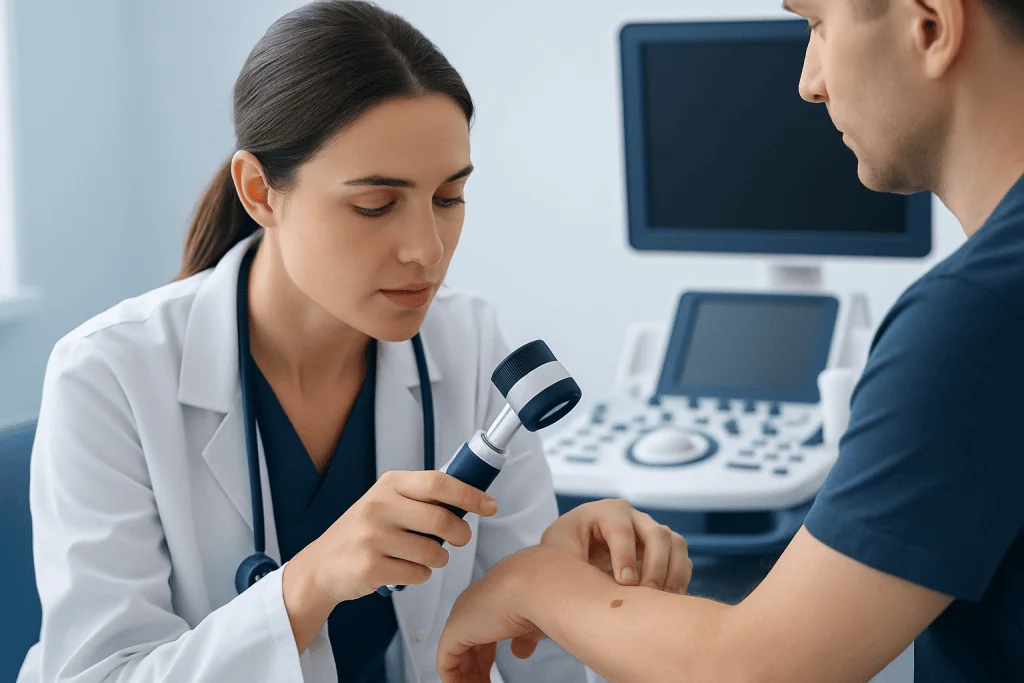
The most useful home guide for recognizing possible melanoma is the ABCDE rule. A stands for asymmetry, B for irregular border, C for color variation, D for diameter or darkening, and E for evolution. Evolution, meaning any noticeable change over time, is often the most important warning sign.
Melanoma does not always follow a classic pattern. A spot may itch, bleed, crust, become tender, or fail to heal. It may look different from the person’s other moles, sometimes called the “ugly duckling” sign. In people with darker skin tones, melanoma may be more likely to appear on the palms, soles, under the nails, or in other less sun-exposed areas, so full skin checks are important for everyone.
- A new mole or spot that appears in adulthood
- A mole that changes in size, shape, height, or color
- A lesion with multiple colors, such as brown, black, red, white, or blue
- A spot with an uneven, notched, or blurred border
- A sore that bleeds, crusts, or does not heal
- A dark streak under a fingernail or toenail not explained by injury
Causes and Risk Factors
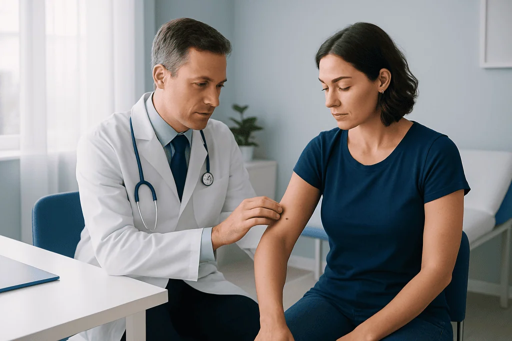
Melanoma develops when DNA changes occur in melanocytes, allowing cells to grow in an uncontrolled way. Ultraviolet radiation from sunlight and tanning devices is a major preventable contributor, especially when exposure causes sunburn. However, melanoma can also occur in areas with limited sun exposure, which shows that genetics and other biological factors can also play a role.
Some people have a higher lifetime risk than others. Risk is increased by a history of blistering sunburns, frequent tanning bed use, fair skin that burns easily, light hair or eyes, many moles, atypical moles, a personal or family history of melanoma, and a weakened immune system. Age can increase risk, but melanoma can occur in younger adults as well.
Having one or more risk factors does not mean a person will develop melanoma, and people without obvious risk factors can still be affected. The practical message is that everyone benefits from sun protection, skin awareness, and timely assessment of changing lesions.
Diagnosis and Biopsy
Diagnosis begins with a careful skin examination. The clinician may ask when the spot appeared, how it has changed, whether it bleeds or itches, and whether the patient has a personal or family history of skin cancer. Dermoscopy, a magnified light-based examination, can help trained clinicians see patterns that are not visible to the naked eye.
If melanoma is suspected, a biopsy is needed to confirm the diagnosis. Whenever possible, the entire suspicious lesion is removed with a small margin of normal-looking skin, a method often called excisional biopsy. In some locations, such as the face, fingers, toes, or very large lesions, a partial biopsy may be chosen to obtain a safe and representative sample.
The pathology report provides key information, including melanoma type, Breslow thickness, ulceration, mitotic activity, margin status, and sometimes genetic features. If the melanoma is deeper or has other higher-risk features, the team may discuss sentinel lymph node biopsy to check whether microscopic cancer cells have reached nearby lymph nodes. Imaging tests are usually reserved for situations where there is concern for spread or for staging more advanced disease.
Staging and What It Means for Treatment
Staging describes how far melanoma has grown or spread. The earliest melanomas are limited to the top layer of the skin, while thicker melanomas may extend deeper. More advanced stages may involve nearby lymph nodes or distant organs. Staging helps doctors estimate risk and choose treatment, but each person’s plan also depends on overall health, melanoma location, pathology details, and patient preferences.
Patients may hear terms such as in situ, localized, regional, or metastatic melanoma. In situ melanoma means the abnormal cells remain in the outermost skin layer. Localized melanoma is invasive but has not been found in lymph nodes or distant sites. Regional melanoma involves nearby lymph nodes or nearby skin or tissue. Metastatic melanoma has spread to distant organs or distant skin sites.
Understanding the stage can reduce uncertainty and help patients ask focused questions. It is reasonable to ask the care team which stage is suspected or confirmed, what tests are needed, whether lymph node evaluation is recommended, and how the results will influence treatment choices.
Treatment Options
Surgery is the main treatment for most early melanomas. After diagnosis, a wider surgical removal is usually performed to remove the melanoma site with a safety margin of normal tissue. The recommended margin depends on the melanoma thickness and location. If a sentinel lymph node biopsy is advised, it is often done around the same time as the wider excision.
For melanoma with a higher risk of recurrence, additional treatment may be recommended after surgery. This is called adjuvant therapy. Depending on the melanoma features, options may include immunotherapy, which helps the immune system recognize and attack cancer cells, or targeted therapy for melanomas with specific gene changes such as BRAF mutations. The choice depends on test results, stage, side effect profile, and the patient’s overall condition.
For advanced or metastatic melanoma, treatment has changed significantly in recent years. Immunotherapy and targeted therapy are commonly used, and some patients may also receive radiation therapy, surgery for selected sites, or participation in a clinical trial. Treatment is personalized, and the team monitors response with physical examinations, imaging when needed, and laboratory tests.
Side effects vary by treatment. Immunotherapy can sometimes cause inflammation in organs such as the skin, bowel, liver, lungs, or hormone glands, while targeted therapies can cause fever, rash, fatigue, or other effects. Patients should report new symptoms promptly, because many side effects are more manageable when addressed early.
Prevention, Self-Care and Follow-Up
Not all melanomas can be prevented, but reducing ultraviolet exposure lowers risk. Sun protection includes seeking shade, wearing protective clothing and a wide-brimmed hat, using broad-spectrum sunscreen, and avoiding tanning beds. Sunscreen should be used as one part of a broader strategy, not as permission to stay in intense sun for long periods.
Skin self-examination helps people notice changes between medical visits. A practical approach is to check the whole skin surface in good light, including the scalp, back, buttocks, soles, between toes, and under nails. A mirror, phone photos, or help from a partner can make it easier to track moles over time.
After melanoma treatment, follow-up schedules depend on stage and recurrence risk. Visits may include skin and lymph node examinations, education about self-checks, and imaging for selected patients. People who have had melanoma have a higher risk of another melanoma, so ongoing dermatology follow-up is an important part of long-term care.
When to See a Doctor
A changing skin spot should be assessed by a qualified doctor, especially if it meets any ABCDE warning sign or looks noticeably different from other moles. Medical evaluation is also important for a sore that does not heal, a lesion that bleeds without clear injury, or a new dark streak under a nail. It is better to have a harmless spot checked than to delay assessment of a melanoma.
People with many atypical moles, a previous melanoma, a strong family history, or a weakened immune system should ask a dermatologist how often professional skin examinations are recommended. Anyone diagnosed with melanoma should feel comfortable asking for a clear explanation of the pathology report, stage, treatment options, expected benefits, possible side effects, and follow-up plan.
For international patients, Acibadem International’s multidisciplinary specialists and JCI-accredited hospitals provide diagnostic evaluation and treatment planning for melanoma and other cancers. As with any cancer diagnosis, decisions should be made with qualified clinicians who can review the individual case in detail.
Frequently asked questions
What does melanoma usually look like?
Melanoma may look like a new or changing mole, an irregular dark spot, or a lesion with more than one color. Some melanomas are pink, red, or skin-colored rather than black. Any spot that changes, bleeds, itches, or looks different from the person’s other moles should be checked.
Is every changing mole melanoma?
No. Many moles change for benign reasons, and some growths that look concerning are not cancer. However, because melanoma is best treated early, a changing mole should be evaluated by a qualified clinician rather than monitored indefinitely at home.
Does a biopsy make melanoma spread?
No. A properly performed skin biopsy does not make melanoma spread. Biopsy is the standard and necessary way to confirm the diagnosis and gather information needed for safe treatment planning.
What happens if melanoma is found early?
Early melanoma is often treated with surgery to remove the lesion and a margin of normal tissue. Depending on the depth and other pathology features, no further treatment may be needed beyond follow-up, or additional lymph node evaluation may be discussed. The doctor will tailor recommendations to the individual pathology report.
When are immunotherapy or targeted therapy used?
Immunotherapy or targeted therapy may be considered for some higher-risk melanomas after surgery or for melanoma that has spread. Targeted therapy usually depends on whether the tumor has a specific gene change, such as a BRAF mutation. These treatments require careful discussion of benefits, risks, monitoring, and possible side effects.
How often should someone check their skin after melanoma?
The follow-up schedule depends on the melanoma stage, personal risk factors, and the treating doctor’s recommendation. Many patients are advised to perform regular self-checks and attend periodic dermatologist visits. Any new or changing spot should be reported without waiting for the next scheduled appointment.
References
- World Health Organization
- American Academy of Dermatology Association
- National Cancer Institute
- European Society for Medical Oncology
- American Cancer Society
This article is for general information only and is not a substitute for professional medical advice. Please consult a qualified doctor about your individual situation.
Melanoma in Turkey — costs, top hospitals & a free quote
JCI-accredited · board-certified surgeons · reply within 24h
Add us as a Preferred Source to see more of our trusted health content across Google Search, AI Overviews and Discover.

Medical Oncology Department
Medical treatment of cancer with chemotherapy, immunotherapy and targeted therapies under a multidisciplinary tumor board.
60 specialists in this unit