Misdiagnosed Clubfoot Ultrasound: Preparation, Procedure and Results

Prenatal ultrasound can identify a possible clubfoot, but it cannot always distinguish a fixed deformity from temporary fetal positioning. A targeted ultrasound and review by maternal-fetal medicine and pediatric orthopedic specialists can clarify an uncertain result.
Key Takeaways
- Prenatal ultrasound can identify a possible clubfoot, but it cannot always distinguish a fixed deformity from temporary fetal positioning.
- A targeted ultrasound and review by maternal-fetal medicine and pediatric orthopedic specialists can clarify an uncertain result.
- Clubfoot is primarily diagnosed after birth through a careful physical examination; imaging is not routinely needed for every baby.
- When true clubfoot is confirmed, early treatment—often the Ponseti method—usually begins in the first weeks of life.
- Parents should seek prompt clinical advice if a prenatal scan suggests clubfoot or a newborn’s foot looks stiff, unusually turned, or difficult to place flat.
A misdiagnosed clubfoot ultrasound is possible because a baby’s position in the womb can make a normal, flexible foot appear turned inward on a scan. A prenatal ultrasound finding should be treated as an important reason for specialist review, while the diagnosis is usually confirmed by a physical examination after birth.
Overview: What a Misdiagnosed Clubfoot Ultrasound Means
A misdiagnosed clubfoot ultrasound usually means that a prenatal scan suggested clubfoot, but the baby was later found not to have a true fixed foot deformity. This can happen when the foot is temporarily held in an inward or downward position in the uterus. Ultrasound is valuable for identifying a possible concern, but it is not the final diagnosis.
Clubfoot, also called congenital talipes equinovarus, is a condition present at birth in which the foot points downward and inward and is difficult to move into a typical position. A true clubfoot is not simply a foot that looks turned on one ultrasound image; it has characteristic stiffness and alignment findings that are best assessed after birth by an experienced clinician.
A possible prenatal finding gives families time to ask questions, arrange appropriate follow-up, and meet a pediatric orthopedic specialist if needed. It does not mean that a baby will necessarily have clubfoot, another health condition, or long-term difficulty walking. If clubfoot is confirmed, early care is highly effective for most children.
How an Ultrasound Suggests Clubfoot
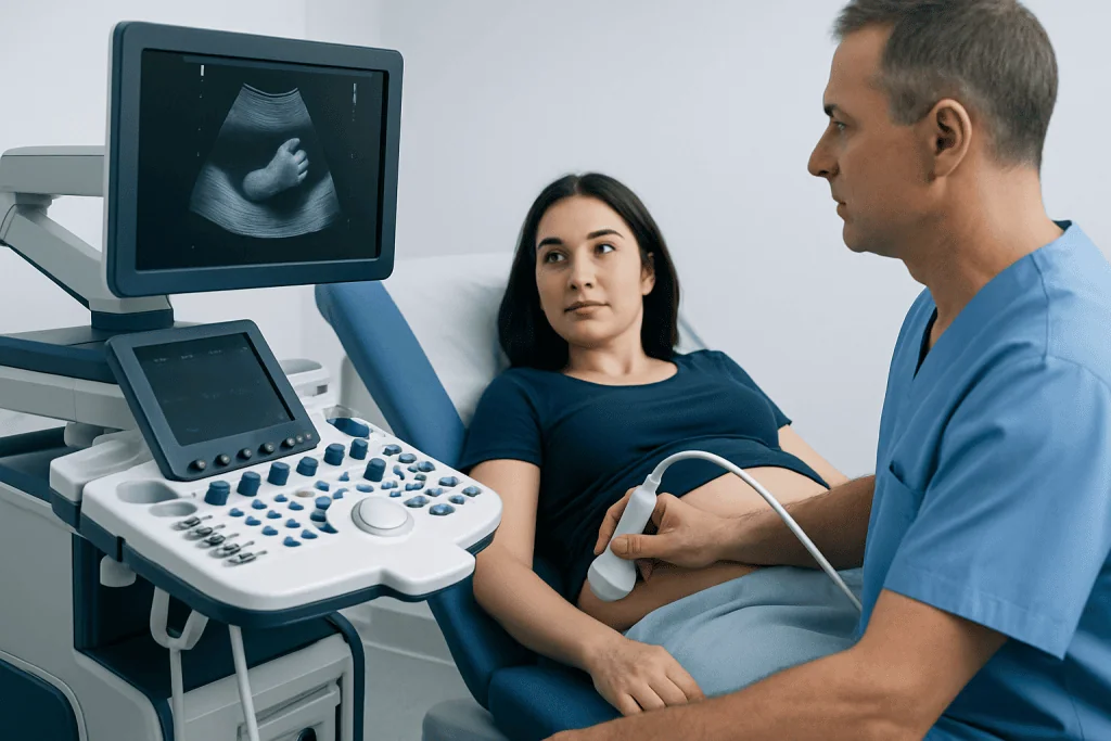
During a routine pregnancy ultrasound, the sonographer views the fetus from different angles and checks development of the limbs, organs, placenta, and amniotic fluid. Clubfoot may be suspected when the sole of the foot appears to face inward rather than toward the lower leg. The finding may involve one foot or both feet.
The quality and certainty of the assessment can be affected by fetal movement, the baby’s posture, gestational age, the amount of amniotic fluid, the position of the placenta, and how clearly the feet can be seen. A foot pressed against the uterine wall or held in a flexed position may look similar to clubfoot in a single view.
When a routine scan raises concern, a clinician may recommend a targeted ultrasound with fetal medicine expertise. This is not painful and does not use radiation. It allows a more detailed look at both feet and at other areas of fetal development that may help place the finding in context.
- Preparation: Most people do not need special preparation. They should follow the imaging center’s instructions about eating, drinking, and arriving for the appointment.
- During the scan: Gel is applied to the abdomen, and a transducer is moved gently across the skin to create images using sound waves.
- Afterward: There is no physical recovery period. The results may be discussed at the visit or reviewed at a follow-up appointment, depending on local practice.
Can an ultrasound be wrong about clubfoot?
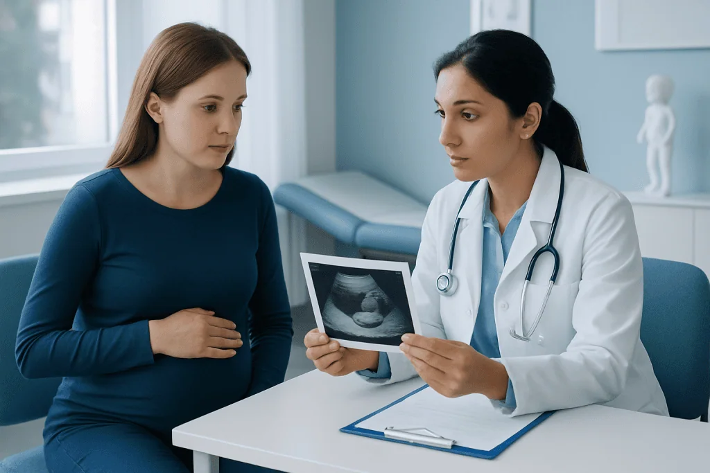
Yes. An ultrasound can be wrong about clubfoot, especially when the baby’s feet cannot be visualized clearly or when a temporary position resembles the typical appearance of clubfoot. This is often described as a false-positive result: the scan raises concern, but the baby does not have a fixed clubfoot after birth.
Ultrasound may also underestimate or miss a foot difference when views are limited, so it should be understood as a screening and assessment tool rather than a stand-alone confirmation. A repeat scan can sometimes provide more information because the fetus may be in a different position at a later visit.
The most useful next step is usually a review of the images and a targeted assessment by a maternal-fetal medicine specialist. If the finding remains likely, consultation with a pediatric orthopedic team before delivery can help families understand what examination and treatment may involve after birth.
A suspected clubfoot should not be interpreted as a parent-caused problem. In most cases, there is no action during pregnancy that can change fetal foot positioning, and the goal is careful assessment and planning rather than blame.
How common is it for an ultrasound to be wrong?
It is not possible to give one reliable percentage for how often an ultrasound is wrong about clubfoot because accuracy varies between studies and depends on the stage of pregnancy, the imaging equipment, fetal position, the experience of the examiner, and whether one or both feet are involved. False-positive prenatal findings are recognized in clinical practice, particularly when the suspected deformity is mild or only one foot is affected.
For this reason, clinicians usually explain that a prenatal ultrasound can identify a possible clubfoot rather than establish the diagnosis with certainty. The chance that the finding represents true clubfoot generally becomes clearer when the foot is assessed in multiple views and when the rest of the fetal anatomy is evaluated.
A detailed follow-up assessment can also identify whether the foot appears isolated or whether there are other findings that deserve further discussion. Most suspected clubfoot findings are isolated, but the care team may recommend genetic counseling or additional testing when ultrasound shows other structural differences, growth concerns, or a relevant family history.
Families may find it helpful to ask what was seen, whether both feet were visualized in more than one position, how confident the imaging specialist is, and what follow-up is recommended. Clear questions can turn an uncertain result into a practical care plan.
What diagnostic procedures should be performed to confirm the diagnosis of clubfoot?
Clubfoot is usually confirmed after birth through a physical examination by a pediatrician and, when appropriate, a pediatric orthopedic specialist. The clinician checks the position of the foot, ankle and heel, whether the foot can be gently moved toward a typical alignment, and whether the calf and lower leg appear affected. A true clubfoot is characteristically stiff and not fully correctable with simple positioning.
Before birth, a targeted fetal ultrasound is the main assessment used to investigate a suspected clubfoot. The specialist reviews the feet in different planes and examines the rest of the fetal anatomy. A fetal echocardiogram, genetic counseling, diagnostic genetic testing, or other assessment may be offered only when there are additional findings or particular risk factors; these tests are not automatically required for every isolated suspected clubfoot.
After birth, X-rays are not routinely needed to diagnose a typical clubfoot because the physical examination is usually more informative in a newborn. Imaging may be considered when the foot appearance is unusual, the diagnosis is uncertain, treatment is not progressing as expected, or the clinician suspects another bone, joint, or neurologic condition.
If clubfoot is confirmed, referral for Ponseti method treatment should be arranged promptly. This structured approach uses gentle manipulation and serial casts, followed by bracing, to gradually guide the foot into a functional position.
How often is clubfoot misdiagnosed?
Clubfoot can be misdiagnosed before birth because prenatal imaging has limitations, but a precise rate is difficult to state for all settings. Reported accuracy differs across clinical studies, and the meaning of “misdiagnosed” may vary: it can refer to a prenatal false-positive finding, a missed prenatal case, or confusion between true clubfoot and another positional foot difference.
After birth, an experienced clinician can usually distinguish true clubfoot from common positional variations. For example, some newborns have feet that look turned inward but are flexible and can be moved easily into a typical position. These flexible postural changes often improve with observation or simple guidance, whereas true clubfoot needs early orthopedic care.
Seeking a pediatric orthopedic opinion is appropriate if there is uncertainty about the newborn examination. A second review can be particularly useful when the prenatal scan and postnatal findings do not match, when one foot is affected, or when the foot is unusually rigid.
Accurate distinction matters because it prevents unnecessary treatment for a flexible positional foot while ensuring that babies with true clubfoot begin care early. The condition is manageable, and the initial assessment should focus on confirming the diagnosis calmly and clearly.
Benefits, Limitations and Possible Next Steps
The main benefit of prenatal ultrasound is preparation. When clubfoot is suspected, the family and clinical team can plan postnatal examination and arrange early access to specialist care if confirmation is needed. Ultrasound is noninvasive, uses no ionizing radiation, and generally involves no recovery time.
The principal limitation is uncertainty. An ultrasound image shows a moment in time and may not show whether the foot is fixed. A suspected finding can cause understandable worry, even when later examination shows normal or flexible foot positioning. Careful communication, repeat imaging when appropriate, and postnatal assessment help reduce this uncertainty.
If true clubfoot is confirmed, treatment commonly starts soon after birth with serial casting and may include a minor tendon procedure in some babies. Long-term brace use is an important part of preventing recurrence. Children are monitored as they grow, and many can participate in everyday activities after successful treatment.
Clubfoot may occasionally occur alongside other conditions, so clinicians consider the baby’s complete examination rather than focusing only on the feet. A pediatric orthopedic specialist can explain whether a finding appears isolated and whether any further evaluation is recommended.
When to Seek Medical Care
Pregnant people should contact their obstetrician, midwife, or fetal medicine team after a scan suggests clubfoot to discuss the result and recommended follow-up. Prompt review is also appropriate if the report mentions other fetal structural findings, although this does not by itself mean that a serious problem is present.
After birth, parents should arrange medical assessment if a baby’s foot remains strongly turned inward or downward, seems stiff, cannot be gently moved toward a typical position, or differs noticeably from the other foot. A pediatrician can assess the baby and refer to pediatric orthopedics when needed.
Parents do not need to wait for pain or delayed walking to seek advice. Clubfoot in newborns is usually not painful, but early assessment supports timely treatment when treatment is needed. Acibadem International’s multidisciplinary specialists and JCI-accredited hospitals can assess and treat clubfoot for international patients.
Families should follow the care plan provided by their own maternity and pediatric teams. This information supports, but does not replace, individual medical advice, examination, or diagnosis.
Frequently asked questions
Can a baby move out of a clubfoot position before birth?
A fetus can change position during pregnancy, and a temporarily turned foot may look concerning on one ultrasound but appear typical on a later scan or after birth. However, true clubfoot is a structural, fixed deformity and does not usually correct simply because the baby changes position in the uterus. Follow-up imaging and postnatal examination help distinguish these situations.
Does a suspected clubfoot on ultrasound mean there are other health problems?
Not necessarily. Many suspected and confirmed cases are isolated, meaning no other developmental differences are found. The care team may recommend a more detailed scan or further assessment if there are additional ultrasound findings, a relevant family history, or other clinical reasons.
Is a second ultrasound necessary after possible clubfoot is found?
A repeat or targeted ultrasound is often recommended when the initial views are limited or when a specialist needs to assess the feet in more detail. Whether it is necessary depends on the original scan findings and the pregnancy care team’s judgment. It is intended to improve assessment, not to cause harm.
Can clubfoot be diagnosed before birth with complete certainty?
Prenatal ultrasound can strongly suggest clubfoot, but complete confirmation usually comes from examining the baby after birth. The physical examination shows whether the foot is fixed and has the characteristic alignment of true clubfoot. This is why prenatal results are commonly described as suspected or possible clubfoot.
What happens if clubfoot is confirmed after delivery?
The baby is referred to a pediatric orthopedic specialist, often within the first weeks of life. Treatment commonly uses gentle weekly manipulation and casts, followed by a brace to maintain correction. The exact plan depends on the foot’s stiffness and the child’s response to treatment.
Should parents worry if only one foot appears affected on ultrasound?
One-sided findings can occur, and they should be reviewed in the same careful way as findings involving both feet. A single affected foot can be harder to interpret on prenatal imaging because fetal posture may influence the appearance. A targeted ultrasound and newborn examination provide the clearest answer.
References
- American Academy of Orthopaedic Surgeons
- Centers for Disease Control and Prevention
- International Ponseti Association
- National Health Service
- Society for Maternal-Fetal Medicine
This article is for general information only and is not a substitute for professional medical advice. Please consult a qualified doctor about your individual situation.
Ultrasound in Turkey — costs, top hospitals & a free quote
JCI-accredited · board-certified surgeons · reply within 24h
Add us as a Preferred Source to see more of our trusted health content across Google Search, AI Overviews and Discover.

Check-up & Preventive Medicine
Comprehensive health screenings designed for early detection and prevention.
2 specialists in this unitMore from the Health Library



Related Specialists

Assoc. Prof. Dr. Bahar Özcabı
Pediatrics
Dr. Orkun Cem Akyazı
Emergency Service
Prof. Dr. İletişim | 444 55 44
Otorhinolaryngology
Prof. Dr. Arel Gereli
Orthopedic Surgery & Traumatology