Mitochondria Diagram: An Evidence-Based Guide for Patients

A mitochondria diagram usually shows the outer membrane, inner membrane, cristae, matrix, and mitochondrial DNA. Mitochondria help cells produce usable energy in the form of ATP.
Key Takeaways
- A mitochondria diagram usually shows the outer membrane, inner membrane, cristae, matrix, and mitochondrial DNA.
- Mitochondria help cells produce usable energy in the form of ATP.
- Different parts of the mitochondrion have different jobs, from transporting molecules to running energy-producing reactions.
- A simple diagram is useful for learning basic biology, but symptoms are needed to evaluate possible mitochondrial disease.
- Persistent muscle weakness, exercise intolerance, seizures, developmental concerns, or unexplained multi-organ symptoms should be assessed by a doctor.
A mitochondria diagram labels the structures inside the cell’s “energy-producing” organelle, including the outer membrane, inner membrane, cristae, matrix, and mitochondrial DNA. Understanding what each part does can help patients make sense of school materials, test discussions, and medical information about energy metabolism and mitochondrial disease.
Overview: what a mitochondria diagram shows
A mitochondria diagram is a labeled picture of a mitochondrion, a small structure inside many human cells that helps produce energy. Most diagrams show the outer membrane, the folded inner membrane, the cristae, the matrix, and small loops of mitochondrial DNA. For patients and families, the main value of the diagram is that it turns an abstract biology concept into a clear visual map.
Mitochondria are often described as the cell’s “powerhouses,” but that phrase only captures part of their role. In addition to helping make adenosine triphosphate, or ATP, they also participate in signaling, heat production, and certain steps of cell growth and cell death. A diagram helps explain where these activities happen and why cells with high energy needs, such as muscle, heart, brain, and liver cells, rely heavily on healthy mitochondria.
People often search for a mitochondria diagram for school, personal learning, or after hearing a doctor mention metabolism or mitochondrial function. While a diagram cannot diagnose illness, it can make it easier to understand medical conversations about inherited mitochondrial disorders, fatigue related to energy metabolism, or why some organs are affected more than others.
The main parts of a mitochondrion
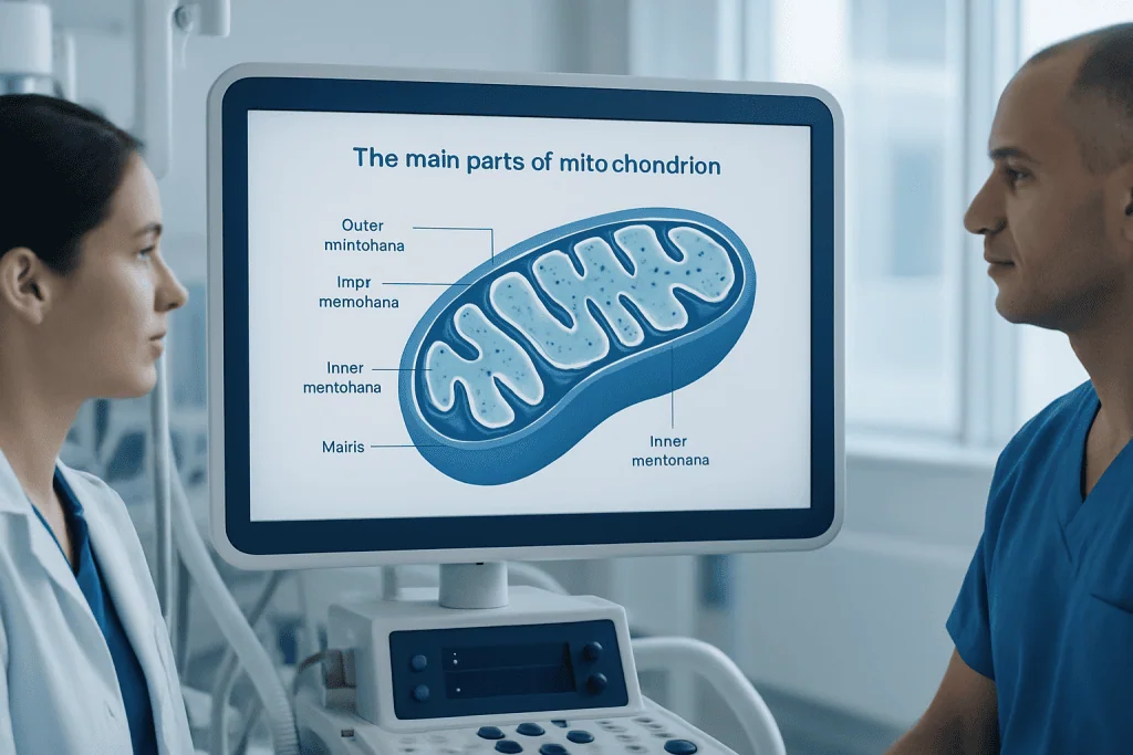
Most evidence-based diagrams label five core structures. The outer membrane is the smooth outer boundary that separates the mitochondrion from the rest of the cell. The inner membrane lies inside it and is highly specialized for energy production. The folds of this inner membrane are called cristae, and these folds increase the available surface area for important chemical reactions.
The space enclosed by the inner membrane is called the matrix. This is where several metabolic reactions take place, including parts of the process that breaks down nutrients to support ATP production. The matrix also contains mitochondrial DNA and ribosomes, which means mitochondria can make some of their own proteins, although they still depend on many proteins made from genes in the cell nucleus.
Some diagrams also label the intermembrane space, which is the narrow region between the outer and inner membranes. This space matters because it helps maintain the proton gradient used to generate ATP. A clear patient-friendly diagram may also show arrows for oxygen use, nutrient breakdown, and ATP output to highlight that mitochondria do not work in isolation but as part of the larger cell.
- Outer membrane: protective outer layer
- Inner membrane: site of key energy-producing proteins
- Cristae: folds that expand working surface area
- Matrix: internal space where metabolic reactions occur
- Mitochondrial DNA: genetic material inside mitochondria
- Intermembrane space: supports ATP generation
How mitochondria make energy

The basic purpose of the mitochondrion is to convert energy from food into ATP, a form of energy cells can use directly. Nutrients from carbohydrates, fats, and sometimes proteins are processed through a series of chemical pathways. Some steps occur outside the mitochondrion, but many of the most important energy-producing steps happen inside it.
Within the mitochondrial matrix, the citric acid cycle helps extract high-energy electrons from nutrients. These electrons then move through the electron transport chain, which is located in the inner membrane. As this happens, protons are pumped across the inner membrane, creating a gradient. The enzyme ATP synthase uses that gradient to make ATP. This process is often called oxidative phosphorylation.
A mitochondria diagram can make this sequence easier to understand because it shows where each step occurs. The matrix is not just empty space, the inner membrane is not just a barrier, and the cristae are not simply folds for decoration. Each structural feature supports a function. When doctors discuss metabolic disorders, inherited enzyme defects, or tissue injury from oxygen shortage, this structure-function relationship becomes especially important.
Why mitochondria matter in health and disease
Because mitochondria are central to energy production, they are especially important in organs that use large amounts of energy. The brain, heart, skeletal muscles, kidneys, eyes, and liver may all be affected if mitochondrial function is impaired. This is one reason mitochondrial disorders can look very different from person to person. One patient may have muscle fatigue, while another may have neurologic symptoms, hearing changes, growth concerns, or heart involvement.
Not every symptom related to fatigue or weakness is caused by mitochondria. Common conditions such as anemia, thyroid disease, sleep problems, infection, depression, medication side effects, and poor nutrition can also reduce energy. Still, when symptoms are persistent, involve several body systems, or begin early in life, doctors may consider whether a mitochondrial condition is part of the picture. Readers learning about this topic may also find it helpful to understand related neurologic conditions in which cellular energy and nerve cell health are active areas of research.
Mitochondrial disease can be inherited or can arise from new genetic changes. Some involve mitochondrial DNA, while others are caused by changes in nuclear genes that affect mitochondrial proteins. A diagram cannot show all of this genetic complexity, but it helps explain why a problem in a tiny cell structure can have whole-body effects. It also explains why specialists often look at symptoms across multiple systems rather than focusing on just one organ.
How a mitochondria diagram is used in medical discussions
In clinical care, a mitochondria diagram is not a diagnostic test. Instead, it is a teaching tool that helps patients understand what clinicians mean when they discuss cellular respiration, metabolic pathways, or inherited mitochondrial disorders. It can also be useful when reviewing lab findings such as lactate levels, genetic test results, muscle biopsy discussions, or imaging findings in the context of unexplained neuromuscular symptoms.
If a doctor suspects a mitochondrial disorder, evaluation often begins with a detailed history and physical examination. The care team may ask about exercise intolerance, developmental milestones, hearing or vision changes, migraines, seizures, muscle weakness, heart symptoms, and family history. Depending on the situation, tests may include blood work, urine studies, heart evaluation, eye examination, hearing testing, brain imaging, electromyography, or genetic testing. In selected cases, genetic testing can help identify a likely inherited cause.
For some patients, specialists in neurology, cardiology, metabolism, genetics, rehabilitation, and nutrition work together to clarify the diagnosis. If neurologic symptoms are present, doctors may also use imaging such as MRI to assess the brain or muscles in the right clinical setting. A diagram can support these conversations by showing where cellular energy problems originate, but diagnosis always depends on symptoms, examination, and appropriate testing.
Treatment, daily management, and self-care
Treatment depends on the underlying problem. There is no single therapy that fixes all mitochondrial conditions, because these disorders vary widely in cause and severity. Management usually focuses on treating symptoms, supporting affected organs, preventing complications, and improving function and quality of life. This may include physical therapy, occupational therapy, nutritional guidance, seizure treatment when needed, hearing or vision support, and careful monitoring of the heart and other organs.
Daily self-care often centers on energy balance. Many people with suspected or confirmed mitochondrial disorders are advised to avoid extreme exhaustion, prolonged fasting, dehydration, and severe illness triggers when possible. Good sleep, regular follow-up, a balanced diet, and a realistic activity plan can help some patients function more steadily. However, self-care should be individualized, especially for children and for adults with complex symptoms.
Patients should be cautious about supplements, restrictive diets, or online claims that promise to “boost mitochondria” quickly. Some supportive treatments are used in selected patients, but they should be recommended by a qualified clinician who understands the person’s diagnosis and overall health. When symptoms affect mobility, exercise tolerance, or daily activities, physical therapy and rehabilitation may be part of a broader care plan.
How to read a mitochondria diagram correctly
A good diagram is accurate, simple, and clearly labeled. Patients should look for labels that identify the outer membrane, inner membrane, cristae, matrix, and mitochondrial DNA. It is also helpful if the diagram shows that ATP production is linked to the inner membrane and that the matrix contains enzymes involved in metabolism. Oversimplified graphics are common online, so using trusted educational sources matters.
One common misunderstanding is that mitochondria only produce energy and do nothing else. Another is that one abnormal blood test or one symptom can confirm a mitochondrial disease. In reality, the body’s energy systems are interconnected, and many non-mitochondrial illnesses can produce similar symptoms. A diagram should support understanding, not replace medical evaluation.
For families, it may help to compare the mitochondrion to a small, organized workspace inside the cell. The membranes define the space, the cristae expand the work surface, and the matrix contains important machinery. This kind of visual explanation can make school biology, medical appointments, and educational materials easier to follow without creating unnecessary worry.
When to seek medical care
Most people looking at a mitochondria diagram are learning basic biology and do not need urgent care. Medical assessment becomes more important when symptoms suggest a possible underlying disorder rather than simple curiosity about cell structure. Examples include ongoing muscle weakness, marked exercise intolerance, developmental delay, seizures, poor coordination, unexplained hearing or vision changes, repeated vomiting, or symptoms affecting more than one organ system.
Adults should seek medical advice for persistent fatigue that is not improving, recurrent fainting, progressive weakness, shortness of breath with mild activity, or unexplained neurologic symptoms. Parents should contact a doctor if a child has developmental regression, feeding difficulty, low muscle tone, unusual fatigue, or episodes of confusion. Emergency care is needed for severe breathing trouble, prolonged seizures, sudden loss of consciousness, or acute stroke-like symptoms.
Assessment is often most helpful when it is organized and multidisciplinary. Near the end of the diagnostic pathway, some patients benefit from consultation in centers with neurology, genetics, imaging, and rehabilitation expertise. Acibadem International’s multidisciplinary specialists and JCI-accredited hospitals diagnose and treat complex neurological and metabolic conditions for international patients when further evaluation is needed.
Frequently asked questions
What is shown in a mitochondria diagram?
A mitochondria diagram usually shows the outer membrane, inner membrane, cristae, matrix, and mitochondrial DNA. Some diagrams also label the intermembrane space and use arrows to show how ATP is produced.
Why are the cristae important?
Cristae are folds of the inner mitochondrial membrane. They increase the surface area available for the proteins involved in energy production, which helps the cell make ATP more efficiently.
Do mitochondria have their own DNA?
Yes, mitochondria contain their own small amount of DNA. However, most proteins needed for mitochondrial function are still made from genes located in the cell nucleus.
Can a mitochondria diagram diagnose mitochondrial disease?
No. A diagram is only an educational image and cannot diagnose any condition. Diagnosis depends on symptoms, medical history, examination, and sometimes laboratory, imaging, or genetic tests.
Which body parts are most affected when mitochondria do not work well?
High-energy organs are often most affected, including the brain, muscles, heart, eyes, liver, and kidneys. The exact pattern can vary widely from one person to another.
Should someone worry if they feel tired and read about mitochondria?
Not necessarily, because tiredness is very common and has many possible causes. If fatigue is persistent, severe, progressive, or comes with weakness, neurologic symptoms, or problems in several body systems, a doctor should evaluate it.
This article is for general information only and is not a substitute for professional medical advice. Please consult a qualified doctor about your individual situation.
Explore treatments in Turkey — costs, top hospitals & a free quote
JCI-accredited · board-certified surgeons · reply within 24h
Add us as a Preferred Source to see more of our trusted health content across Google Search, AI Overviews and Discover.

More from the Health Library

Motion Sickness — Explained by Medical Evidence, Not Myths

Taro Vegetable: What Patients Need to Know

Serotonin Tinnitus Severity Study: What Patients Need to Know

Manic Depression — Explained by Medical Evidence, Not Myths

Manic Episode: What Patients Need to Know