MR Elastography: A Noninvasive Scan for Liver Stiffness Explained
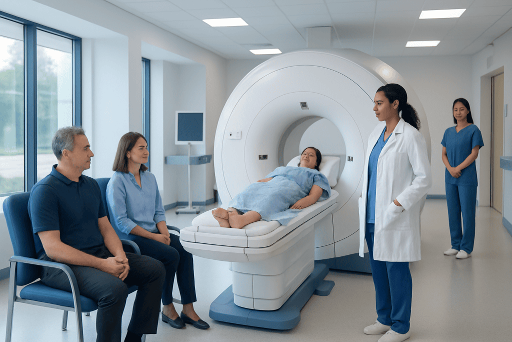
MR elastography uses MRI technology to measure how stiff liver tissue is. It can help identify liver fibrosis and track chronic liver disease over time.
Key Takeaways
- MR elastography uses MRI technology to measure how stiff liver tissue is.
- It can help identify liver fibrosis and track chronic liver disease over time.
- The test is noninvasive and is often used when doctors want more information than standard imaging provides.
- MR elastography does not replace every liver biopsy, but it may reduce the need for one in some patients.
- Preparation is usually simple, but people with certain implants or severe claustrophobia should discuss this in advance.
MR elastography is an advanced imaging test that measures liver stiffness without needles or surgery. It can help doctors detect and monitor liver fibrosis and other chronic liver conditions in a safe, noninvasive way.
Overview of MR Elastography
MR elastography is a specialized form of MRI that measures the stiffness of tissues, most commonly the liver. In liver disease, stiffness often increases when scar tissue builds up. Because of this, the scan can help doctors assess fibrosis, which is the medical term for scarring that develops over time in response to inflammation or injury.
Unlike a liver biopsy, MR elastography does not require a needle sample. Instead, it combines standard MRI images with gentle mechanical vibrations that pass through the liver. A computer then analyzes how these waves move through the tissue. Stiffer tissue changes the wave pattern, which allows the radiologist to estimate liver stiffness.
This test is especially useful in people with chronic liver disease, fatty liver disease, hepatitis, or other conditions that may lead to fibrosis or cirrhosis. It is also helpful for monitoring disease over time, since it can be repeated without exposing the patient to radiation. For many people, MR elastography offers a more comfortable way to gather important information about liver health.
How the Scan Works
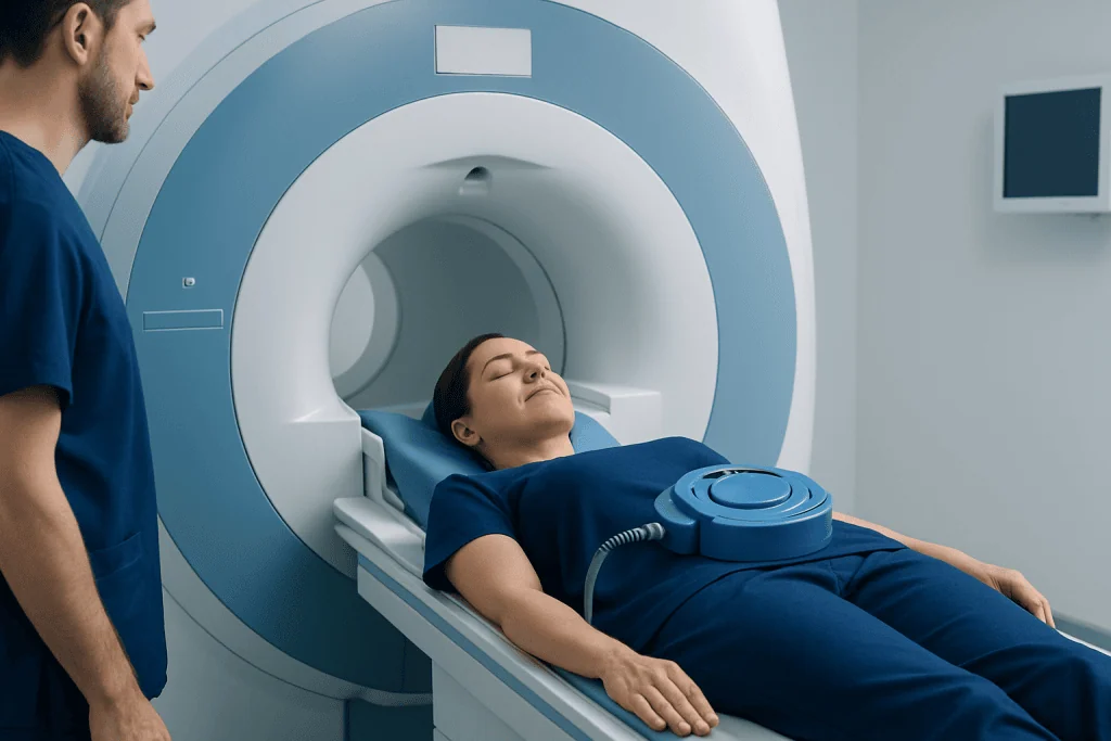
During MR elastography, a small device is placed over the right upper abdomen, where the liver sits. This device creates low-frequency vibrations that are usually felt as a mild tapping or humming sensation. These vibrations travel through the liver while the MRI machine captures images.
Special software converts these images into color-coded maps that show tissue stiffness. Areas with higher stiffness may suggest more advanced fibrosis, although the scan results always need to be interpreted in the context of symptoms, blood tests, and other imaging findings. The goal is not simply to label tissue as normal or abnormal, but to help doctors understand the stage and pattern of liver disease.
Because the test uses MRI technology, it does not involve ionizing radiation. It also examines a larger portion of the liver than a biopsy sample can. This can be useful because liver fibrosis may not be evenly distributed, and a small tissue sample may not always represent the entire organ accurately.
Why Doctors Order MR Elastography

Doctors may recommend MR elastography when they want to evaluate possible liver fibrosis without immediately turning to biopsy. It is commonly used in people with abnormal liver blood tests, fatty liver disease, chronic viral hepatitis, autoimmune liver disease, or suspected cirrhosis. It may also be used after treatment to monitor whether liver disease is stable, improving, or progressing.
The scan can be particularly helpful when standard ultrasound or routine MRI cannot answer the full clinical question. It adds functional information by estimating stiffness rather than only showing anatomy. In this way, MR elastography complements other tools such as blood work, ultrasound, and conventional MRI.
MR elastography may be considered as part of the evaluation for conditions linked to liver scarring, including fatty liver disease and chronic hepatitis. In some cases, it is performed alongside broader liver imaging such as MRI scan to give a more complete picture of liver structure and tissue quality.
Although the liver is the most common organ assessed, elastography techniques can also be explored in other organs in selected settings. Still, for most patients, the main reason for MR elastography is to detect or follow liver fibrosis in a noninvasive and repeatable way.
What to Expect Before, During, and After the Test
Preparation for MR elastography is usually straightforward. Many centers ask patients not to eat for a few hours before the scan, though instructions can vary. Patients should tell their care team about pacemakers, implanted devices, metal fragments, pregnancy, kidney problems, or a history of reacting to contrast dye, if contrast is being considered for any additional MRI sequences.
During the test, the patient lies on an MRI table that moves into the scanner. The vibration device is secured over the abdomen, and the person is asked to lie still and sometimes briefly hold their breath while images are taken. The MRI machine can be noisy, so ear protection is typically provided. The scan itself is painless, though some people find the confined space uncomfortable.
After the exam, most people can return to normal activities right away. If no sedative was used, there is usually no recovery time. A radiologist reviews the images and produces a report for the referring doctor, who then explains what the measured stiffness may mean in the context of the patient’s overall liver health.
People who feel anxious in enclosed spaces should mention this before the appointment. The team may suggest strategies to make the experience easier, and in some cases a wider scanner or pre-planned support can help. If broader evaluation is needed, doctors may pair the examination with other imaging such as ultrasound or additional liver-focused tests.
Benefits and Limitations
The main benefit of MR elastography is that it is noninvasive. It can provide valuable information about liver stiffness without a needle, incision, or hospital stay. It also evaluates a relatively large volume of liver tissue, which may improve reliability when fibrosis is patchy. For many patients, it offers a practical way to monitor chronic disease over time.
Another advantage is that MR elastography can often be added to a routine liver MRI exam, allowing structural and stiffness information to be reviewed together. This can be useful when doctors are also looking for signs of inflammation, fatty change, or other abnormalities. In some settings, it may help reduce the need for biopsy, especially when the clinical picture is clear.
At the same time, the test has limitations. It does not by itself diagnose the cause of liver stiffness. Increased stiffness can be influenced not only by fibrosis but also by inflammation, congestion, or technical factors. For this reason, doctors interpret the results carefully and do not rely on a single number alone.
MR elastography may also be less suitable for people who cannot undergo MRI safely, such as those with certain incompatible implants. Very severe iron overload, difficulty holding still, or specific body-related technical issues can also affect image quality. In some situations, a doctor may still recommend blood tests, additional imaging, or a biopsy to confirm the diagnosis.
Understanding the Results
MR elastography results are usually reported as a measurement of liver stiffness. In general, lower stiffness suggests healthier, more flexible liver tissue, while higher stiffness may point to increasing fibrosis. However, the meaning of a result depends on the person’s age, medical history, symptoms, blood tests, and the reason the scan was ordered.
Doctors often use the findings to estimate whether fibrosis is absent, mild, moderate, or advanced. This can help guide next steps, such as lifestyle changes, treatment for an underlying liver condition, or closer follow-up. The scan is especially useful when repeated over time, because trends can show whether the liver is becoming more or less stiff.
It is important for patients not to interpret a stiffness value on their own. Results should be reviewed by a qualified specialist, such as a gastroenterologist, hepatologist, or radiologist. If the scan suggests significant scarring, the doctor may recommend more testing to clarify the stage of disease and check for complications.
Prevention, Follow-Up, and When to See a Doctor
MR elastography itself does not prevent liver disease, but it can support earlier detection and better follow-up. Many liver conditions become more manageable when identified before advanced scarring develops. Depending on the cause, prevention and self-care may include maintaining a healthy weight, limiting alcohol, controlling diabetes, managing cholesterol, getting recommended vaccines, and following treatment plans for viral or autoimmune liver disease.
People should speak with a doctor if they have ongoing fatigue, abdominal swelling, yellowing of the skin or eyes, unexplained itching, persistent abnormal liver blood tests, or known risk factors for chronic liver disease. A clinician can decide whether MR elastography, standard imaging, blood tests, or referral to a liver specialist is the next best step.
For individuals already diagnosed with liver disease, regular follow-up is important even when symptoms are mild or absent. Chronic liver conditions can progress quietly, and noninvasive monitoring tools may help reduce uncertainty. A personalized plan is usually based on the underlying diagnosis, the degree of fibrosis, and how stable the condition has been over time.
In centers with advanced imaging programs, multidisciplinary teams can help interpret MR elastography as part of a broader liver assessment. Acibadem International’s multidisciplinary specialists and JCI-accredited hospitals diagnose and treat liver conditions for international patients, using modern imaging and coordinated follow-up when appropriate.
Frequently asked questions
Is MR elastography the same as a regular MRI?
No. MR elastography uses an MRI scanner, but it adds gentle vibrations and special image processing to measure tissue stiffness. A regular MRI mainly shows structure, while MR elastography also gives functional information about how stiff the liver is.
Does MR elastography hurt?
MR elastography is generally painless. Most people only notice a mild vibration or tapping sensation on the abdomen during the scan. The main challenge for some patients is staying still in the MRI machine.
Can MR elastography replace a liver biopsy?
In some cases, it may reduce the need for a biopsy, but it does not replace biopsy in every situation. A biopsy may still be needed if the diagnosis is unclear, if doctors need to look at liver cells directly, or if other test results do not match.
How long does the test take?
The elastography portion is relatively short, but the total appointment depends on whether other MRI images are taken at the same time. Many patients spend around 30 to 60 minutes in the imaging department. The care team can explain the expected timing before the exam.
Who should not have MR elastography?
Some people cannot have MRI-based tests safely, especially if they have certain non-compatible implanted devices or metal fragments in the body. Others may need special planning if they are pregnant, have severe claustrophobia, or have trouble lying still. A doctor or radiology team will review safety questions in advance.
What conditions can increase liver stiffness?
Fibrosis and cirrhosis are common causes of increased liver stiffness, but they are not the only ones. Inflammation, congestion, and some other liver changes can also affect the measurement. That is why doctors interpret the scan together with blood tests, symptoms, and medical history.
References
- World Health Organization
- National Institute of Diabetes and Digestive and Kidney Diseases
- Radiological Society of North America
- American College of Radiology
- European Association for the Study of the Liver
This article is for general information only and is not a substitute for professional medical advice. Please consult a qualified doctor about your individual situation.
Oncology care in Turkey — second opinion and treatment plan
JCI-accredited · board-certified surgeons · reply within 24h
Add us as a Preferred Source to see more of our trusted health content across Google Search, AI Overviews and Discover.