MRI Children’s Hospital: Preparation, Procedure and Results

MRI uses strong magnets and radio waves rather than ionising radiation. Children must remain still during image acquisition; some may benefit from child-life support, distraction techniques or sedation.
Key Takeaways
- MRI uses strong magnets and radio waves rather than ionising radiation.
- Children must remain still during image acquisition; some may benefit from child-life support, distraction techniques or sedation.
- Fasting instructions are especially important when sedation or anaesthesia is planned.
- Parents can often stay nearby or in the scan room after safety screening, although local policies vary.
- Results are interpreted by a radiologist and discussed by the clinician who requested the scan.
An MRI at a children's hospital is a radiation-free imaging examination that helps clinicians view organs, bones, joints, blood vessels and the brain in detail. Preparation depends on the body area being scanned and whether a child may need sedation, so families should follow the hospital's specific instructions.
MRI at a Children's Hospital: An Overview
An MRI children’s hospital appointment is a specialised imaging visit designed around the physical and emotional needs of infants, children and teenagers. Magnetic resonance imaging (MRI) uses a powerful magnetic field and radio waves to produce detailed pictures of internal structures. It does not use X-rays or ionising radiation.
A clinician may request MRI to investigate symptoms, assess an injury, monitor a known condition or guide treatment planning. Depending on the reason for imaging, the scan may focus on the brain, spine, joints, abdomen, pelvis, heart or blood vessels. The MRI team adapts communication, comfort measures and monitoring to the child’s age and needs.
Many children complete an MRI while awake with reassurance, music, videos or other distraction. However, because clear images require stillness, some younger children and children who are very anxious, in pain or unable to lie still may need sedation or general anaesthesia. The imaging department will explain the safest approach in advance.
How MRI Works and Who May Need It
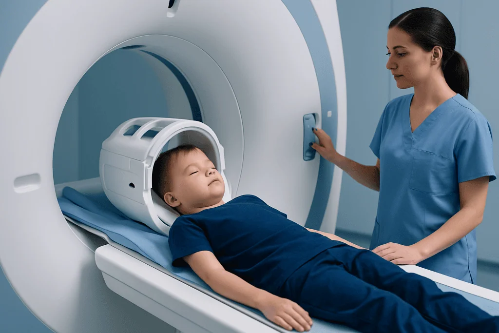
The MRI scanner is a large, tube-shaped machine containing a strong magnet. During the examination, the machine sends radiofrequency energy into the body and detects signals from tissues. A computer converts these signals into detailed images that a radiologist can interpret.
MRI can be particularly useful when clinicians need a detailed view of soft tissues that may not be as clearly seen with other imaging tests. In children, it may be used to assess headaches with concerning features, seizures, developmental concerns, spinal symptoms, sports injuries, abdominal or pelvic conditions, inflammation, tumours, or congenital differences. The scan is requested when its likely clinical value outweighs the practical demands of the examination.
Before scheduling, the team checks for anything that could be affected by the magnet. This includes certain implanted medical devices, cochlear implants, programmable shunts, metal fragments from an injury, braces or retainers, and some medication patches. Most surgical implants are MRI-compatible under specified conditions, but the radiology team must verify this rather than making assumptions.
- MRI may be performed with or without contrast material.
- Contrast can help show blood vessels, inflammation and some abnormalities more clearly.
- A parent or guardian should share information about kidney problems, allergies, prior contrast reactions and all implanted devices.
How Should I Prepare My Child for a Pediatric MRI?

Preparation begins with the instructions from the imaging centre, as requirements vary according to the child’s age, the body part being examined, use of contrast and whether sedation or anaesthesia is planned. Families should ask when to arrive, whether fasting is required, which medicines should be taken, and whether previous imaging or medical device information should be brought to the appointment.
Parents can help by explaining the scan in calm, age-appropriate language. It may be helpful to say that the machine takes pictures while the child lies very still, that it makes loud tapping or knocking sounds, and that the team can see and hear them throughout. Avoid presenting the scan as a punishment or promising that it will be quick, since scan length can vary.
Comfortable clothing without metal fasteners may simplify changing, although the department may provide a gown. Leave jewellery, hair clips, watches, coins, electronic devices and other metal objects at home where possible. A familiar comfort item may be allowed if it has been checked for safety. Younger children may also benefit from practice games that involve lying still for short periods.
If sedation is being considered, strict fasting instructions are essential. The care team may arrange a pre-procedure assessment to review the child’s health, airway, medications and prior experiences with sedation or anaesthesia. Families should tell the team if the child develops fever, cough, vomiting, breathing difficulty or another new illness before the appointment, as the plan may need to change.
What Happens if I Drink Water Before an MRI?
For an MRI without sedation or anaesthesia, drinking water is usually not a problem and may be encouraged in some situations. However, the correct advice depends on the type of MRI and the hospital’s protocol. Some abdominal, pelvic or urinary tract scans may involve specific drinking, fasting or bladder-filling instructions to improve the images.
When a child will receive sedation or general anaesthesia, drinking water can be subject to timed restrictions. These restrictions reduce the risk of stomach contents entering the lungs while the child is sleepy or unconscious. Clear liquids may sometimes be permitted until a certain time, but parents should follow only the instructions provided by the anaesthesia or radiology team.
If a child has eaten or drunk something that may not fit the instructions, parents should tell staff honestly before the scan. The team will decide whether it is safe to proceed, whether the schedule needs to be adjusted, or whether another plan is appropriate. It is better to ask than to withhold this information.
Step-by-Step: What Happens During a Pediatric MRI?
On arrival, staff confirm the child’s identity, the requested examination and relevant medical history. A safety questionnaire is completed to identify metal, implanted devices, prior surgery, allergies and other factors that may affect MRI safety. If contrast is planned, the team discusses why it is being used and checks for any relevant medical concerns.
The child changes into MRI-safe clothing if needed and removes metal items. If sedation or anaesthesia is planned, a specialist team monitors the child before, during and after the scan. For some contrast-enhanced examinations, a small intravenous line is placed. The child is then positioned on a padded table, sometimes with a special receiving coil placed around the body area being scanned.
The table moves slowly into the scanner. The child must remain still while sequences of images are taken. The machine can be noisy, so earplugs or headphones are provided. Staff communicate through an intercom and can pause if needed. Scan duration varies widely, but the team usually explains the expected length before beginning.
After imaging is complete, the child leaves the scanner and any intravenous line is removed unless it is needed for other care. If there was no sedation, most children can return to their usual activities immediately unless their clinician gives different advice. MRI is part of MRI diagnostic imaging, which may be used alongside examination and other tests to clarify a child’s health needs.
Can I Sit with My Child During an MRI?
In many paediatric MRI services, a parent or guardian may be able to stay in the scan room or nearby to provide reassurance. This depends on the child’s clinical needs, the scanner layout, local infection-control policies and whether sedation or anaesthesia is used. The accompanying adult must complete the same MRI safety screening as the child.
Anyone entering the MRI room must remove metal and disclose implants, devices, prior metal injuries or possible pregnancy. Objects that seem harmless, including phones, keys, bank cards, watches and some clothing accessories, can become hazardous near the magnet or be damaged by it. Staff decide who may enter and where they can sit or stand safely.
When a parent cannot remain in the room, staff can often support the child with clear explanations, headphones, visual entertainment or age-appropriate coping strategies. Child-life professionals, nurses, radiographers and anaesthesia staff may all contribute to making the experience calmer and more predictable.
Benefits, Risks and Recovery After MRI
The main benefit of MRI is its ability to provide highly detailed images without ionising radiation. This can help clinicians identify or rule out certain causes of symptoms, evaluate an injury or condition over time, and plan the next steps in care. A normal result can also be useful, although it is always interpreted in the context of the child’s symptoms and examination.
MRI itself is generally considered safe when screening procedures are followed. The principal risks relate to the strong magnet, which is why metal and implanted devices must be assessed carefully. Some children find the confined space, noise or need to stay still uncomfortable. Contrast material and sedation can cause side effects or uncommon reactions, so these are considered only when clinically appropriate and managed by trained teams.
Children who have an MRI while awake often resume school, play and normal eating and drinking soon afterwards. After sedation or anaesthesia, they need observation until they are alert and stable. They may be sleepy, irritable, nauseated or less coordinated for the rest of the day, and should be supervised according to the discharge advice provided.
A radiologist reviews the images and sends a report to the requesting clinician. Results may be available promptly in urgent situations, while routine reports can take longer depending on the examination and service. The clinician who ordered the MRI explains what the findings mean and whether follow-up, monitoring or treatment is needed.
Is There Anything I Shouldn't Do Before an MRI Scan?
Families should not ignore or guess at fasting instructions, particularly if sedation or anaesthesia is planned. They should also avoid bringing metal items into the MRI area and should not use medication patches, cosmetics, hair products or clothing with metallic components without checking if they are safe for the specific examination. Some products may contain metallic particles or interfere with image quality.
It is important not to conceal information about implants, surgery, metal injuries, pregnancy in an accompanying adult, kidney disease, allergies or prior reactions to contrast. Parents should also notify the department about a new illness before the appointment. The team may advise proceeding, changing the plan or postponing the scan based on the child’s safety.
Children should not stop prescribed medicines unless a doctor specifically tells them to do so. Parents should ask in advance about routine medicines, inhalers, diabetes treatments and any medicines that need to be taken with food. Written instructions from the hospital take priority because they are tailored to the child and the planned procedure.
When to Seek Medical Care
Parents should seek urgent medical assessment rather than waiting for a planned MRI if a child has severe difficulty breathing, loss of consciousness, a seizure that is prolonged or different from usual, sudden weakness, severe head injury, confusion, persistent vomiting with severe headache, or other rapidly worsening symptoms. Emergency services should be contacted according to local guidance when symptoms appear life-threatening.
For non-emergency concerns, contact the child’s doctor if symptoms are new, persistent, worsening or affecting daily function. The doctor can assess whether MRI is appropriate or whether another test, specialist consultation or treatment is needed. An MRI is a diagnostic tool, not a replacement for clinical evaluation.
Acibadem International’s multidisciplinary specialists and JCI-accredited hospitals can assess children who need diagnostic imaging and coordinate care for international patients. Families should discuss the most suitable imaging pathway with a qualified paediatric clinician.
Frequently asked questions
Does a pediatric MRI use radiation?
No. MRI uses a magnetic field and radio waves to create images, rather than X-rays or ionising radiation. This is one reason MRI can be useful when detailed imaging is needed in children.
How long does an MRI scan take for a child?
The duration depends on the body area being scanned, the number of image sequences and whether contrast is needed. Some examinations are relatively short, while more detailed studies can take longer. The imaging team can give an estimate before the appointment.
Will my child need sedation for an MRI?
Not every child needs sedation. It may be considered when a child is too young, anxious, uncomfortable or unable to remain still long enough for clear images. The decision is based on the child, the type of scan and the safety assessment.
Can my child eat before an MRI?
Children having an MRI without sedation may often eat normally, unless they receive different instructions for their specific scan. If sedation, anaesthesia or certain abdominal examinations are planned, fasting may be required. Parents should follow the hospital’s written instructions exactly.
Is MRI contrast safe for children?
MRI contrast is used only when it is expected to add useful diagnostic information. The care team reviews the child’s health history, including kidney problems and prior reactions, before using it. Reactions are uncommon, but staff are prepared to recognise and manage them.
When will we receive MRI results?
A radiologist reviews the images and sends a report to the clinician who requested the scan. Timing varies by urgency and local workflow. The requesting clinician explains the results in relation to the child’s symptoms, examination and care plan.
References
- American College of Radiology
- Radiological Society of North America
- U.S. Food and Drug Administration
- Society for Pediatric Radiology
- National Institute of Biomedical Imaging and Bioengineering
This article is for general information only and is not a substitute for professional medical advice. Please consult a qualified doctor about your individual situation.
Joint and spine care in Turkey — expert assessment & treatment
JCI-accredited · board-certified surgeons · reply within 24h
Add us as a Preferred Source to see more of our trusted health content across Google Search, AI Overviews and Discover.