MRI for Vision Problems: When Brain and Orbit Imaging Is Recommended
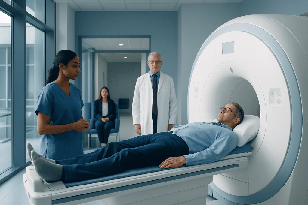
MRI can show the brain, optic nerves, eye sockets, and surrounding tissues in detail. It is often used when vision symptoms may be linked to neurological or orbital conditions.
Key Takeaways
- MRI can show the brain, optic nerves, eye sockets, and surrounding tissues in detail.
- It is often used when vision symptoms may be linked to neurological or orbital conditions.
- Common reasons for MRI include sudden vision loss, double vision, optic nerve swelling, or unexplained visual field changes.
- MRI does not use ionizing radiation, but some patients may need contrast dye for clearer images.
- The need for brain MRI, orbit MRI, or both depends on the symptoms and examination findings.
- A neuroophthalmologist or radiologist helps decide the most appropriate imaging plan.
MRI for vision problems may be recommended when symptoms suggest that the optic nerves, brain, or tissues around the eyes need closer evaluation. It helps doctors look for causes that may not be visible during a standard eye exam alone.
Overview: Why MRI May Be Used for Vision Problems
MRI for vision problems is a detailed imaging test that helps doctors examine structures involved in seeing, including the brain, optic nerves, eye muscles, and the soft tissues within the eye sockets, also called the orbits. It is often requested when symptoms suggest that the cause of a vision change may be coming from the nervous system or from tissues behind the eyes rather than from the front surface of the eye alone.
A routine eye examination can identify many common causes of blurred vision, such as refractive error, cataract, or dry eye. However, some symptoms point to a deeper problem that cannot be fully assessed with external eye tests. In these situations, MRI can provide more information by creating detailed images of the brain and the area around the eyes.
Doctors may order an MRI of the brain, an MRI of the orbits, or both. Brain MRI helps evaluate visual pathways, the pituitary region, and areas that control eye movement. Orbit MRI focuses more closely on the optic nerves, eye muscles, and surrounding tissues. The exact imaging plan depends on a person’s symptoms, medical history, and findings on examination.
Symptoms That May Lead to Brain or Orbit MRI
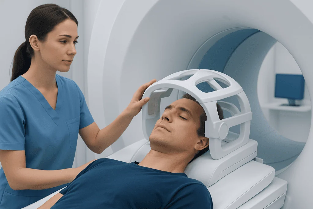
Not every vision complaint needs MRI. In many people, common eye conditions are the cause. Imaging is more likely to be recommended when symptoms are unusual, sudden, progressive, or associated with other neurological signs. The goal is to identify whether there is inflammation, compression, reduced blood flow, demyelination, or another structural issue affecting vision.
Examples of symptoms that may prompt MRI include sudden vision loss in one eye, unexplained double vision, loss of side vision, pain with eye movement, or episodes of temporary vision dimming. MRI may also be helpful when an eye doctor sees swelling of the optic nerve, abnormal eye movements, unequal pupils, or other findings suggesting involvement beyond the eyeball itself.
- Sudden or unexplained loss of vision
- Double vision, especially if it is new or persistent
- Visual field loss, such as missing side vision
- Optic disc swelling or suspected optic neuritis
- Bulging of one or both eyes
- Eye movement problems or drooping eyelid
- Vision changes together with headache, weakness, numbness, or balance problems
When symptoms are linked to conditions such as optic neuritis or concern for compression of the visual pathways, MRI can be an important next step in evaluation. It helps guide both diagnosis and treatment planning.
What MRI Can Show in the Brain and Orbits

MRI uses a strong magnetic field and radio waves to create detailed images of internal tissues. For vision-related concerns, it can show the optic nerves from the back of the eyes toward the brain, the optic chiasm where visual pathways cross, the brain tissue involved in vision, and the muscles and fat inside the orbits. This level of detail can help detect problems that may not appear on other tests.
Brain MRI may reveal inflammation, tumors, stroke-related changes, demyelinating disease such as multiple sclerosis, pituitary gland abnormalities, or pressure effects on the visual pathways. Orbit MRI is especially useful for conditions affecting the optic nerves, the eye muscles, and tissues behind the eyes, including inflammation, thyroid eye disease, infection, and masses.
Sometimes a contrast agent is used during MRI to make certain tissues easier to see. Contrast can help highlight active inflammation, breakdown of the blood-brain barrier, or abnormal blood vessels. Doctors decide whether contrast is needed based on the question they are trying to answer and on the patient’s kidney function, allergy history, and general health.
If doctors are assessing structures in more detail, they may combine MRI findings with other tests such as visual fields, optical coherence tomography, blood tests, or a neurological examination. In selected cases, related imaging such as 3 Tesla MRI imaging may offer especially detailed views of delicate structures.
Common Conditions MRI May Help Evaluate
There are many reasons a person with visual symptoms might need imaging. MRI is not a diagnosis by itself, but it can support or clarify what doctors suspect based on symptoms and examination. In neuroophthalmology, it is often used to investigate whether a problem is affecting the optic nerves, cranial nerves, brain, or orbit.
One common indication is suspected optic neuritis, which can cause blurred vision, reduced color vision, and pain with eye movement. MRI can show inflammation of the optic nerve and may also look for brain lesions that suggest demyelinating disease. MRI may also be used when doctors are concerned about a tumor, a mass pressing on the optic pathways, or inflammation in the orbit.
Double vision is another common reason for imaging. MRI may help assess the nerves and muscles responsible for eye movement, and it may be ordered if there is concern for a neurological cause. It can also help evaluate conditions such as brain tumors when symptoms include visual changes, headache, or imbalance.
Other situations include unexplained visual field defects, papilledema related to raised pressure inside the skull, pituitary lesions, thyroid eye disease, vascular abnormalities, or symptoms that overlap with multiple sclerosis or other inflammatory disorders. The imaging protocol is tailored to the suspected condition rather than using the same scan for everyone.
How Doctors Decide Whether MRI Is Recommended
The decision to request MRI is based on the whole clinical picture. Doctors consider how quickly symptoms developed, whether one or both eyes are affected, whether there is pain, and whether there are associated neurological symptoms. Findings from the eye exam are especially important, including pupil reactions, eye movements, the appearance of the optic nerve, and visual field testing.
If a problem appears to involve the front of the eye only, such as the cornea, lens, or retina, MRI is usually not the first test. But if symptoms or exam findings suggest a problem behind the eye or within the brain, MRI becomes more useful. A neuroophthalmologist may recommend brain MRI, orbit MRI, or both, often with special sequences designed to assess the optic nerves and eye muscles.
Doctors also think about which test is most appropriate and safest. In some situations, CT may be preferred, such as after trauma or when bone detail is needed quickly. In others, MRI is favored because it offers better soft tissue detail and avoids ionizing radiation. If there is concern about blood vessels, tests such as MR angiography may be added to assess the circulation related to the brain and eyes.
What to Expect During the MRI Scan
An MRI scan is painless, but it does require lying still inside the scanner for a period of time. The machine makes loud tapping or knocking sounds during imaging, so ear protection is usually provided. Depending on the type of study, the scan may take around 20 to 45 minutes, and sometimes longer if both the brain and orbits are being examined or if contrast is used.
Before the scan, patients are asked about metal implants, pacemakers, prior surgeries, kidney disease, and possible pregnancy. It is important to mention any history of claustrophobia, as the care team may offer strategies to make the experience more comfortable. In some settings, open or wider-bore MRI systems may be available, or a doctor may discuss whether medication is appropriate.
If contrast is needed, it is usually given through a vein in the arm. Most people tolerate it well, but the team will review any previous reactions and whether recent kidney testing is needed. After the scan, patients can usually return to normal activities unless they received sedating medication.
For people being assessed at specialized centers, imaging is often interpreted together with eye examination findings and visual testing. Near the end of the diagnostic pathway, centers such as Acibadem International may coordinate neuroophthalmology, neurology, and radiology input for international patients in JCI-accredited hospitals when complex vision symptoms need multidisciplinary evaluation.
After MRI: Understanding Results and Next Steps
MRI results are interpreted by a radiologist and then reviewed with the doctor who ordered the test. In neuroophthalmology, imaging findings are most useful when they are matched with the patient’s symptoms and eye examination. A scan may confirm a suspected diagnosis, narrow the list of possibilities, or show that a different explanation should be considered.
Sometimes MRI identifies a clear cause of vision symptoms, such as optic nerve inflammation, a lesion affecting the visual pathways, or enlargement of tissues in the orbit. In other cases, the scan may be normal even though symptoms are real. A normal MRI can still be valuable because it helps rule out major structural causes and guides the next stage of testing.
Treatment depends on the underlying condition rather than the MRI alone. Some causes may need medication, monitoring, hormone assessment, or care from neurology, endocrinology, or ophthalmology. Others may require more specialized evaluation, and in selected cases additional imaging such as functional MRI or follow-up MRI may be advised if symptoms evolve over time.
When to Seek Medical Care for Vision Changes
Any sudden or significant change in vision should be assessed promptly by a qualified doctor. This is especially true if visual symptoms are accompanied by eye pain, severe headache, weakness, numbness, speech difficulty, confusion, or loss of balance. These combinations can point to urgent neurological or vascular causes that need timely care.
Double vision that starts suddenly, loss of vision in one eye, a curtain-like shadow over vision, or swelling around the eye should not be ignored. Even when symptoms are mild, recurrent, or brief, a professional evaluation is still important because some conditions can come and go before becoming more noticeable.
People should also follow up if an eye doctor recommends imaging, even if vision seems to improve. MRI is often ordered not because the situation is immediately dangerous, but because careful evaluation can prevent delays in diagnosing treatable conditions. Early assessment helps doctors choose the most appropriate care and provide reassurance when serious causes are excluded.
Frequently asked questions
Why would someone need an MRI for vision problems?
An MRI may be recommended when vision symptoms suggest a problem involving the optic nerves, brain, or tissues behind the eyes. It is especially useful when a regular eye exam does not fully explain symptoms such as sudden vision loss, double vision, or visual field changes.
What is the difference between a brain MRI and an orbit MRI?
A brain MRI looks at the brain and the visual pathways that connect the eyes to the brain. An orbit MRI focuses more closely on the eye sockets, optic nerves, eye muscles, and nearby soft tissues. Some patients need one study, while others benefit from both.
Does MRI for vision problems use radiation?
No. MRI does not use ionizing radiation. Instead, it uses magnets and radio waves to create detailed images, which makes it different from CT scanning.
Will contrast dye always be needed?
Not always. Doctors order contrast when it may help show inflammation, abnormal blood vessels, tumors, or other changes more clearly. Whether contrast is used depends on the clinical question and on the patient’s medical history.
Can an MRI detect optic neuritis?
MRI can often help show inflammation of the optic nerve and may support the diagnosis of optic neuritis. It may also be used to look for related changes in the brain that could affect treatment planning and follow-up.
What if the MRI is normal but vision symptoms continue?
A normal MRI does not mean symptoms are unimportant. It may simply mean there is no major structural abnormality visible on the scan. Doctors may then recommend other eye tests, blood work, follow-up visits, or repeat imaging depending on the situation.
References
- American Academy of Ophthalmology
- American Academy of Neurology
- Radiological Society of North America
- National Eye Institute
- American College of Radiology
This article is for general information only and is not a substitute for professional medical advice. Please consult a qualified doctor about your individual situation.
Explore treatments in Turkey — costs, top hospitals & a free quote
JCI-accredited · board-certified surgeons · reply within 24h
Add us as a Preferred Source to see more of our trusted health content across Google Search, AI Overviews and Discover.

More from the Health Library



Related Specialists

Deniz Beyza Kılıç
Physical Medicine & Rehabilitation
Prof. Dr. Feyzullah Çetinkaya
Pediatric Allergy
Prof. Dr. Tugan Beşe
Gynecology & Obstetrics
Dr. İbrahim Keskin
Emergency Service