MRI Scan: An Evidence-Based Guide for Patients
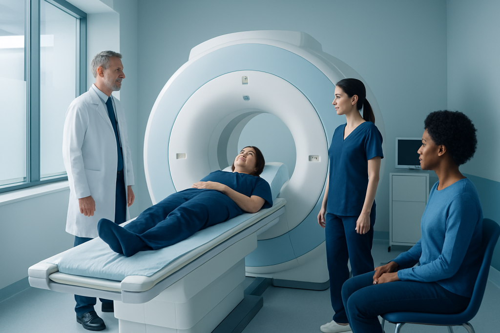
An MRI scan creates highly detailed images using magnets and radio waves, not X-rays. It is commonly used for the brain, spine, joints, abdomen, pelvis, heart, and blood vessels.
Key Takeaways
- An MRI scan creates highly detailed images using magnets and radio waves, not X-rays.
- It is commonly used for the brain, spine, joints, abdomen, pelvis, heart, and blood vessels.
- Most people can have an MRI safely, but metal implants, devices, and contrast allergies need review beforehand.
- The test is painless, though it can be noisy and may feel uncomfortable for people with claustrophobia.
- Preparation depends on the body area being examined and whether contrast dye is needed.
- A radiologist interprets the images, and the results are discussed with the referring doctor.
An MRI scan is a noninvasive imaging test that uses a strong magnet and radio waves to create detailed pictures of organs, joints, the brain, spine, and other tissues. It does not use ionizing radiation and is widely used to help diagnose symptoms, guide treatment decisions, and monitor many medical conditions.
What an MRI scan is and why it is used
An MRI scan, short for magnetic resonance imaging, is a medical imaging test that produces detailed pictures of structures inside the body. It uses a powerful magnet, radio waves, and computer processing rather than ionizing radiation. This makes it especially useful when doctors need clear images of soft tissues such as the brain, spinal cord, muscles, ligaments, organs, and blood vessels.
Doctors request MRI scans for many reasons. The test can help evaluate headaches, back or neck pain, joint injuries, numbness, dizziness, abdominal or pelvic symptoms, and suspected inflammation or tumors. It is also used to monitor known conditions over time and to plan certain procedures or surgeries.
Compared with some other imaging methods, MRI often provides more detail for soft tissues. For example, it is commonly used in the evaluation of brain tumors, spinal disc problems, sports injuries, and some liver or pelvic conditions. In some cases, it may be used alongside ultrasound, CT, or X-ray so the medical team can build a fuller picture of what is happening.
How MRI works and what it can show
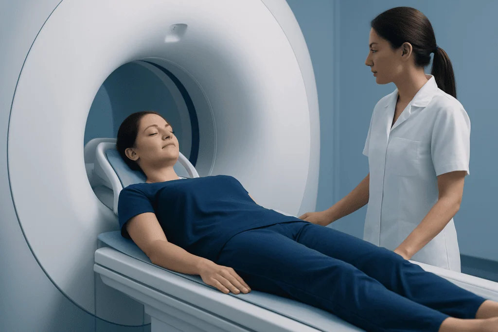
MRI works by aligning hydrogen atoms in the body’s tissues with a strong magnetic field. Radiofrequency pulses briefly disturb that alignment, and as the atoms return to their original state, they produce signals. A computer converts those signals into detailed cross-sectional images. Different scan settings can highlight water, fat, blood flow, inflammation, or tissue damage in different ways.
This flexibility is one reason MRI is so valuable. It can show normal anatomy, swelling, tears, bleeding, infection, and changes linked to chronic disease. In the brain, it can help assess stroke, multiple sclerosis, and tumors. In the spine, it can show discs, nerves, and the spinal cord. In joints, it helps evaluate cartilage, ligaments, tendons, and bone marrow.
There are also specialized types of MRI. Magnetic resonance angiography can look at blood vessels. Cardiac MRI can evaluate heart structure and function. Functional MRI may map brain activity in selected situations. Depending on the reason for the test, the doctor may recommend a routine MRI or a more targeted study such as brain MRI scan or spine MRI.
When doctors may recommend an MRI scan

An MRI scan is ordered when symptoms, examination findings, or other test results suggest a problem that needs closer imaging. It is often chosen when soft tissue detail matters or when a doctor wants to avoid radiation exposure, especially in situations where repeated imaging may be needed.
Common reasons include persistent headaches, seizures, unexplained weakness, numbness, balance problems, hearing changes, joint pain after injury, chronic back pain, or suspicion of internal organ disease. MRI is also used in cancer care to define the extent of disease, assess response to treatment, or monitor for recurrence. In vascular medicine, it may be used to look at arteries and veins.
Not every symptom requires MRI. For some problems, a physical examination, blood tests, ultrasound, CT scan, or plain X-ray may be more appropriate as a first step. The choice depends on the body area, the urgency of the problem, and what clinical question the doctor is trying to answer. When needed, MRI can be part of a broader imaging pathway that may include MR angiography or other targeted studies.
Preparing for the scan and what happens during the test
Preparation for an MRI scan is usually simple, but it varies by the body area being examined. Many scans do not require fasting, while some abdominal studies may. If contrast material is planned, the care team may review kidney function, previous reactions to contrast agents, and current medications. Patients should follow the instructions given by the imaging center or doctor.
Before the scan, patients are asked detailed safety questions about metal implants, pacemakers, surgical clips, hearing devices, metal fragments, and pregnancy. Jewelry, watches, hairpins, credit cards, and other removable metal objects must be taken off. In some cases, a gown is provided to avoid interference from zippers or metallic fibers in clothing.
During the test, the patient lies on a narrow table that moves into the scanner. The machine makes loud tapping or thumping sounds, so ear protection is usually offered. The scan itself is painless, but staying still is important because movement can blur the images. Some MRI exams take 15 to 30 minutes, while more complex studies may take longer. If anxiety or claustrophobia is a concern, patients should mention this beforehand so supportive options can be discussed.
- Bring a list of implants, surgeries, and medications.
- Arrive early in case forms or safety screening are needed.
- Tell the team about claustrophobia, pregnancy, or kidney disease.
- Ask whether eating or drinking restrictions apply to the planned scan.
MRI with contrast, safety, and possible risks
Some MRI scans are performed with a contrast agent, usually gadolinium-based, injected into a vein. Contrast can help highlight blood vessels, inflammation, infection, tumors, or areas where the blood-brain barrier has changed. Not every MRI needs contrast, and the decision depends on what the doctor is looking for.
For most people, MRI is considered very safe. It does not use radiation, and complications from the scan itself are uncommon. The main safety concern is the strong magnet, which is why all metal devices and implants must be carefully reviewed. Many modern implants are MRI-conditional, meaning the scan may still be possible under specific settings, but this must be confirmed in advance.
Contrast reactions are uncommon and are often mild when they do occur. In people with significant kidney disease, the team may take extra precautions before giving contrast. Patients should also mention prior allergic reactions, asthma, implanted devices, tattoos with metallic pigments, or any history of metal injury to the eyes. If there is uncertainty, the radiology team may recommend additional checks before proceeding.
Children, older adults, and pregnant patients may still be able to have an MRI when medically appropriate. In pregnancy, the decision is based on clinical need, timing, and whether contrast is necessary. A qualified doctor and radiology team can explain the expected benefits and any precautions for the individual situation.
Understanding the results and next steps
After the scan, a radiologist reviews the images and prepares a report for the referring doctor. The report describes what was seen and whether any findings may explain the symptoms. MRI results often need to be interpreted together with the patient’s medical history, examination, and other tests. A finding on MRI does not always mean there is a serious problem, and some abnormalities may be incidental and unrelated to symptoms.
Depending on the result, the next step may be reassurance, follow-up imaging, blood tests, physical therapy, medication, or referral to a specialist. For example, MRI findings can help guide care in neurology, orthopedics, oncology, and gastroenterology. In selected cases, MRI can support decisions about procedures such as MRI-guided biopsy when tissue diagnosis is needed.
If the report is unclear, patients should ask their doctor to explain the findings in plain language. Helpful questions include what the scan showed, whether it explains the symptoms, whether anything urgent was found, and what treatment or follow-up is recommended. Understanding the clinical context is often more important than focusing on technical words in the report.
When to seek medical care
An MRI scan itself is usually planned rather than urgent, but the symptoms that lead to imaging can sometimes require prompt medical evaluation. A person should seek timely medical care for severe or sudden headache, new weakness on one side of the body, trouble speaking, seizures, loss of vision, chest pain, significant shortness of breath, or sudden loss of bladder or bowel control. These symptoms may need emergency assessment rather than waiting for routine imaging.
Medical advice should also be sought if there is worsening back pain with numbness or weakness, persistent joint swelling after injury, unexplained weight loss, ongoing abdominal pain, or neurological symptoms that are progressing. In these situations, a doctor can decide whether MRI is the right next test and how quickly it should be arranged.
After an MRI with contrast, patients should contact a healthcare professional if they develop concerning symptoms such as rash, breathing difficulty, or significant swelling, although these reactions are uncommon. Near the end of the care pathway, some people may choose assessment at centers with coordinated imaging and specialty care. Acibadem International’s multidisciplinary specialists and JCI-accredited hospitals diagnose and treat conditions that may require MRI evaluation for international patients.
Frequently asked questions
Is an MRI scan the same as a CT scan?
No. An MRI scan uses magnets and radio waves, while a CT scan uses X-rays. MRI often gives better detail for soft tissues, while CT is commonly used when speed is important or for certain bones, lungs, and emergencies.
Does an MRI scan use radiation?
No. MRI does not use ionizing radiation. This is one reason it is often preferred when repeated imaging may be needed, although the best test still depends on the medical question.
How long does an MRI scan take?
Many MRI scans take about 15 to 45 minutes, but some can be shorter or longer depending on the body part and whether contrast is used. More specialized studies may require extra sequences and additional time.
Is an MRI scan painful?
The scan itself is painless. The main discomfort usually comes from lying still, the loud noises, or feeling confined inside the scanner. Patients who are anxious or claustrophobic should tell the care team in advance.
Can a person with a pacemaker or metal implant have an MRI scan?
Sometimes, yes, but it depends on the exact device or implant. Many newer devices are MRI-conditional, which means scanning may be possible under strict conditions. The radiology team must review the device details before the appointment.
Why might contrast be needed for an MRI scan?
Contrast can make certain tissues or blood vessels easier to see and can help doctors look for inflammation, infection, tumors, or active disease. Not all MRI scans need contrast, and the decision is made based on the clinical question.
What should a patient do before an MRI scan?
They should follow the instructions from the imaging center, especially about food, drink, and medications. It is also important to report implants, prior surgeries, kidney problems, pregnancy, and any past reactions to contrast agents.
References
- Radiological Society of North America
- American College of Radiology
- National Institute of Biomedical Imaging and Bioengineering
- National Health Service
- World Health Organization
This article is for general information only and is not a substitute for professional medical advice. Please consult a qualified doctor about your individual situation.
Joint and spine care in Turkey — expert assessment & treatment
JCI-accredited · board-certified surgeons · reply within 24h
Add us as a Preferred Source to see more of our trusted health content across Google Search, AI Overviews and Discover.

More from the Health Library

Dermatillomania: An Evidence-Based Guide for Patients

T4: A Complete Medical Overview

Burn Scars: An Evidence-Based Guide for Patients

Elbow Flexion: A Complete Medical Overview

Lime Powder — Explained by Medical Evidence, Not Myths