MRI Tech School: Preparation, Procedure and Results

MRI technologists use magnetic fields and radio waves, not ionizing radiation, to create detailed medical images. MRI tech school commonly includes science prerequisites, accredited training, clinical placements and certification requirements.
Key Takeaways
- MRI technologists use magnetic fields and radio waves, not ionizing radiation, to create detailed medical images.
- MRI tech school commonly includes science prerequisites, accredited training, clinical placements and certification requirements.
- The profession requires technical precision, communication skills, safety awareness and comfort supporting people in confined or noisy environments.
- MRI results are interpreted by a radiologist; the timing of results depends on the examination and the healthcare setting.
- MRI technologists may have strong earning potential, but salaries vary substantially by region, credentials, work setting and experience.
MRI tech school is an educational pathway for people who want to operate MRI equipment, support patients and produce detailed images for medical diagnosis. Programs combine anatomy and imaging science with supervised clinical experience, while patients also benefit from understanding how MRI examinations are prepared, performed and reported.
MRI Tech School: What It Prepares Students to Do
MRI tech school prepares students for a career as a magnetic resonance imaging technologist, often called an MRI technologist or MRI tech. These healthcare professionals operate MRI scanners, position patients safely, select imaging protocols and obtain high-quality images that help doctors assess organs, joints, blood vessels, the brain and other body structures.
MRI uses a powerful magnet and radiofrequency energy to create detailed images. Unlike X-rays and CT scans, a standard MRI examination does not use ionizing radiation. The technologist does not diagnose a condition; instead, a radiologist reviews the images and sends a report to the clinician who ordered the scan.
The MRI tech school process usually combines classroom learning with hands-on clinical education. Students learn patient care, sectional anatomy, physics, imaging principles, MRI safety, infection prevention and professional ethics. They also practice communicating calmly and clearly with people who may be in pain, anxious, or unable to remain still for long periods.
How difficult is MRI tech school?
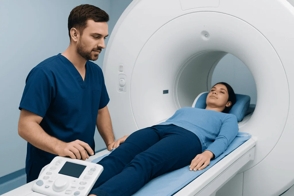
MRI tech school can be challenging, but it is achievable for students who are prepared for science-based coursework, structured clinical training and careful attention to safety. The academic component commonly includes anatomy and physiology, medical terminology, patient care, physics concepts and imaging principles. MRI-specific learning also involves understanding how the scanner works and why metallic objects require strict screening.
Many students find the clinical portion demanding in a different way. Clinical placements require punctuality, professionalism, communication and the ability to apply classroom knowledge while caring for real patients. Students must learn to follow detailed protocols, recognize when to ask for help and work effectively with radiologists, nurses and other imaging professionals.
Success is not based only on technical ability. Empathy, organization, clear communication and composure are important because MRI examinations may be difficult for people with claustrophobia, pain, mobility limitations or concerns about contrast material. A program that provides supervised experience and clear safety education can help students build confidence gradually.
What to know before becoming an MRI tech?
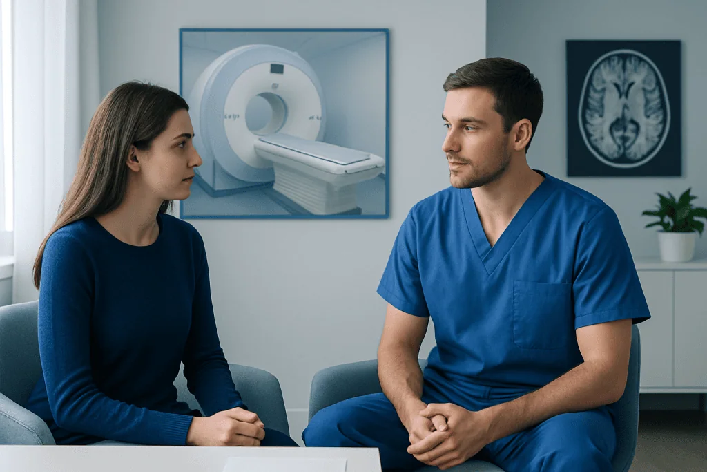
Before becoming an MRI tech, it is helpful to understand that training and licensing or certification requirements vary by country, state and employer. In the United States, many MRI technologists first qualify in radiography or another imaging discipline and then complete MRI-focused education and certification. Some programs offer a more direct route into MRI, depending on local regulations and accreditation standards.
MRI tech school prerequisites, sometimes searched as MRI tech school pre reqs, often include a high school diploma or equivalent and college-level courses such as anatomy and physiology, mathematics, English composition and medical terminology. Programs may also ask applicants to complete background checks, health screening, immunization documentation, cardiopulmonary resuscitation training or observation hours before clinical placement.
Prospective students should compare programs carefully. Useful questions include whether the program is appropriately accredited, how many clinical hours are offered, whether students have access to current MRI equipment, what certification pathway it supports and whether graduates meet local eligibility requirements. Speaking directly with the school and the relevant professional licensing or certification body can clarify the pathway.
- Expect to work around strong magnetic fields and follow strict safety procedures every day.
- Be prepared to assist patients with transfers, positioning and comfort measures within the limits of training and workplace policy.
- Understand that some shifts may include evenings, weekends or urgent examinations, especially in hospitals.
- Choose a program that emphasizes patient-centered care as well as imaging quality.
MRI Procedure Preparation: How an MRI Examination Works
For patients, MRI procedure preparation begins with a safety questionnaire. The care team needs to know about implanted devices, previous surgery, metal fragments, medication patches, pregnancy, kidney problems and prior reactions to contrast material. Many implants are MRI-compatible under specific conditions, but this must be confirmed before entering the scan room.
During the examination, the patient lies on a padded table that moves into the MRI scanner. A receiving device called a coil may be placed around the area being scanned. The machine produces loud tapping or knocking sounds, so ear protection is provided. The patient can usually communicate with the technologist through an intercom and may receive instructions to remain still or briefly hold their breath.
Some examinations use a contrast agent, commonly gadolinium-based, to improve visibility of certain tissues or blood vessels. The clinician and imaging team determine whether contrast is appropriate based on the reason for the scan, medical history and kidney function when relevant. MRI may be used to investigate symptoms or monitor known concerns, including selected brain, spine, musculoskeletal and vascular conditions; for example, imaging can form part of assessment for brain tumors or multiple sclerosis when a clinician considers it appropriate.
MRI prep requirements differ by the body area and whether contrast, sedation or a specialized scan is planned. Patients should follow the instructions provided by their imaging center rather than relying on general advice alone. They may be asked to avoid food for a period before certain scans, remove jewelry and cosmetics containing metallic particles, and arrange an escort if sedation is used.
How to prepare for MRI results?
Preparing for MRI results can make the follow-up conversation more useful and less stressful. Patients may wish to write down the symptoms that led to the scan, when they began, what makes them better or worse, and any previous imaging or treatment. It can also help to bring a current medication list and relevant medical records to the appointment where results will be discussed.
After the scan, a radiologist interprets the images and prepares a report. The ordering clinician places the findings in context with the person’s symptoms, examination and other test results. An MRI finding is not always a diagnosis by itself, and some changes may be incidental, meaning they are not related to the reason for the scan.
People can ask practical questions such as: What did the MRI show? Does it explain the symptoms? Are more tests needed? What treatment or monitoring is recommended? If the result identifies a condition affecting the spine, specialist evaluation and options such as spine surgery may be considered only when clinically appropriate; many findings are managed with observation, rehabilitation, medication or other non-surgical approaches.
Result timing varies. Urgent findings are generally communicated promptly according to local procedures, while routine reports may take longer. If a patient has not received expected information, contacting the ordering clinician or imaging center is reasonable.
Benefits, Risks and Recovery After MRI
The main benefit of MRI is its ability to provide detailed images of soft tissues without ionizing radiation. It can help clinicians evaluate the brain, spinal cord, muscles, ligaments, joints, abdominal organs and blood vessels. Accurate imaging may guide next steps, such as monitoring, medication, rehabilitation or referral to a specialist.
MRI is generally well tolerated, and most people can return to normal activities immediately after a standard scan. There is no recovery period from the magnetic field itself. If contrast material is used, patients are usually observed briefly according to local practice and should report symptoms that occur after leaving. If sedation is needed because of severe anxiety or inability to remain still, recovery instructions may include rest, avoiding driving and having a responsible adult accompany the patient home.
Important MRI risks are largely related to the strong magnet, contrast use and the need to lie still in an enclosed space. Undisclosed metal can become hazardous or interfere with imaging, which is why screening is essential. Some people experience anxiety, noise discomfort or warmth. Serious reactions to modern MRI contrast agents are uncommon, but anyone with kidney disease, previous contrast reactions or pregnancy should discuss this with the care team in advance.
For people whose symptoms suggest a condition requiring a broader plan, MRI is only one part of care. Depending on the diagnosis, teams may discuss options such as physical therapy and rehabilitation alongside other treatments.
Can MRI techs make 200k a year?
It is possible for a small number of MRI technologists to reach very high annual earnings, but an income of 200,000 dollars per year is not typical for the profession. Pay depends on location, demand for specialized imaging staff, experience, certification, shift differentials, overtime, travel assignments and the employer’s pay structure.
Hospital-based roles may offer different compensation from outpatient imaging centers, academic institutions or temporary staffing assignments. Working nights, weekends or emergency coverage may increase earnings in some settings, although these schedules can also affect work-life balance. Compensation can change over time, so students should use current local labor-market information rather than relying on broad claims online.
Career decisions are often more sustainable when based on interest in patient care, safety-focused technology and clinical teamwork, not salary alone. Continuing education, reliable clinical skills and recognized credentials can support career progression, while local rules determine the qualifications needed for independent practice.
When to Seek Medical Care
Medical care should be sought promptly for sudden or severe symptoms such as weakness on one side of the body, difficulty speaking, new loss of vision, a seizure, severe headache unlike previous headaches, chest pain, serious injury or trouble breathing. These symptoms may require urgent assessment and should not wait for a routine MRI appointment or result.
People should contact the imaging center or ordering clinician before an MRI if they have an implanted device, possible metal in the eye or body, severe kidney disease, pregnancy, a previous contrast reaction or intense claustrophobia. The team can review safety information and discuss whether a different approach, additional preparation or another imaging method is suitable.
Acibadem International’s multidisciplinary specialists and JCI-accredited hospitals support international patients who need diagnostic assessment and treatment planning. A qualified clinician can explain whether MRI is appropriate, interpret results in context and recommend the next safe step.
Frequently asked questions
How long does MRI tech school take?
The length varies by the entry route, program type and local requirements. Some students complete a radiography qualification before specializing in MRI, while others enter an MRI-focused program. Clinical training and certification preparation are important parts of the overall timeline.
Do MRI techs need to be radiologic technologists first?
Not always. In some regions and programs, MRI is an advanced credential obtained after radiography training, while other pathways permit direct MRI education. Students should confirm the rules of the licensing authority, credentialing organization and employers in the place where they intend to work.
What are common MRI tech school prerequisites?
Common prerequisites include anatomy and physiology, medical terminology, mathematics, communication coursework and health screening requirements for clinical practice. Individual schools may also require observation hours, immunizations, background checks or entrance assessments. Requirements should be checked directly with each program.
Does an MRI scan hurt?
An MRI scan is usually painless, although staying still may be uncomfortable if a person has pain or limited mobility. The scanner is loud, and some people find the enclosed space difficult. The technologist can provide ear protection, communication support and positioning aids when appropriate.
How long does it take to get MRI results?
The timing depends on the urgency of the examination, the imaging center and how quickly the radiologist can finalize the report. The ordering clinician usually discusses the findings and what they mean for the individual. Urgent results are handled through local clinical pathways.
Can someone have an MRI with a pacemaker or metal implant?
Some pacemakers, joint replacements, surgical clips and other implants can be scanned under specific conditions, while others may not be suitable. The exact device model and implantation details must be reviewed before the scan. Patients should never assume an implant is safe without confirmation from the MRI team.
References
- Radiological Society of North America
- American College of Radiology
- American Registry of Radiologic Technologists
- National Institute of Biomedical Imaging and Bioengineering
- U.S. Food and Drug Administration
This article is for general information only and is not a substitute for professional medical advice. Please consult a qualified doctor about your individual situation.
Joint and spine care in Turkey — expert assessment & treatment
JCI-accredited · board-certified surgeons · reply within 24h
Add us as a Preferred Source to see more of our trusted health content across Google Search, AI Overviews and Discover.