

Nerves in Eyes Swollen: Common Causes, Related Conditions, and When to See a Doctor

A swollen optic nerve is a clinical finding, not a diagnosis by itself. Papilledema specifically means optic disc swelling caused by raised pressure inside the skull.
Key Takeaways
- A swollen optic nerve is a clinical finding, not a diagnosis by itself.
- Papilledema specifically means optic disc swelling caused by raised pressure inside the skull.
- Optic neuritis is inflammation of the optic nerve and often causes pain with eye movement or changes in color vision.
- An eye examination, vision testing, imaging, and sometimes blood tests or a lumbar puncture may be needed to identify the cause.
- Sudden vision loss, severe headache, new neurological symptoms, or symptoms after head injury require urgent medical care.
“Nerves in eyes swollen” commonly describes swelling at the optic nerve head, the area where the optic nerve enters the back of the eye. This finding can have causes ranging from inflammation and infection to raised pressure around the brain, so a timely examination by an eye specialist is important even when vision seems normal.
What Does “Nerves in Eyes Swollen” Mean?
People may use the phrase “nerves in eyes swollen” after being told that the optic nerve looks swollen during an eye examination. The optic nerve is not visible in its full length, but its front portion can be seen at the back of the eye as the optic disc. When this area appears elevated, blurred, or congested, clinicians may describe it as optic disc swelling.
Optic disc swelling can affect one eye or both. It is important because the optic nerve carries visual information from the eye to the brain. Swelling does not always mean that permanent damage will occur, but it can signal a condition that needs prompt assessment to preserve vision and address the underlying cause.
The terms used in this area can be confusing. Papilledema is optic disc swelling caused specifically by increased pressure inside the skull. Optic neuritis means inflammation of the optic nerve. Other conditions can resemble true swelling, including an anatomical variation called optic disc drusen. A careful assessment helps distinguish these possibilities.
Symptoms That May Occur
Some people with optic nerve swelling have no obvious symptoms and learn about it during a routine eye test. Others notice blurred or dim vision, reduced side vision, brief episodes of vision darkening, double vision, or difficulty seeing colors as vividly as before. Symptoms can develop gradually or more suddenly depending on the cause.
With optic neuritis, common features include reduced vision in one eye, discomfort or pain that is worse when moving the eye, and colors appearing faded. In contrast, papilledema may initially leave central vision relatively clear but can cause short episodes of visual dimming, often with changes in posture, coughing, or straining.
Headache, nausea, vomiting, ringing in the ears that seems to follow the heartbeat, weakness, numbness, imbalance, fever, or neck stiffness may provide important clues about the cause. These symptoms do not automatically indicate a serious condition, but they should be shared clearly with a clinician. The presence or absence of pain alone cannot reliably determine the cause.
- Blurred, dim, or reduced vision
- Brief episodes of vision loss or greying-out
- Pain with eye movement
- Changes in color vision or contrast
- Headache, nausea, double vision, or pulsating ear noise
Common Causes and Related Conditions

Raised pressure inside the skull is an important cause of swelling in both optic discs. This can occur for several reasons, including idiopathic intracranial hypertension, a condition in which pressure rises without a mass lesion being identified. It may also result from problems that affect the normal circulation or drainage of cerebrospinal fluid, bleeding, a brain mass, certain infections, or blood clots in veins that drain the brain.
Inflammation of the optic nerve, known as optic neuritis, is another cause. It may occur after some infections, alongside autoimmune or inflammatory conditions, or as part of disorders affecting myelin, the protective covering around nerves. Not every case has an identifiable trigger. Optic neuritis often affects one eye, but the pattern varies by person and cause.
Other possibilities include reduced blood supply to the optic nerve, sometimes called ischemic optic neuropathy; severe high blood pressure; infections involving the eye or nervous system; infiltrative conditions; medication-related effects; and injury. Diabetes, vascular risk factors, sleep apnea, smoking, and some medicines may be relevant in selected cases, although they do not explain every presentation.
Finally, not every elevated-looking optic disc is truly swollen. Optic disc drusen are small deposits within the optic nerve head that can create a similar appearance, sometimes called pseudopapilledema. Differentiating true swelling from a look-alike is essential because the investigations and urgency may be different.
How Doctors Evaluate Optic Nerve Swelling
Assessment usually starts with a detailed history. The clinician will ask when symptoms began, whether one or both eyes are affected, whether pain occurs with eye movement, and whether there are headaches or neurological symptoms. Medical conditions, recent infections, pregnancy status where relevant, medicines, supplements, and family history can also help guide evaluation.
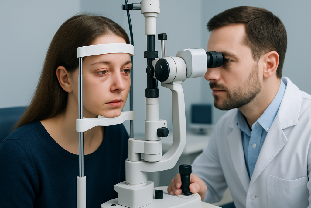
A comprehensive eye examination may include checking visual acuity, color vision, pupils, eye movements, and peripheral vision. The clinician examines the optic disc after dilating the pupils when appropriate. Optical coherence tomography, or OCT, can measure layers of the retina and optic nerve, while visual field testing can detect changes that may not be noticed in daily life. Eye photographs may be used to monitor change over time.
If true optic disc swelling is suspected, further tests may be needed promptly. Depending on the clinical picture, these can include blood pressure measurement, blood tests, magnetic resonance imaging or other brain imaging, and imaging of the blood vessels. After imaging has excluded certain causes, a lumbar puncture may be recommended to measure cerebrospinal fluid pressure and test the fluid when infection or inflammation is a concern.
These tests are selected individually rather than routinely for every person. Their purpose is to identify the cause safely, distinguish urgent conditions from less concerning look-alikes, and create an appropriate treatment plan.
Treatment Depends on the Underlying Cause
There is no single treatment for a swollen optic nerve because management is directed at the cause. If increased pressure inside the skull is identified, treatment may involve addressing the source of the pressure, using medicines that reduce cerebrospinal fluid production in suitable patients, and monitoring vision closely. In some circumstances, procedures or surgery may be needed to protect vision or treat the underlying problem.
For optic neuritis, a specialist considers the severity of vision loss, examination findings, imaging results, and the possibility of related inflammatory or infectious conditions. Some people improve over time, while others may benefit from corticosteroid treatment or other targeted therapies. Steroids are not appropriate for every type of optic nerve problem and should only be used under medical supervision.
When the cause is high blood pressure, infection, impaired blood flow, or a medication effect, treatment focuses on that condition. Rapid control of severely elevated blood pressure, appropriate antimicrobial treatment when infection is confirmed or strongly suspected, and review of contributing medicines can be important parts of care.
Follow-up eye testing is often needed even after treatment begins. Vision, visual fields, and optic nerve appearance may change at different rates, and regular review allows the care team to adjust the plan. Acibadem International’s multidisciplinary specialists and JCI-accredited hospitals evaluate and treat optic nerve conditions for international patients when coordinated eye and neurological care is needed.
Self-Care and What Not to Do
Optic nerve swelling should not be managed with home remedies alone. Rest, hydration, and reducing screen exposure may ease general eye strain or headache discomfort, but they do not treat swelling of the optic nerve. It is safer to arrange a professional assessment than to wait for symptoms to settle, particularly if there are changes in vision.
People should not start, stop, or change prescribed medicines without speaking to the clinician who manages their care. This includes corticosteroids, blood pressure medicines, antibiotics, weight-loss products, vitamin A-containing supplements, and hormonal medicines. Some medicines and supplements may be relevant to raised intracranial pressure or other causes, but decisions should be individualized.
Keeping a short symptom record can be helpful. It may include the timing of visual changes, headache pattern, eye pain, recent illnesses, and all medicines or supplements used. If vision is impaired, avoiding driving and arranging support for travel to appointments can reduce risk.
Healthy habits such as managing blood pressure, avoiding smoking, attending diabetes care appointments, and following advice about sleep apnea can support overall eye and vascular health. However, these steps do not replace diagnostic testing when optic nerve swelling is suspected.
When to Seek Medical Care
Any new diagnosis or suspicion of optic nerve swelling should be assessed promptly by an ophthalmologist, optometrist with urgent referral pathways, or another qualified clinician. Even without pain or major visual symptoms, swelling can occasionally reflect raised pressure or another condition that requires timely treatment.
Urgent or emergency assessment is appropriate for sudden or rapidly worsening vision loss, a severe new headache, persistent vomiting, confusion, fainting, weakness, numbness, trouble speaking, seizures, fever with neck stiffness, or symptoms after a head injury. New double vision or severe eye pain should also be assessed without delay.
People who are already being monitored should contact their care team sooner if vision becomes dimmer, color vision changes, headaches worsen, or new neurological symptoms appear. Early review is especially important during pregnancy, after a recent infection, or when there is a history of inflammatory neurological disease.
Most causes can be evaluated systematically, and many can be treated or monitored effectively. The key step is not to assume that a swollen-looking optic nerve is simply eye strain or fatigue; an appropriate examination can clarify what is happening and guide the next steps.
Frequently asked questions
Can swollen nerves in the eyes go away?
Optic nerve swelling may improve when its underlying cause is identified and treated. Recovery depends on the cause, how quickly treatment begins, and whether the optic nerve has been significantly affected. Follow-up examinations are important because visual recovery and optic disc appearance may not improve at the same pace.
Is optic nerve swelling always an emergency?
Not every case is an emergency, but it should be evaluated promptly because some causes require urgent treatment. Sudden vision loss, severe headache, neurological symptoms, fever with neck stiffness, or symptoms following head injury need emergency assessment. A clinician can determine the appropriate level of urgency after examining the eyes and reviewing symptoms.
What is the difference between papilledema and optic neuritis?
Papilledema is optic disc swelling caused by increased pressure inside the skull, and it often affects both eyes. Optic neuritis is inflammation of the optic nerve and commonly causes reduced vision, pain with eye movement, or altered color vision, often in one eye. Both need medical assessment, but their causes and treatments differ.
Can high blood pressure cause swollen optic nerves?
Severely elevated blood pressure can damage retinal blood vessels and may be associated with optic disc swelling. This can be a sign of a hypertensive emergency and needs immediate medical attention. Routine blood pressure checks are therefore an important part of evaluating optic nerve swelling.
Can an eye exam detect pressure in the brain?
An eye examination can show signs that raise concern for increased pressure inside the skull, including papilledema. However, the eye exam alone cannot confirm the cause or directly measure pressure in the brain. Brain imaging and, in selected situations, a lumbar puncture may be needed.
Can screen time cause optic nerve swelling?
Screen use can contribute to dry eyes, eye strain, and headaches, but it does not usually cause true optic nerve swelling. If an examination has suggested a swollen optic nerve, it should not be attributed to screen time without a proper medical evaluation. Taking screen breaks may improve comfort but does not replace necessary testing.
References
- American Academy of Ophthalmology
- National Eye Institute
- Mayo Clinic
- National Institute of Neurological Disorders and Stroke
- Merck Manual Professional Edition
This article is for general information only and is not a substitute for professional medical advice. Please consult a qualified doctor about your individual situation.
Explore treatments in Turkey — costs, top hospitals & a free quote
JCI-accredited · board-certified surgeons · reply within 24h
Add us as a Preferred Source to see more of our trusted health content across Google Search, AI Overviews and Discover.
Check your numbers in seconds
BMI, calories, due date, blood pressure and 30+ more clinical calculators — free, instant, doctor-reviewed ranges.

More from the Health Library



Related Specialists

Prof. Dr. Sefa Müezzinoğlu
Orthopedic Surgery & Traumatology
Dr. Numan Doğu Güner
Urology
Prof. Dr. Abdullah Büyükçelik
Medical Oncology
Assoc. Prof. Dr. Emre Özgü
Gynecology & Obstetrics