Non Ossifying Fibroma MRI: Preparation, Procedure and Results
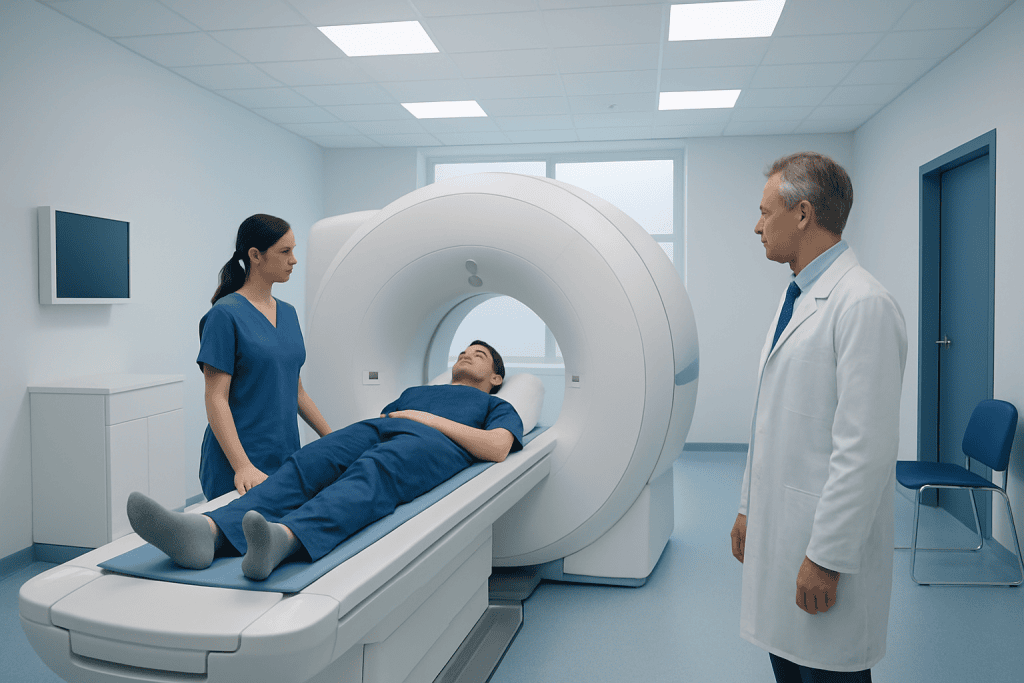
MRI can clarify the size, location and internal features of a non-ossifying fibroma, but an X-ray is often sufficient for a typical lesion. Non-ossifying fibromas are benign and do not usually become cancerous.
Key Takeaways
- MRI can clarify the size, location and internal features of a non-ossifying fibroma, but an X-ray is often sufficient for a typical lesion.
- Non-ossifying fibromas are benign and do not usually become cancerous.
- Most lesions need observation rather than treatment, although larger lesions may increase the chance of a bone fracture.
- MRI uses a magnetic field and radio waves, not ionizing radiation.
- New pain, limping, swelling or an injury followed by severe pain should be assessed by a clinician.
A non ossifying fibroma MRI is a detailed, radiation-free scan used when an X-ray does not fully explain a bone lesion, symptoms are present, or clinicians need to assess bone strength. Most non-ossifying fibromas are harmless developmental lesions in children and adolescents and gradually resolve as the person grows.
Non Ossifying Fibroma MRI: Preparation, Procedure and Results
A non ossifying fibroma MRI is an imaging examination that provides detailed pictures of a bone lesion and the surrounding bone marrow, muscles and soft tissues. It is usually requested when the appearance on an X-ray is not fully typical, when there is persistent pain, or when a clinician needs to determine whether a larger lesion may weaken the bone.
Non-ossifying fibroma, also called NOF, is a common benign developmental bone lesion seen mainly in children and adolescents. It most often affects the long bones around the knee, including the femur and tibia. MRI does not use radiation and can help the care team distinguish an NOF from other bone findings while planning appropriate follow-up.
For many people, the result is reassuring: the lesion has typical benign features and can be monitored over time. MRI findings are interpreted together with age, symptoms, examination findings and X-rays, rather than in isolation.
How MRI Works and Who May Need It
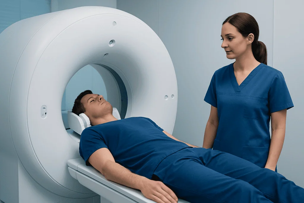
MRI uses a strong magnet and radiofrequency waves to create cross-sectional images of the body. Different MRI sequences highlight fluid, fat, bone marrow and soft tissue in different ways. Although MRI is excellent for examining marrow and surrounding tissues, a plain X-ray often shows the classic shape and border of a non-ossifying fibroma particularly well.
An MRI may be appropriate when a lesion is unusually large, has features that are not fully characteristic on X-ray, causes ongoing symptoms, or is located where bone stability is a concern. It may also be used after a fracture through an area containing an NOF, to assess the extent of injury and guide orthopedic care.
People with certain implanted devices, metal fragments, some older aneurysm clips, or severe kidney impairment should tell the imaging team beforehand. Most MRI scans for suspected NOF are performed without contrast. If contrast is considered helpful, the radiology team will explain why and check for relevant health concerns.
Preparing for a Non Ossifying Fibroma MRI
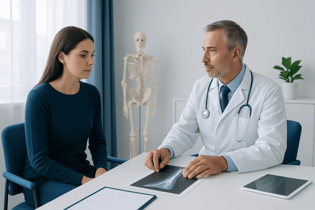
Preparation is usually simple. Unless sedation or contrast has been planned, people can generally eat, drink and take regular medicines as usual. The imaging center will provide specific instructions if a child needs sedation because they cannot remain still during the scan; fasting may then be required for safety.
Before entering the scanner room, all metal items must be removed, including jewelry, watches, hair accessories, hearing aids and removable dental appliances. Patients should tell staff about any implants, prior surgery, medical devices, pregnancy, allergies, kidney disease, or possible metal exposure at work or from an earlier injury.
MRI scanners can be noisy and enclosed. Ear protection is supplied, and many centers offer music or a call button for communication. Parents or carers may be able to remain nearby for a child, depending on local safety policies.
What Happens During the Procedure and Recovery
At the appointment, a radiographer confirms safety information and positions the affected limb on the scanning table. A specialized coil may be placed around the area to improve image quality. The person lies still while the table moves into the scanner; only the body part being examined may need to be centered in the magnet.
The scan commonly takes about 20 to 45 minutes, depending on the body area and the number of image sequences needed. The machine makes rhythmic knocking and tapping sounds, but MRI is painless. It is important to remain as still as possible because movement can blur the images.
There is usually no recovery time after a standard MRI, and normal activities can resume immediately. If sedation was used, the person needs observation until fully awake and should follow the discharge instructions regarding eating, activity and supervision. A radiologist reviews the images and sends a report to the referring clinician, who discusses the results and next steps.
What Is the MRI Appearance of Non-Ossifying Fibroma?
On MRI, a non-ossifying fibroma is typically located near the outer portion of a long bone, often close to a growth plate in younger people. It is usually well defined and contained within the bone, with a thin rim of denser bone around it. The lesion does not generally extend aggressively into surrounding soft tissues.
The signal appearance varies with the stage of the lesion and the mixture of fibrous tissue, mineralization and healing changes. It commonly appears relatively low in signal on T1-weighted images and variable, often low to intermediate, on T2-weighted images. MRI may also show whether the lesion occupies enough of the bone width to raise concern about structural strength.
Radiologists look for reassuring features such as a sharp margin, intact outer bone layer and absence of a soft-tissue mass. If the images are not typical, further assessment may include comparison with X-rays, follow-up imaging, or referral to an orthopedic specialist with experience in bone lesions.
Is Non-Ossifying Fibroma Serious?
Non-ossifying fibroma is not cancer and is usually not serious. It commonly causes no symptoms and is discovered incidentally after an X-ray is taken for a different reason, such as a sports injury. The lesion is considered a developmental variation in bone growth rather than a malignant tumor.
The main potential complication is a pathologic fracture, meaning a break in bone weakened by a large lesion. This is uncommon, but the likelihood may be higher when an NOF occupies a substantial part of a weight-bearing bone. Pain after a fall, sudden inability to bear weight, or a new deformity requires prompt medical assessment.
For a typical, small lesion without symptoms, reassurance and periodic clinical or X-ray follow-up may be all that is needed. Decisions are individualized according to the lesion’s size, location, imaging appearance, activity level and symptoms.
What Can Be Mistaken for a Non-Ossifying Fibroma?
Several benign bone lesions can sometimes resemble a non-ossifying fibroma, especially when the imaging appearance is not classic. Depending on the patient’s age and the bone involved, the differential diagnosis may include fibrous cortical defect, fibrous dysplasia, simple bone cyst, aneurysmal bone cyst, chondroblastoma or other fibrous lesions.
Less commonly, infection, trauma-related changes or a more aggressive tumor may need to be considered when there are unusual imaging features, marked pain, systemic symptoms or a lesion that does not match the expected pattern. This does not mean these alternatives are likely; it means careful image interpretation is important when findings are uncertain.
MRI helps evaluate the marrow and surrounding soft tissues, while X-rays provide important information about the lesion’s border and effect on the bone cortex. A musculoskeletal radiologist and orthopedic specialist can combine these findings to decide whether observation, repeat imaging or further tests are appropriate.
Can Nonossifying Fibroma Heal on Its Own?
Yes. Most non-ossifying fibromas gradually heal or become less visible on their own as a child reaches skeletal maturity. Over time, the lesion often becomes more mineralized and is replaced by normal bone. This process can take years and is usually monitored with occasional X-rays when follow-up is needed.
Surgery is not routinely required. If a lesion is large, painful, associated with a fracture or considered to create a significant fracture risk, an orthopedic surgeon may discuss treatment such as curettage, in which the lesion is removed from the bone, and bone grafting or other stabilization when necessary.
The benefits of MRI are detailed assessment without radiation and better evaluation of possible bone weakness or alternative diagnoses. Limitations include cost, noise, the need to remain still, and occasional need for sedation in young children. Contrast reactions are uncommon when contrast is used, and the imaging team takes safety precautions before administering it.
When to Seek Medical Care
Medical review is appropriate for persistent or worsening bone pain, a limp, swelling near a bone, reduced ability to use a limb, or pain that wakes a child from sleep. Evaluation is also important after an injury if there is severe pain, inability to bear weight, visible deformity or concern for a fracture.
Most NOFs are managed by pediatric orthopedics or orthopedic specialists in collaboration with radiologists. Acibadem International’s multidisciplinary specialists and JCI-accredited hospitals evaluate bone lesions and coordinate imaging, orthopedic review and follow-up for international patients.
Families should avoid assuming that every bone lesion is an NOF based on symptoms or an online image. A qualified clinician should interpret the scan and explain whether activity changes, follow-up imaging or treatment is needed.
Frequently asked questions
Do all non-ossifying fibromas need an MRI?
No. Many non-ossifying fibromas have a typical appearance on X-ray, and no MRI is necessary. MRI is most useful when the lesion is large, painful, atypical in appearance, linked to a fracture, or needs more detailed assessment.
Does a non ossifying fibroma MRI use radiation?
No. MRI uses magnetic fields and radio waves rather than ionizing radiation. This can be particularly helpful when imaging children and adolescents, although the scan may take longer than an X-ray.
Will a non ossifying fibroma show up on MRI?
Yes. MRI can show the lesion, its border, its effect on nearby bone marrow and whether there is surrounding soft-tissue change. X-rays remain important because they often demonstrate the typical location and shape of an NOF clearly.
Can a child play sports with a non-ossifying fibroma?
Many children can continue normal activities when the lesion is small and the bone is not significantly weakened. Activity advice should come from the treating clinician, particularly for large lesions in weight-bearing bones or after pain or a fracture.
Is biopsy needed for non-ossifying fibroma?
Biopsy is usually not needed when the lesion has classic clinical and imaging features. It may be considered only when imaging is uncertain or there are atypical features that require a definitive diagnosis.
How long does it take to get MRI results for an NOF?
Timing varies by imaging center and clinical urgency. A radiologist reviews the scan and sends a report to the referring clinician, who explains the result in the context of the person’s symptoms, X-rays and examination.
References
- American Academy of Orthopaedic Surgeons
- Radiopaedia
- Royal College of Radiologists
- National Institute of Arthritis and Musculoskeletal and Skin Diseases
This article is for general information only and is not a substitute for professional medical advice. Please consult a qualified doctor about your individual situation.
Explore treatments in Turkey — costs, top hospitals & a free quote
JCI-accredited · board-certified surgeons · reply within 24h
Add us as a Preferred Source to see more of our trusted health content across Google Search, AI Overviews and Discover.

More from the Health Library



Related Specialists

Prof. Dr. Şule Arslan
Physical Medicine & Rehabilitation
Prof. Dr. İbrahim Kaya
Orthopedic Surgery & Traumatology
Dr. Şahiner Sezgin
Internal Medicine
Assoc. Prof. Dr. Yüksel Erdal
Neurology