Normal Mammogram Images: A Complete Medical Overview
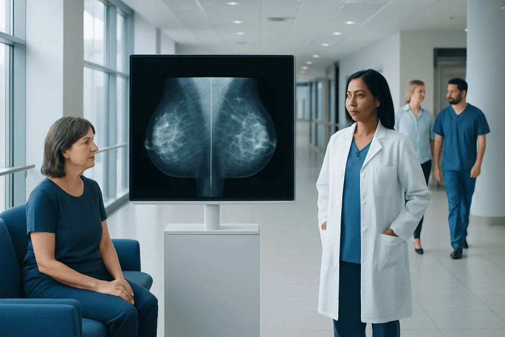
A normal mammogram means the radiologist did not identify a suspicious abnormality requiring further assessment at that time. Mammograms look different from person to person because normal breast tissue can contain varying amounts of fat and dense glandular tissue.
Key Takeaways
- A normal mammogram means the radiologist did not identify a suspicious abnormality requiring further assessment at that time.
- Mammograms look different from person to person because normal breast tissue can contain varying amounts of fat and dense glandular tissue.
- Breast density can make small changes harder to see on a mammogram and may influence recommendations for additional screening.
- A normal screening result does not replace medical evaluation for a new lump, nipple change, skin change, or persistent breast symptom.
- Comparing current images with prior mammograms helps radiologists identify whether an area is stable or newly changed.
Normal mammogram images generally show breast tissue without findings that appear suspicious for cancer. However, a normal result should always be understood alongside the radiology report, breast density, personal risk factors, and any new breast symptoms.
Overview: What Normal Mammogram Images Mean
Normal mammogram images are X-ray pictures of the breasts in which the radiologist does not see a finding that looks suspicious enough to require additional imaging or biopsy. The report may be described as negative, normal, or as showing benign changes. It does not mean that every breast looks identical or that there is no normal variation in breast tissue.
Mammography is used mainly for breast cancer screening and for investigating symptoms or clinical findings. Images are usually taken from two standard views of each breast, allowing the radiologist to examine the breast tissue from different angles. Digital mammography and tomosynthesis, also called 3D mammography, can provide detailed views that may improve assessment in some people.
A normal result is reassuring, but it is a point-in-time assessment rather than a lifetime guarantee. Mammograms can miss some cancers, particularly in dense breasts, and a person should still report any new or persistent breast changes to a healthcare professional.
How Radiologists Read Mammogram Images
On a mammogram, fat usually appears darker, while denser tissue such as glands, milk ducts, connective tissue, and many benign or abnormal findings appears lighter or white. Because cancers can also appear white, the radiologist carefully evaluates the shape, edges, location, and pattern of denser areas rather than relying on color alone.
Normal images may show a mixture of fatty and fibroglandular tissue, lymph nodes in expected locations, and symmetrical structures. It is common for the two breasts not to be perfectly identical. What matters is whether there is a new asymmetry, a distorted area, a suspicious mass, or a concerning pattern of calcifications.
Comparison with earlier mammograms is particularly valuable. A stable finding that has looked the same over several years is often reassuring, whereas a new or changing area may need closer evaluation. For this reason, patients should tell the imaging center where previous mammograms were performed whenever possible.
- Negative result: no suspicious finding is identified.
- Benign result: a non-cancerous finding, such as a typical calcification pattern, may be noted.
- Incomplete result: extra images or ultrasound are needed before a final assessment can be made.
Breast Density and Normal Variation

Breast density describes the proportion of fibroglandular tissue compared with fatty tissue seen on a mammogram. It is determined by the radiologist, not by how the breasts look or feel. Breasts may be described as almost entirely fatty, having scattered areas of density, heterogeneously dense, or extremely dense.
Dense tissue is common and normal, especially in younger people and in those influenced by hormonal factors. It can make mammograms more difficult to interpret because both dense tissue and many cancers appear light on the image. Dense breasts are also associated with a modestly higher breast cancer risk, although density alone does not determine whether someone will develop cancer.
A report may include information about breast density and, depending on the person’s overall risk, may suggest discussing supplemental screening with a doctor. This could include breast ultrasound, MRI, or other imaging in selected situations. Additional tests are not automatically needed for every person with dense breasts; the decision should consider family history, genetic factors, prior chest radiation, and other individual risks.
What a Mammogram Report Can Include
The final report records the imaging findings and usually provides a BI-RADS assessment category, a standardized system developed by the American College of Radiology. For routine screening, BI-RADS 1 means negative and BI-RADS 2 means benign findings. Both are generally considered reassuring results, with a recommendation to continue appropriate routine screening.
A BI-RADS 0 assessment does not mean cancer. It means the radiologist needs additional mammographic views, ultrasound, prior images, or other information to complete the evaluation. Call-backs after screening are common and often lead to benign explanations such as overlapping tissue, cysts, or harmless calcifications.
The report may also mention findings such as benign calcifications, cysts, fibroadenomas, surgical clips, or stable post-treatment changes. If a patient has questions about wording in a report, the referring clinician or breast imaging team can explain what it means in the context of the individual’s health history.
For people who have symptoms or an abnormal clinical examination, a normal screening mammogram may not be the final step. Diagnostic mammography, targeted ultrasound, or other testing may be recommended to investigate the specific concern. Information about breast cancer can also help patients understand why a complete assessment is important when symptoms persist.
Why a Normal Mammogram May Still Need Follow-Up
A normal mammogram is highly useful, but no screening test detects every cancer. Very small cancers may not yet be visible, and dense breast tissue can obscure an abnormality. Some breast cancers also do not form a clear mass or may develop between regularly scheduled mammograms.
Follow-up recommendations depend on whether the mammogram was performed for routine screening or because of a symptom. A person with a new palpable lump, focal breast pain, nipple discharge, skin thickening, or a change in breast shape should seek clinical advice even if a recent mammogram was normal. A clinician may recommend a focused examination and additional imaging based on the location and nature of the change.
People at increased risk may need a screening plan that differs from the usual population schedule. Risk can be increased by certain inherited gene changes, a strong family history, personal history of breast cancer or high-risk lesions, or radiation therapy to the chest at a young age. A doctor can help assess risk and discuss whether breast MRI or other screening approaches are appropriate.
Preparing for Mammography and Supporting Clear Images
For the clearest possible images, patients should follow the instructions provided by their imaging center. It is usually helpful to avoid deodorant, powder, lotion, or perfume on the chest and underarm area on the day of the examination, as some products can leave particles that resemble calcifications on an image.
During mammography, the breast is briefly compressed between imaging plates. Compression spreads the tissue, reduces movement, and lowers the radiation dose needed to produce a clear image. The pressure can be uncomfortable for some people, but it is temporary. Patients should tell the technologist if they have pain, mobility limitations, breast implants, recent surgery, or concerns about positioning.
Scheduling at a time when the breasts are less tender may improve comfort for people who menstruate. Bringing details of prior mammograms, breast procedures, relevant family history, and current symptoms also helps the imaging team interpret the study accurately. Routine screening intervals should be decided with a qualified clinician according to age, risk, local guidance, and personal preferences.
When to Seek Medical Care
Medical care should be sought promptly for a new breast lump or thickening, a lump in the armpit, bloody or spontaneous nipple discharge, nipple inversion that is new, skin dimpling, persistent redness, or an unexplained change in breast size or shape. These symptoms often have non-cancerous causes, but they should be assessed rather than monitored indefinitely at home.
Persistent pain in one specific area of the breast, especially when associated with a palpable change, also deserves medical advice. A normal mammogram should not delay evaluation of a symptom that is new, worsening, or not resolving. Urgent assessment is appropriate if there are signs of infection, such as redness, warmth, swelling, fever, or feeling unwell.
For international patients who need evaluation of breast symptoms or an imaging finding, Acibadem International’s multidisciplinary specialists and JCI-accredited hospitals can provide diagnostic assessment and treatment planning. Decisions about further testing, including breast biopsy when clinically indicated, should be made with a breast specialist and radiology team.
Frequently asked questions
What do normal mammogram images look like?
Normal mammogram images vary because each person has a different balance of fatty and dense breast tissue. In general, the radiologist sees no suspicious mass, distortion, or calcification pattern that requires further investigation. A normal image may still include harmless, benign findings.
Does a normal mammogram mean there is no breast cancer?
A normal mammogram means no suspicious abnormality was seen at the time of imaging, which is reassuring. However, mammography cannot identify every cancer, particularly in dense breasts. New or persistent breast symptoms should still be evaluated by a healthcare professional.
What is the difference between BI-RADS 1 and BI-RADS 2?
BI-RADS 1 means the mammogram is negative, with no abnormal finding identified. BI-RADS 2 means a benign, non-cancerous finding is present, such as typical benign calcifications. Both categories usually lead to a recommendation for routine screening at the appropriate interval.
Why was I called back after my mammogram if it may be normal?
A call-back means the radiologist needs additional information, such as extra views, ultrasound, or comparison with prior images. This is not a cancer diagnosis. Many call-backs show overlapping normal tissue or benign changes after more detailed imaging.
Can dense breasts have a normal mammogram?
Yes. Dense breasts can have a normal mammogram, and density itself is common and not a diagnosis. Because density can reduce the visibility of some abnormalities, a doctor may discuss whether additional screening is appropriate based on the person’s full risk profile.
Should I have an ultrasound after a normal mammogram?
Not everyone needs an ultrasound after a normal mammogram. It may be used when there is a specific symptom, a finding needing clarification, dense breast tissue combined with other risk factors, or a clinician’s recommendation. The most suitable approach depends on individual risk and imaging results.
References
- American College of Radiology
- U.S. Food and Drug Administration
- National Cancer Institute
- American Cancer Society
- World Health Organization
This article is for general information only and is not a substitute for professional medical advice. Please consult a qualified doctor about your individual situation.
Oncology care in Turkey — second opinion and treatment plan
JCI-accredited · board-certified surgeons · reply within 24h
Add us as a Preferred Source to see more of our trusted health content across Google Search, AI Overviews and Discover.